Evaluation of the traditional and revised world
health organization classifications of dengue cases
in Brazil
Fa´bio Rocha Lima,IMariana Garcia Croda,IIDaniella Araujo Muniz,IIsabella Trausula Gomes,IKarla Roberta de Moraes Soares,IMonique Rodrigues Cardoso,IRaquel Luciana Angela Marques Tauro,IJulio CrodaI
IFederal University of Grande Dourados, Faculty of Health Sciences, Dourados/MS, Brazil.IIFederal University of Grande Dourados, University Hospital,
Dourados/MS, Brazil.
OBJECTIVE: Dengue is a worldwide public health problem with approximately 50 million cases reported annually. The World Health Organization proposed a revised classification system in 2008 to more effectively identify the patients who are at increased risk of complications from dengue. Few studies have validated this new classification system in clinical practice. We conducted a cross-sectional study of patients hospitalized for dengue in Dourados, Mato Grosso do Sul, Brazil, to evaluate the capacity of the two classification systems for detecting severe cases of dengue.
MATERIALS AND METHODS:We conducted a cross-sectional study of survey data from the medical records of patients admitted to the University Hospital of the Federal University of Grande Dourados under clinical suspicion of dengue during an epidemic from September 2009 to April 2010.
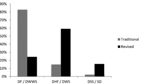
RESULTS:The distribution of patients according to the traditional classification system was as follows: dengue fever, 150/181 (82.9%); dengue hemorrhagic fever, 27/181 (14.9%); and dengue hemorrhagic shock, 4/181 (2.2%). Using the revised classification system, the distribution was as follows: dengue without warning signs, 45/181 (24.3%); dengue with warning signs, 107/181 (59.1%); and severe dengue, 29/181 (15.6%). Of the 150 patients classified as having dengue fever, 105 (70%) were reclassified as having dengue with warning signs or severe dengue.
CONCLUSION:These data demonstrate that the revised classification system has greater discriminatory power for detecting patients at risk of progression to severe disease and those needing hospitalization.
KEYWORDS: Dengue; Dengue Hemorrhagic Fever; Dengue Shock; Severe Dengue; Case Classification.
Lima FR, Croda MG, Muniz DA, Gomes IT, Soares KR, Cardoso MR, et al. Evaluation of the traditional and revised world health organization classifications of dengue cases in Brazil. Clinics. 2013;68(10):1299-1304.
Received for publication onApril 4, 2013;First review completed onApril 26, 2013;Accepted for publication onMay 2, 2013
E-mail: [email protected]
Tel.: 55 67 3410-2321
& INTRODUCTION
Dengue is an important arthropod-borne viral disease found worldwide. Dengue is caused by four serotypes of the genusFlavivirus(DENV-1, DENV-2, DENV-3, and DENV-4) and is transmitted via the bite of theAedes aegyptimosquito, an urban vector that has adapted to human dwellings (1-7). The worldwide incidence of dengue has increased over the past three decades. There are approximately 50 million symptomatic infections per year, and approximately two
million cases require hospitalization, indicating that dengue is a serious global public health concern (8-11).
There is no vaccine or specific treatment for preventing the natural progression of the disease, and it is important that physicians have the appropriate tools to diagnose severe forms of dengue rapidly and accurately. This diagnosis requires a clinical classification system that provides simple and quick detection of patients who may develop more severe disease. It is especially important to detect plasma leakage, which is treated with intravenous rehydration, to reduce the mortality of dengue (8,10,12).
The 1997 World Health Organization (WHO) classifica-tion subdivides dengue into dengue fever (DF), dengue hemorrhagic fever (DHF), and dengue shock syndrome (DSS). The criteria for classifying a patient as having DHF are very rigid; the patient must exhibit four criteria: fever, hemorrhagic manifestations, thrombocytopenia, and evi-dence of third-space fluid loss (serous effusions, hemocon-centration, or hypoproteinemia) (4,6,12).
Copyrightß2013CLINICS– This is an Open Access article distributed under the terms of the Creative Commons Attribution Non-Commercial License (http:// creativecommons.org/licenses/by-nc/3.0/) which permits unrestricted non-commercial use, distribution, and reproduction in any medium, provided the original work is properly cited.
No potential conflict of interest was reported.
Several limitations to the practical applicability of these criteria have been noted, particularly in the evaluation of severe cases. Many patients who develop a severe clinical presentation do not meet the criteria for DHF and are ultimately classified as having dengue fever. In 2008, the WHO proposed a new classification system, based on a multicenter study in Asian and Latin American countries, that provided a list of clinical signs suggestive of a severe disease outcome (warning signs). This system classified dengue into dengue without warning signs (DWWS), dengue with warning signs (DWS), and severe dengue (SD) (4,6,12,13).
The WHO dengue classification of 1997 aims to stratify dengue patients according to the pathophysiological level of disease progression, particularly with respect to the occurrence of plasma leakage. The WHO dengue classifica-tion of 2008 uses clinical signs and symptoms to stratify dengue patients according to disease severity (4,6,12,13).
Based on this perspective, a cross-sectional survey was conducted to evaluate the ability of the two classification systems to detect severe cases of dengue based on the medical records of dengue patients who were admitted to the University Hospital of the Federal University of Grande Dourados during the dengue epidemic in the summers of 2009-2010. This study is the first in Brazil to evaluate the revised WHO classification criteria.
& MATERIAL AND METHODS
Data were collected through patient notification obtained from the National Notifiable Diseases Information System (SINAN) and the records of suspected cases of dengue at the University Hospital of the Federal University of Grande Dourados during September 2009 and April 2010 in Dourados, Mato Grosso do Sul. Indigenous patients and patients whose records could not be located were excluded. From the collected records, a survey of the following epidemiological data was conducted: gender, age, and city of residence. The clinical data obtained included reports of fever, headache, myalgia, arthralgia, retro-orbital pain, skin rash, vomiting, diarrhea, epistaxis, petechiae, gingival bleed-ing, metrorrhagia, hematuria, warning signs, abdominal pain, persistent vomiting, postural hypotension, hepatome-galy, gastrointestinal hemorrhaging, drowsiness/irritability, oliguria, hypothermia, respiratory distress, shock, hypoten-sion, converging blood pressure, cold extremities, cyanosis, rapid and thready pulse, slow capillary refill, and serous effusions (pleural and peritoneal). The laboratory data included a complete blood count (hematocrit, hemoglobin, leukocyte count, and platelet count), liver enzymes (AST and ALT), and liver function tests (albumin, PT, and aPTT). The clinical outcomes included death, admission to the ICU, and the need for blood transfusions (packed red blood cells, plasma, and platelets). The clinical and laboratory data were collected within the first 24 hours of admission.
Dengue patients were classified retrospectively based on warning signs and disease severity. The warning signs were as follows: abdominal pain or tenderness, persistent vomiting, clinical fluid accumulation, mucosal bleeding, lethargy/rest-lessness, liver enlargement greater than 2 cm, and an increase in the hematocrit concurrent with a rapid decrease in platelet count. The criteria used for SD were as follows: shock, fluid accumulation with respiratory distress, severe bleeding, AST or ALT greater than or equal to 1,000, impaired consciousness, and severe involvement of the heart and other organs.
These data were tabulated and analyzed to evaluate the primary variables associated with ICU admission and to compare the traditional and revised WHO clinical classifica-tions regarding the development and clinical severity of disease. ICU admission was used as a proxy for disease severity.
The variables were entered into the database of the Epi-Data program, version 3.0, and analyzed using the SAS statistical software, version 9.1.
The distribution pattern of each variable was evaluated, and the statistical methods were selected based on the specific distribution pattern. The variables that exhibited an asym-metric distribution were evaluated using non-paraasym-metric tests, whereas those that exhibited a normal distribution were evaluated using parametric tests. Categorical or dichotomous data were analyzed using the chi-square test or Fisher’s exact test when the number of subjects per cell was less than 5.
The study was approved by the committee for ethics in research of the Federal University of Grande Dourados under number 01304112.9.0000.5160.
& RESULTS
Between September 2009 and April 2010, 7,827 dengue cases were reported to the SINAN (3,913 cases per 100,000 inhabitants), and 495 (6.32%) patients were hospitalized. A total of 211 patients were admitted to the University Hospital of the Federal University of Grande Dourados. Twenty-four cases were excluded: 22 patient records could not be located, and 2 patients were indigenous people. The study included 187 patients from SINAN who were admitted to the University Hospital of the Federal University of Grande Dourados. Of these, 77 (41.4%) were male. The patients ranged in age from 1 to 90 years, with an average age of 37.1 years. The majority of patients (90.8%) lived in the city of Dourados (Table 1). During the epidemic, not all cases were confirmed; 160/187 (86%) patients underwent serological examination; 12% were negative, 57% were positive, and 31% were inconclusive for dengue. The most common classical symptom of dengue was fever, which was present in 173 patients (98.3%), followed by myalgia (84.5%) and headache (78.1%). Warning signs were present in 108 patients (60.7%). Of these warning signs, the most frequent was abdominal pain, which was present in 92 patients (53.5%). Nineteen patients (11.4%) presented with signs of shock (Table 1).
Based on the traditional classification, the distribution of the patients in the study was 82.9% DF, 14.9% DHF, and 2.2% DSS. The revised classification placed DWWS at 24.3%, DWS at 59.1%, and SD at 15.6% (Figure 1).
variables associated with a specific outcome, such as death or the need for a blood transfusion (Table 5).
& DISCUSSION
This study was conducted during a major dengue epidemic in the state of Mato Grosso do Sul from September 2009 to October 2010. During the first 13 weeks
of the epidemic in 2010, there were 59,199 reported cases of dengue statewide (13.2% of the cases reported nationally). Mato Grosso do Sul had the 4thhighest number of cases of any state in the country and the 2nd highest incidence
(2,507.8 cases per 100,000 inhabitants) (14).
The frequencies of the classical signs and symptoms, hemorrhagic manifestations, and warning signs of dengue fever observed in this study were similar to those of
Table 1 -The distribution of demographic, clinical, and laboratory data and the clinical outcomes of patients hospitalized with suspected dengue at the University Hospital of the Federal University of Grande Dourados from September 2009 to April 2010 (N = 187).
Variable DF DHF and SSD p-value
Demographic data
Gender - Male, N (%) 63/150 (42.0%) 11/31 (35.5%) 0.5017
Age, mean+/- SD 37.8+/- 22.1 33.4+/- 27.6 0.5124a
Clinical data
Fever, N (%) 140/142 (98.6%) 30/30 (100%) 0.5132
Myalgia, N (%) 115/134 (85.8%) 19/24 (79.2%) 0.4029
Headache, N (%) 96/122 (78.7%) 20/26 (76.9%) 0.8426
Vomiting, N (%) 66/114 (57.9%) 19/25 (76.0%) 0.0926
Arthralgia, N (%) 51/97 (52.16%) 6/19 (31.6%) 0.0941
Skin rash, N (%) 44/92 (47.8%) 9/19 (47.4%) 0.9710
Retro-orbital pain, N (%) 42/95 (44.2%) 3/16 (18.8%) 0.0962b
Diarrhea, N (%) 34/98 (34.7%) 11/24 (45.8%) 0.3107
Hemorrhagic signs
Petechiae, N (%) 63/124 (50.8%) 17/31 (54.8%) 0.6878
Epistaxis, N (%) 21/116 (18.1%) 11/30 (36.7%) 0.0285
Gingival bleeding, N (%) 27/121 (22.3%) 6/29 (20.7%) 0.8496
Metrorrhagia, N (%) 15/114 (13.2%) 3/29 (10.3%) 1.0000b
Gastrointestinal hemorrhaging, N (%)
Hematuria, N (%) 7/113 (6.2%) 2/28 (7.1%) 1.0000b
Warning signs, N (%) 80/145 (55.2%) 28/31 (90.3%) 0.0003
Abdominal pain, N (%) 69/140 (49.3%) 23/30 (76.7%) 0.0063
Serous effusions, N (%) 14/136 (10.3%) 13/29 (44.8%) ,0.0001
Respiratory distress, N (%) 11/131 (8.4%) 9/26 (34.6%) 0.0002
Shock, N (%) 12/134 (9.0%) 7/31 (22.6%) 0.0322
Hypotension, N (%) 11/129 (8.5%) 6/30 (20.0%) 0.0670
Drowsiness/irritability, N (%) 6/131 (4.6%) 10/30 (33.3%) ,0.0001
Postural hypotension, N (%) 5/239 (3.9%) 4/36 (15.4%) 0.0438b
Persistent vomiting, N (%) 4/131 (3.1%) 3/29 (13.8%) 0.0366b
Hepatomegaly, N (%) 5/132 (3.8%) 3/29 (10.3%) 0.1562b
Hypothermia, N (%) 2/130 (1.5%) 4/30 (13.3%) 0.0954b
Cyanosis, N (%) 2/127 (1.6%) 3/30 (10.0%) 0.0484b
Oliguria, N (%) 2/130 (1.5%) 3/29 (10.3%) 0.0426b
Rapid and thready pulse, N (%) 2/127 (1.6%) 2/30 (6.7%) 0.1652b
Cold extremities, N (%) 1/127 (0.8%) 2/30 (6.7%) 0.0937b
Slow capillary refill, N (%) 1/127 (0.8%) 1/30 (3.3%) 0.3466b
Converging blood pressure, N (%) 0/127 (0%) 0/30
Laboratory data
Platelet count, mean+/- SD 81,9+/- 87,0 43,8+/- 62,6 0.0219a
Hematocrit, mean+/- SD 37.9+/- 5.0 39.1+/- 6.7 0.2600a
Hemoglobin, mean+/- SD 12.4+/- 2.0 12.8+/- 2.8 0.6041a
Leukocyte count, mean+/- SD 2,1+/- 1,7 2,3+/- 1,5 0.6409a
AST, mean+/- SD 99.1+/- 63.9 1011.0+/- 3226.7 0.0058a
ALT, mean+/- SD 70.5+/- 5.6 431.8+/- 178.7 0.0002a
Albumin, mean+/- SD 3.5+/- 0.5 2.9+/- 0.6 0.0010a
TP, mean+/- SD 14.6+/- 2.3 18.6+/- 8.5 0.0026a
aPTT, mean+/- SD 45.4+/- 13.2 58.1+/- 27.0 0.0254a
Clinical outcome
Death, N (%) 1/140 (0.7%) 2/28 (7.1%) 0.0724b
ICU admission, N (%) 12/144 (8.3%) 14/31 (45.2%) ,0.0001
Length of hospitalization (days), mean+/- SD 3.5+/- 3.5 8.8+/- 10.2 ,0.0001a Length of hospitalization in ICU (days), mean+/- SD 5.5+/- 9.3 10.2+/- 12.4 0.3333a
Blood transfusion, N (%) 13/147 (8.8%) 11/29 (37.9%) ,0.0001
Packed red blood cells, N (%) 5/147 (3.4%) 6/29 (20.7%) 0.0004
Platelets, N (%) 8/147 (5.4%) 8/29 (27.6%) 0.0002
Plasma, N (%) 3/147 (2.0%) 4/29 (13.8%) 0.0149b
previously published studies (15-17). Laboratory data, such as platelet counts, were frequently recorded (73% of cases) and were consistent with the frequencies reported by Cortin˜as et al. (70.5%), Barniol et al. (72%), and Moura˜o et al. (89.9%) (6,15,16).
Using the traditional 1997 WHO classification, the majority of patients were classified as having dengue fever (82.9%) (18). These data, although slightly higher, were similar in pattern to those reported by Moura˜o et al. (66.3%), Barniol et al. (67.1%), and Narvaez et al. (70.8%) (4,6,16). The use of the traditional classification system as a criterion for admission to the hospital was not useful. The majority of patients admitted to the hospital were classified as DF. Notably, 8.3% of the DF patients required ICU admission, indicating that the traditional classification was unable to identify the patients who developed more severe disease.
When the two classifications were compared, the majority of the DF patients (70%) were reclassified as DWS or SD. Similar data were reported in the studies by Barniol et al. (57.6%) and Narvaez et al. (93.4%) (4,6). In the study by Narvaez et al., 6.6% of patients were reclassified as having dengue without warning signs (6).
The criteria for DHF were very rigid in the traditional 1997 WHO classifications. Patients could be classified as DHF if they presented with all of the following signs (20): fever for two to seven days, thrombocytopenia (#100,000/
mm3), hemorrhagic manifestations (positive tourniquet test, hemorrhagic skin, and mucosal bleeding), and plasma leakage due to increased capillary permeability (a 20% increase in hematocrit values over the baseline at admission, a 20% decrease in hematocrit values after the appropriate treatment, and the presence of pleural effusion, ascites, or hypoproteinemia).
The requirement of meeting all of the criteria caused difficulty in detecting severe cases. Several authors reported difficulties in determining the criteria and classifying patients as DHF (4,19-21). In many situations, it is difficult to demonstrate hemoconcentration based on a 20% increase in hematocrit values; intravenous fluid replacement may alter hematocrit levels and hamper the use of this criterion. Many countries do not have a normal hematocrit value for their population, making it difficult to determine the 20% limit. Some researchers use hematocrit values to evaluate hemoconcentration during convalescence. This evaluation involves a retrospective diagnosis, which is not intended to predict the risk of developing severe disease (6).
Another limitation was the need to perform laboratory tests to diagnose DHF; these tests included hematocrit, platelet count, serum albumin, chest radiography, and abdominal ultrasound tests. The requirement for these tests could complicate the diagnosis in countries in which access to these tests is limited (22). These data demonstrate the low
Figure 1 - Distribution of patients with suspected dengue who were admitted to HU/UFGD from September 2009 to April 2010
according to the traditional and revised WHO classifications (N = 187). DF (dengue fever), DHF (dengue hemorrhagic fever), DSS (dengue shock syndrome), DWWS (dengue without warning signs), DWS (dengue with warning signs), SD (severe dengue).
Table 2 -Correlations between the traditional and revised WHO classifications of patients with suspected dengue who were hospitalized at the University Hospital of the Federal University of Grande Dourados from September 2009 to April 2010.
DWWS DWS SD TOTAL p-value
DF 45 (30%) 90 (60%) 15 (10%) 150 ,0.0001 DHF 0 (0%) 17 (63%) 10 (37%) 27
DSS 0 (0%) 0 (0%) 4 (100%) 4
TOTAL 45 107 29 181
DF (dengue fever), DHF (dengue hemorrhagic fever), DSS (dengue shock syndrome), DWWS (dengue without warning signs), DWS (dengue with warning signs), SD (severe dengue).
Table 3 -Correlations between the traditional and revised WHO classifications of patients with suspected dengue who were admitted to the Intensive Care Unit at the University Hospital of the Federal University of Grande Dourados from September 2009 to April 2010.
DWWS DWS/SD TOTAL
DF 1 (8.3%) 11 (91.7%) 12 (46.2%)
DHF/DSS 0 (0%) 14 (100%) 14 (53.8%)
TOTAL 1 (8.3%) 25 (96.2%) 26 (100.0%)
discriminatory power of the traditional classification in detecting severe cases of dengue. Previous studies have demonstrated that the level of detecting severe cases of dengue is 39% for the traditional classification and 92% for the revised classification (6,23).
The increased discriminatory power of the revised classification can be explained by the requirement for a single criterion (warning sign) to classify a patient as being at risk of progressing to severe disease (4). The presence of shock, independent of thrombocytopenia or hemoconcen-tration, is sufficient to classify a patient as having SD (4,23). The classical signs and symptoms of dengue were not significantly correlated with the need for ICU admission. The majority of the warning signs that determine disease severity (with the exception of uncontrollable vomiting, hepatomegaly, and serous effusions) were strongly asso-ciated with ICU admission. These warning signs were significantly correlated with disease that resulted in death or the need for a blood transfusion.
By using the ICU as a proxy for the severity of illness, we demonstrated that the new classification is more sensitive (96.2%) than the traditional classification, but there is a significant loss of specificity (28.2%). Although patients admitted to the ICU have a higher frequency of warning signs, an indication for ICU admission should not be based
Table 4 -Correlations between the traditional and revised WHO classifications of patients with suspected dengue who were admitted to the general ward at the University Hospital of the Federal University of Grande Dourados from September 2009 to April 2010.
DWWS DWS/SD TOTAL
DF 42 (31.8%) 90 (68.2%) 132 (88.6%)
DHF/DSS 0 (0%) 17 (11.4%) 17 (11.4%)
TOTAL 42 (28.2%) 107 (71.8%) 149 (100.0%)
DF (dengue fever), DHF (dengue hemorrhagic fever), DSS (dengue shock syndrome), DWWS (dengue without warning signs), DWS (dengue with warning signs), SD (severe dengue).
Table 5 -Clinical outcomes of patients with suspected dengue who were hospitalized at HU/UFGD from September 2009 to April 2010 based on demographic and clinical data compared with the need for ICU admission (N = 187).
Variable
Patients admitted to the ICU
Patients not admitted
to the ICU p-value
Demographics
Gender - Male, N (%) 12/27 (44.4%) 92/104 (60.1%) 0.1281
Clinical data
Fever, N (%) 24/24 (100%) 143/146 (97.9%) 0.4786
Headache, N (%) 14/19 (73.7%) 99/127 (77.95%) 0.6782
Myalgia, N (%) 11/16 (68.7%) 120/140 (85.7%) 0.0797
Arthralgia, N (%) 3/10 (30.0%) 52/104 (50.0%) 0.3244a
Retro-orbital pain, N (%) 1/8 (11.1%) 46/102 (45.1%) 0.0760a
Skin rash, N (%) 5/13 (38.5%) 49/98 (49.0%) 0.4756
Vomiting, N (%) 15/18 (83.3%) 66/118 (55.9%) 0.0274
Diarrhea, N (%) 8/17 (47.1%) 36/103 (35.0%) 0.3372
Epistaxis, N (%) 4/21 (19.0%) 15/122 (20.5%) 0.8792
Petechiae, N (%) 10/23 (43.5%) 68/130 (52.3%) 0.4349
Gingival bleeding, N (%) (%) 3/23 (13.0%) 30/125 (24.0%) 0.2907a
Metrorrhagia, N (%) 1/16 (5.9%) 8/68 (11.8%) 0.4687a
Hematuria, N (%) 2/22 (9.1%) 7/117 (6.0%) 0.6339a
Warning Signs*, N (%) 24/26 (92.3%) 82/146 (56.1%) 0.0005
Abdominal pain, N (%) 17/23 (73.9%) 74/143 (51.7%) 0.0474
Uncontrollable vomiting, N (%) 2/19 (10.5%) 5/137 (3.5%) 0.2036a
Postural hypotension, N (%) 4/17 (23.5%) 5/135 (3.7%) 0.0097a
Hepatomegaly, N (%) 1/19 (5.3%) 2/138 (4.4%) 1.0000a
Gastrointestinal hemorrhaging, N (%) 10/22 (45.5%) 17/139 (12.2%) 0.0001
Drowsiness, N (%) 8/21 (38.1%) 8/137 (5.8%) ,0.0001
Oliguria, N (%) 2/19 (10.5%) 3/137 (2.2%) 0.1126a
Hypothermia, N (%) 5/20 (25.0%) 1/137 (0.7%) 0.0021a
Respiratory distress, N (%) 8/18 (44.4%) 12/136 (8.8%) ,0.0001
Shock, N (%) 8/23 (34.8%) 10/140 (7.1%) ,0.0001
Hypotension, N (%) 7/22 (31.8%) 9/135 (6.7%) 0.0003
Converging blood pressure, N (%) 0/21 (0%) 0/134 (0%)
Cold extremities, N (%) 3/21 (14.3%) 0/134 (0%) 0.0022a
Cyanosis, N (%) 3/21 (14.3%) 2/134 (1.5%) 0.0181a
Rapid and thready pulse, N (%) 4/21 (19.0%) 0/134 (0%) ,0.0001a
Slow capillary refill, N (%) 2/21 (9.5%) 00/134 (0%) 0.0176a
Serous effusion, N (%) 13/20 (65.0%) 19/47 (40.4%) 0.0674
Pleural effusion, N (%) 7/16 (43.7%) 7/32 (21.9%) 0.1160
Ascites, N (%) 8/18 (44.4%) 15/32 (46.9%) 0.8685
Clinical outcome
Death, N (%) 3/22 (13.6%) 0/148 (0%) 0.0019a
Blood transfusion, N (%) 15/23 (65.2%) 8/153 (5.23%) ,0.0001a
Blood transfusion - packed erythrocytes, N (%) 9/23 (39.1%) 2/153 (1.2%) ,0.0001a
Blood transfusion - platelets, N (%) 10/23 (43.5%) 6/153 (3.9%) ,0.0001
Blood transfusion - plasma, N (%) 6/23 (26.1%) 0/153 (0%) ,0.0001a
on the presence of warning signs alone. Further studies with a larger sample size are necessary to assess the associated variables that could predict disease severity and ICU admission. Prospective studies involving outpatients should be performed to identify whether the new classification is important in indicating hospitalization in different settings. This study has some limitations and potential confound-ing factors that are inherent in a retrospective design based on a secondary database. The results of this study are limited by the absence of important variables that may not be recorded in medical charts, including symptoms, physical exams, laboratory findings, and the use of intravenous rehydration. Another relevant limitation is using ICU admission as a proxy for disease severity. ICU admission may not represent all severe cases due to a lack of beds in an ICU. A less common limitation is that ICU admission could be related to the age or comorbidities of a patient.
We conclude that the revised WHO classification is more effective than the traditional classification for identifying severe cases of dengue. The revised classification exhibits greater practical applicability in developing countries such as Brazil because it is less dependent on complementary exams. The ability to appreciate and recognize the warning signs of the revised classification is essential for clinicians to identify patients at risk of developing severe disease and to determine an appropriate course of action on a per-case basis. Future research to evaluate the warning signs of patient outcomes and the cost-effectiveness of the new classification system is necessary to ascertain whether the new classification system requires further modification or whether elements of the two classification systems can be combined.
& AUTHOR CONTRIBUTIONS
Lima FR, Croda MG, and Croda J conceived and designed the study. Croda MG, Muniz DA, Gomes IT, Cardoso MR, and Tauro RL collected data from medical records. Lima FR and Croda J performed and interpreted the data analysis. Lima FR and Croda J were responsible for writing the manuscript. Croda MG and Croda J critically revised the manuscript. Croda J is the guarantor of the paper.
& REFERENCES
1. Diaz-Quijano FA. Preditores de sangrado espontaneo en dengue: una revision sistematica de la literature [Predictors of spontaneous bleeding in dengue patients: a systematic review of the literature]. Invest Clin 2008;49(1):111-22.
2. Gibbons RV, Vanghn DW. Dengue: an escalating problem. BMJ. 2002;324(7353):1563-7, http://dx.doi.org/10.1136/bmj.324.7353.1563. 3. Kyle JL, Harris E. Global Spread and Persistence of Dengue. Annu Rev
Microbiol. 2008;62:71-92, http://dx.doi.org/10.1146/annurev.micro.62. 081307.163005.
4. Narvaez F, Gutierrez G, Perez MA, Elizondo D, Nun˜ez A, Balmaseda A, et al. Evaluation of the traditional and revised WHO classifications of
dengue disease severity. PLoS Negl Trop Dis. 2011;5(11):e1397, http:// dx.doi.org/10.1371/journal.pntd.0001397.
5. Diaz-Quijano FA, Villar-Centeno LA, Martinez-Veja RA. Complicaciones asociadas a la trombocitopenia profunda em pacientes com dengue [Complications associated to severe thrombocytopenia in patients with dengue]. Rev Med Chil. 2006;134(2):167-73.
6. Barniol J, Gaczkowski R, Barbato EV, Cunha RV, Salgado D, Martinez E, et al. Usefulness and applicability of the revised dengue case classifica-tion by disease: multicentre study in 18 countries. BMC Infectious Dis. 2011;11:106, http://dx.doi.org/10.1186/1471-2334-11-106.
7. Gu´sman MG, Kouri G. Dengue: an update. Lancet Infect Dis. 2002;2:33-42, http://dx.doi.org/10.1016/S1473-3099(01)00171-2.
8. Araujo JMGA, Schatzmayr HG, Filippis AMB, Santos FB, Cardoso MA, Britto C, et al. A retrospective survey of dengue virus infection in fatal cases from an epidemic in Brazil. J Virol Methods. 2009;155(1):34-8, http://dx.doi.org/10.1016/j.jviromet.2008.09.023.
9. Castro RAC, Castro JA, Barez MYC, Frias MV, Dixit J, Genereux M. Thrombocytopenia associated with dengue hemorrhagic fever responds to intravenous administrations of anti-d (RH0-D) immune globulin.
Am J Trop Med Hyg. 2007;76(4):737-42.
10. Monteiro ESC, Coelho ME, Cuha IS, Cavalcante MAS, Carvalho FAA. Aspectos epidemiolo´gicos e vetoriais da dengue na cidade de Teresina, Piauı´ - Brasil, 2002 a 2006 [Epidemiological and vector-related indicators of dengue fever in Teresina city, Piauı´ State, Brazil, from 2002 to 2006]. Epidemiol. Serv. Sau´de [serial on the Internet]. 2009;18(4):365-74. 11. Pontes RJS, Ruffino-Netto A. Dengue em localidade urbana da regia˜o
sudeste do Brasil: aspectos epidemiolo´gicos [Dengue in the urban locality of southeastern Brazil: epidemiological aspects]. Rev Sau´de Pu´blica. 1994;28(3):218-27.
12. Alexander N, Balmaseda A, Coelho ICB, Dimaano E, Hien TT, Hung NT, et al. Multicentre prospective study on dengue classification in four Southeast Asian and three Latin American countries. Trop Med and Inter Health. 2011;16(8):936-48, http://dx.doi.org/10.1111/j.1365-3156.2011. 02793.x.
13. WHO Dengue guidelines for diagnosis, treatment, prevention and control. Third edition. Geneva: World Health Organization, 2009. 14. Ministe´rio da Sau´de - Secretaria da Vigilaˆncia em Sau´de - Informe
epidemiolo´gico da dengue - ana´lise de situac¸a˜o e tendeˆncia - 2010 [Ministry of Health - Bureau of Health Surveillance - Epidemiological report on dengue - situation and trend analysis - 2010].
15. Cortinas MG, Gonzalez DV, Cordero JC, Olivers MLL. Dengue hemorra´gico - Estudio clinico de 200 pacientes. [Dengue hemorrhagic fever - A clinical study of 200 patients] Rev Cubana Med. 1999;38(1);13-8. 16. Moura˜o MPG, Lacerda MVG, Macedo VO, Santos JB. Thrombocytopenia in patients with virus infection in the Brazilian Amazon. Platelets. 2007;18(8):605-12, http://dx.doi.org/10.1080/09537100701426604. 17. Srikiatkhachorn A, Gibbons RV, Green S, Libraty DH, Thomas SJ, Endy
TP. Dengue hemorrhagic fever: The sensitivity and specificity of the World Health Organization for identification of severe cases of dengue in Thailand, 1994-2005. Clin Infect Dis. 2010;50(8):1135-43, http://dx.doi. org/10.1086/651268.
18. WHO Dengue haemorrhagic fever: diagnosis, treatment, prevention and control. 2ndedition. Geneva: World Health Organization, 1997.
19. Samsi TK, Wulur H, Sugianto D, Bartz CR, Tan R. Some clinical and epidemiological observations on virologically confirmed dengue hemor-rhagic fever. Paediatr Indones. 1990;30(11-12):293-303.
20. Lucas GN, Amerasinghe A, Sriranganathan S. Dengue hemorrhagic fever in Sri Lanka. Indian J Pediatr. 2000;67(7):503-4.
21. Srivastava VK, Suri S, Bhasin A, Srivstava L, Bharadwaj M. An epidemic of dengue hemorrhagic fever and dengue shock syndrome in Delhi: a clinical study. Ann Trop Paediatr. 1990;10(4):329-34.
22. Rigau-Pe´rez JG. Severe dengue: the need for new case definitions. Lancet Infect Dis. 2006;6(5):297-302, http://dx.doi.org/10.1016/S1473-3099(06) 70465-0.