Yeasts from
Macrobrachium amazonicum
: a focus on antifungal
susceptibility and virulence factors of
Candida
spp.
Raimunda S.N. Brilhante1, Manoel A.N. Paiva1,2,3, C ´elia M.S. Sampaio2,3, Carlos E.C. Teixeira1, D ´ebora S.C.M. Castelo-Branco1, Joa˜o J.G. Leite1, Camila A. Moreira1, Liliane P. Silva3, Rossana A. Cordeiro1, Andr ´e J. Monteiro4, Jos ´e J.C. Sidrim1& Marcos F.G. Rocha1,2
1
Department of Pathology and Legal Medicine, Medical Mycology Specialized Center, College of Medicine, Post-Graduation Program in Medical Microbiology, Federal University of Cear ´a, Fortaleza, Cear ´a, Brazil;2College of Veterinary Medicine, Post-Graduation Program in Veterinary Sciences,
State University of Cear ´a, Fortaleza, Cear ´a, Brazil;3Laboratory of Carciniculture of the State University of Cear ´a, Fortaleza, Cear ´a, Brazil; and 4
Department of Statistics and Applied Mathematics, Federal University of Cear ´a, Fortaleza, Cear ´a, Brazil
Correspondence:Raimunda S.N. Brilhante, Rua Baraˆo de Canind ´e, 210, Montese. CEP 60.425-540, Fortaleza, CE, Brazil. Tel.:155 85 3295 1736; fax:155 85 3366 8303; e-mail: brilhante@ufc.br
Received 21 June 2010; revised 20 December 2010; accepted 27 December 2010. Final version published online 14 February 2011.
DOI:10.1111/j.1574-6941.2011.01050.x
Editor: Philippe Lemanceau
Keywords
Yeast diversity; antifungal susceptibility; Macrobrachium amazonicum; virulence factors.
Abstract
In the present study, it was sought to compare yeast microbiota of wild and captive Macrobrachium amazonicum and evaluate the antifungal susceptibility and pro-duction of virulence factors by the recovered isolates ofCandidaspp. Additionally, cultivation water was monitored for the presence of fungi. Overall, 26 yeast isolates belonging to three genera and seven species were obtained, out of which 24 were Candidaspp., withCandida famataas the most prevalent species for both wild and captive prawns. From cultivation water, 28 isolates of filamentous fungi were obtained, withPenicilliumspp.,Cladosporium spp. andAspergillusspp. as the most frequent genera. Eight out of 24 Candida spp. isolates were resistant to azole derivatives, out of which four were recovered from wild-harvested prawns. As for production of virulence factors, three (12.5%) and eight (33.3%) isolates presented phospholipase and protease activity, respectively. This is the first comparative study between wild and captive prawns and the first report on yeast microbiota of M.amazonicum. The most relevant finding was the high percentage of resistant Candidaspp., including from wild individuals, which suggests the occurrence of an environmental imbalance in the area where these prawns were captured.
Introduction
Macrobrachium amazonicumis a continental prawn (Martin & Davis, 2001) that is widely distributed in South America, from the Orenoco basin, through the Amazon River, to the Paraguay basin (Holthuis, 1952). Among all Brazilian native species, the Amazon river prawn (M. amazonicum) is the preferred one for cultivation, due to its rapid growth and easy maintenance in captivity (Moraes-Valenti & Valenti, 2010).
There are reports that emphasize the importance of yeasts and filamentous fungi for aquaculture, as pathogens of different groups of economic interest (Gatesoupe, 2007). Candida species represent the greatest number of isolates from prawn farming, with Candida sake as the most common one (Johnson & Bueno, 2000). Additionally, yeasts have been found in water and sediment from lakes and ponds inhabited by these crustaceans (Luet al., 1998).
Potentially pathogenic yeast species have been isolated from freshwater environments (Medeiroset al., 2008) and animals (Brito et al., 2009; Brilhante et al., 2010). It is noteworthy that among these fungi, resistant strains have also been found. In a research work on lake water from Southeastern Brazil, resistance phenomena to itraconazole and amphotericin B in 50% of the isolated yeasts and 6% of the isolatedCandidaspp., respectively, were observed (Me-deiroset al., 2008). Additionally, the occurrence of resistance to ketoconazole, fluconazole and itraconazole inCandida albicansandCandida tropicalisobtained from dogs (Brito et al., 2009) and to fluconazole and itraconazole amongC. albicansrecovered from cockatiels (Nymphicus hollandicus) (Sidrimet al., 2010) was observed.
Until now, no studies comparing microbiota composi-tion, antifungal susceptibility profile and virulence factors of yeasts recovered from M. amazonicum from natural
MICR
environment and captivity have been performed. Therefore, this study sought to compare yeast microbiota of captive and wildM. amazonicum and evaluate in vitroantifungal susceptibility and phospholipase and protease activities of recoveredCandidaspp. In addition, the presence of fungi in cultivation water was investigated.
Materials and methods
Wild-harvested prawns
Adults ofM. amazonicum were harvested from Catu Lake (03149044,800S and 381290900W), in the municipality of
Aquir-az, 35 km from Fortaleza, Cear´a, Northeastern Brazil. The animals were captured by covos, a traditional trap used to capture this species of prawn, and were properly taken to Laboratory of Crustacean Culture of the State University of Cear´a, where healthy individuals, presenting all thoracic and abdominal appendices, were randomly selected for this study.
Captive prawns
Experiment set-up and obtention of larvae
Larvae, juveniles and adultsM.amazonicumthat were culti-vated at the Laboratory of Carciniculture of the State Uni-versity of Cear´a were used in this study. For the obtention of larvae, ovigerous females, in good health conditions, present-ing all thoracic and abdominal appendices and eggs at the final stage of embryonary development, were used (Valentiet al., 1998). These females were collected from Catu Lake, as described for wild-harvested prawns, and then maintained in a 60-L glass fiber tank, containing water with salinity of 4000 mg L 1, under controlled temperature and abundant aeration, to which a biofilter was attached. These females were not fed while they were kept in the eclosion tank.
Set-up and maintenance of larviculture
Newly hatched larvae ofM.amazonicumwere stored in three rectangular tanks, each with a capacity of 70 L, filled with 60 L of water with different salinity, equipped with closed water circulation and heating systems. During larviculture, the experiments were performed in three different tanks, each one of them with different salinities: 2000, 4000 and 6000 mg L 1for tanks T1, T2 and T3, respectively. A density of 20 larvae L 1 was used. Water temperature, pH and ammonia and nitrite concentrations were monitored daily.
Larvae were fedad libitumwithArtemiasp. nauplii, in the morning, from stages II to IX. Starting at stage V, larvae were also fed in the afternoon. After observing the first post-larva, polyvinyl chloride pipe substrates were placed into the tanks, in order to avoid cannibalism.
Larval health
Every 3 days, four larvae ofM.amazonicumwere taken from each tank and placed into a clock glass, containing water from the respective salinity, in order to observe larval stage and larval health status. Such evaluation was based on the criteria described previously forMacrobrachium rosenbergii larvae (Tayamen & Brown, 1999). Briefly, natatory behavior, presence of parasites and/or necrosis, intestinal conditions, pigmentation and phototaxis were evaluated.
Set-up and maintenance of grow-out
The post-larvae obtained during larviculture were stored in three different tanks, T4, T5 and T6, which contained the animals from the tanks with salinities of 2000 mg L 1(T1), 4000 mg L 1(T2) and 6000 mg L 1(T3), respectively. The new tanks were circular, with a capacity of 400 L, filled to 360 L, equipped with closed water circulation and heating systems and populated at a density of 2 post-larvae L 1. Only fresh water was used in the grow-out tanks, after filtering by activated charcoal filter. Water temperature, pH and ammo-nia and nitrite concentrations were monitored daily.
During 3 weeks, post-larvae were fedad libitumwithArtemia sp. nauplii and pelleted ration (Coyleet al., 2010) only in the morning. After this period, an all-ration diet was instituted and offered in the morning and in the afternoon.
Collection of biological material for fungal isolation
After randomly selecting healthy wild adults from Catu Lake, the digestive tracts of 10 individuals were removed by making a dorsal transverse incision, and were placed in sterile slants containing sterile saline and were treated as one single sample. Overall, 18 collections were performed, with a total of 180 adult prawns.
After starting larviculture, 10 larvae from each tank (T1, T2 and T3) were collected, once a week, for 3 weeks, totaling 30 larvae per tank. After collection, larvae were ground and suspended in 1 mL of sterile saline (NaCl 0.9%). During the grow-out phase, 10 individuals (juveniles and/or adults) were collected, weekly, from each tank (T4, T5 and T6) and were treated as described for larvae, until reaching 2 cm in length. When the cultivated animals were bigger than 2 cm, their digestive tracts were removed, as described for wild-harvested adults. Overall, 15 collections were performed during this phase, with a total of 150 individuals per tank. After each collection, microbiological processing was im-mediately performed, as described in the following section.
walls). Then, samples were taken to the laboratory, where they were homogenized in vortex and an aliquot of 100mL was streaked onto microbiological media. All samples from animal and water were taken to the Specialized Medical Mycology Center (CEMM) of Federal University of Cear´a, in Fortaleza, Cear´a, Brazil.
Microbiological processing
Yeast isolation
For each sample, two culture media were used for primary isolation: 2% Sabouraud agar with chloramphenicol (0.5 g L 1), and birdseed (Guizotia abyssinica) agar. Larvae and post-larvae suspensions were streaked onto the media, using a microbiological loop.
Digestive tracts were opened and homogenized in sterile Petri dishes, and approximately 1 g was added to a saline solution (NaCl 0.9%) containing chloramphenicol (0.4 g L 1). The suspension was homogenized in a vortex for 3 min, and then left to decant for 30 min at 251C. Afterwards, aliquots of
100mL from the supernatant of each sample were streaked onto both media (Brilhanteet al., 2010). Petri dishes contain-ing the cultured media were incubated at 251C for 10 days, and were observed daily.
Identification
Initially, colonies that were suggestive of yeasts were observed microscopically (40), after Gram staining, in order to verify the presence of blastoconidia, hyphae or pseudohyphae, as well as to discard the occurrence of bacterial contamination. Then, yeasts were identified by performing specific morpho-logical and biochemical tests and, when necessary, through VITEK 2 (bioM´erieuxs) (Brilhanteet al., 2010).
Briefly, identification of Candida species was based on phenotypical characteristics, such as macromorphology, colony observation, and micromorphology, after growth on Cornmeal-Tween 80 agar. Additionally, biochemical tests were performed, such as carbohydrate and nitrogen assim-ilation and urease production (De Hooget al., 2002). All Candidaspp. isolates were grown on chromogenic medium (HiCrome Candida Differential Agar, HiMedia Labora-tories, Mumbai, India), for the identification of mixed colonies (Brilhanteet al., 2010).
Cryptococcusspp. isolates were initially grown onto corn-meal Tween-80 agar and on Christensen’s urea agar for microscopic and biochemical evaluation, which suggested the genus. Afterwards, an automated analysis was performed using VITEK 2 (bioM´erieuxs) in order to determine the species.
For Rhodotorula spp., colonies were initially identified based on their color. Then, the microorganism was sub-mitted to urease production tests and grown on 2% malt
extract agar for morphologic evaluation. Sugar assimilation tests were performed for each isolate and these were crucial for species identification (De Hooget al., 2000).
Identification of fungi from cultivation water
In order to identify filamentous fungi obtained from culti-vation water, slide cultures on potato dextrose agar were performed. Micromorphological analysis was interpreted according to the identification keys (De Hooget al., 2000). Additionally, yeasts that were obtained from water were identified as described above.
In vitro antifungal susceptibility test
Twenty-four isolates ofCandidaspp. were tested: threeC. albicans(one from larvae, one from the digestive tract of captive adults and another from the digestive tract of wild-harvested adults), threeC.tropicalis(one from the digestive tract of captive adults and two from the digestive tract of wild-harvested adults), four Candida parapsilosis (three from the digestive tract of captive adults and one from the digestive tract of wild-harvested adults), 10Candida famata (two from the digestive tract of captive adults, two from the cultivation water and six from the digestive tract of wild-harvested adults) and four Candida guilliermondii (one from the cultivation water and three from the digestive tract of wild-harvested adults).
The minimum inhibitory concentration (MIC) for these microorganisms was determined by a broth microdilution method as described by Clinical and Laboratory Standard Institute (CLSI, 2002) and in other researches of our group (Britoet al., 2009; Sidrimet al., 2010). As quality control for each test performed, C. parapsilosis ATCC 22019 was in-cluded. The strains were tested against four drugs: ampho-tericin B, caspofungin, itraconazole and fluconazole. Stock solutions of amphotericin B (Sigma Chemical Corporation) and itraconazole (Janssen Pharmaceutica, Belgium) were prepared with dimethyl sulfoxide and caspofungin (Merck Sharp & Dohme, Brazil) and fluconazole (Pfizer, Brazil) were diluted with distilled water.
considered resistant to amphotericin B, caspofungin, itraco-nazole and flucoitraco-nazole, respectively.
Inocula of all tested isolates were prepared from 1-day-old cultures grown on potato dextrose agar at 351C. Sterile
0.9% saline (5 mL) was added to sterile glass slants and a sample of the colony was added to the saline solution, adjusting its concentration to 0.5 on McFarland Scale (CLSI, 2002). Afterwards, inocula were diluted 1 : 100 and then 1 : 20, in RPMI 1640 medium, withL-glutamine (HiMedia
Laboratories), buffered to pH 7 with 0.165 M morpholine-propanesulfonic acid. The final concentration of the inocula was 0.5–2.5103cells mL 1(CLSI, 2002; Britoet al., 2009). Susceptibility testing was performed on 96-well micro-dilution trays, which were properly prepared and incubated at 351C, for 48 h (Britoet al., 2009). For each isolate, drug-free and yeast-drug-free controls were included and all the isolates were tested in duplicate. For the azole derivatives and caspofungin, the MIC was defined as the lowest drug concentration inhibiting 80% growth when compared with the growth in the control well, and for amphotericin B the MIC was the lowest concentration at which no growth was observed (CLSI, 2002; Britoet al., 2009; Sidrimet al., 2010).
Minimum fungicidal concentration (MFC)
After obtaining MIC values, microdilution trays were agi-tated in order to homogenize the inocula and 100-mL aliquots of each isolate, from three consecutive wells with increasing drug concentration, starting at the MIC, were subcultured into slants containing 2% Sabouraud dextrose agar, which were incubated for 48 h at 351C. The MFC was defined as the lowest drug concentration at which subcul-tures presented negative results or produced less than three colonies, indicating the death of 499% of the original inoculum (Tawaraet al., 2000).
Phospholipase production
The same isolates that were tested for antifungal suscept-ibility were evaluated for phospholipase activity. The test was performed according to the previously described meth-odology (Price et al., 1982), with some modifications. Briefly, the medium used was 2% Sabouraud dextrose agar, to which 1 mol L 1 sodium chloride, 0.05 mol L 1 calcium
chloride and 8% sterile egg yolk emulsion, at a concentra-tion of 30%, were added. The emulsion was heated up to 401C and incorporated into the sterile medium, after it
reached a temperature of 501C. Then, the medium was
poured into 90-mm Petri dishes, forming a 4-mm film. Yeast inocula were prepared in sterile saline at a final concentra-tion of 4 on McFarland scale. A 5-mL drop of each inoculum was placed on a 5-mm sterilized filter paper disk, which was
then placed on the agar. The plates were incubated at 351C, for 7 days (Sidrimet al., 2010).
Phospholipase activity (Pz) was determined by calculat-ing the ratio between the diameter of the fungal colony and the total diameter, including the colony and the precipita-tion zone. Thus, when Pz = 1, the isolate is phospholipase negative; when 14PzZ0.64, the isolate is positive for phospholipase activity; and when Pzo0.64, the isolate is strongly positive for this enzyme (Sidrimet al., 2010).
Protease production
The 24 isolates ofCandidaspp. were also screened for protease production. The protease test was performed according to a previously described methodology (Charney & Tomarelli, 1947; Cenci et al., 2008) with modifications. Briefly, yeast inocula were prepared from 1-day-old cultures in a sterile saline solution, reaching a final concentration of 4 on McFar-land scale. Then, the inocula were cultured in RPMI medium in a 1 : 1 dilution, by adding 2 mL of each inoculum to 2 mL of RPMI. After 48 h of incubation in a shaker at 150 r.p.m., the inocula were centrifuged at 805gfor 15 min, and the super-natant was divided into three slants containing 1 mL each. The content from one of the slants was considered the reference substance (blank) and the content from the other two slants was used for testing in duplicate. To the blank content, 1 mL of trichloroacetic acid and 1 mL of azoalbumine were added, while only 1 mL of azoalbumine was added to the other slants. All slants were incubated in 371C bath for 48 h, after which
the reaction was stopped by adding 1 mL of trichloroacetic acid to the tested slants. Then, the tubes were centrifuged for 30 min at 805g, and 2 mL of the supernatant were added to 2 mL of 5% NAOH for posterior spectrophotometric analysis at an absorbance of 530 nm.
Statistical analysis
In order to compare yeast prevalence, Fisher’s exact test was applied. Pearson’s correlation coefficient was used to calcu-late the correlation between MICs and MFCs. Paired-samples Student’s t-test was used to compare MICs with MFCs. For all cases, a significance level of 5% was adopted for significant conclusions.
Results
From 126 samples of wild-harvested adults, captive animals and tank water, 26 yeast isolates belonging to three genera and seven species were recovered. Fourteen (54%), eight (31%) and four (15%) isolates were obtained from wild-harvested prawns, captive prawns and cultivation water, respectively (Table 1).
sixC. famata, twoC. tropicalis, oneC. parapsilosis, threeC. guilliermondiiand oneCryptococcus laurentii(Table 1).
Out of eight yeast isolates from captive prawns, one was obtained from larvae (1/26; 3.85% – C. albicans) and no isolates were recovered from post-larvae. On the other hand, seven isolates were obtained from the digestive tract of adult prawns (7/26; 26.92% – oneC. albicans, twoC. famata, one C. tropicalis and three C. parapsilosis) (Table 1). Candida famata(P= 0.0311) andC.guilliermondii(P= 0.0599) were more frequently isolated from wild-harvested prawns, when compared with those from captivity.
No yeast isolates were recovered from larviculture water with salinities of 2000 and 6000 mg L 1 (T1 and T3,
respectively) or from grow-out tanks T4 and T5. Three isolates (3/26; 11.54% – one C.famata, oneC.guilliermondii and one Rhodotorula mucilaginosa) were obtained from water with a salinity of 4000 mg L 1. (T2) and one water
sample from grow-out tank T6 was positive for yeast growth (1/26; 3.85% –C.famata) (Table 1).
It was possible to recover filamentous fungi from 42.6% (23/54) of cultivation water samples. These fungi belonged to five genera, with a total of 28 isolates (Table 2). Four (4/ 28; 14.29%) and two (2/28; 7.14%) isolates were obtained from larviculture tanks T2 and T3, respectively. No filamen-tous fungi were recovered from the larviculture tank with a salinity of 2000 mg L 1 (T1). Nine (9/28; 32%), six (6/28; Table 1. Yeast species isolated fromMacrobrachium amazonicumand water from cultivation
Yeast species
Collection site
Total
Wild prawns Captive prawns Water
Larvae Juvenile and adult Larviculture Grow-out
T2 T4 T5 T2 T6
n % n % n % n % n % n % n %
Candida albicans 1 3.85 1 3.85 – – 1 3.85 – – – – 3 11.54
Candida famata 6 23.08 – – 1 3.85 1 3.85 1 3.85 1 3.85 10 38.46
Candida guilliermondii 3 11.54 – – – – – – 1 3.85 – – 4 15.38
Candida parapsilosis 1 3.85 – – – – 3 11.54 – – – – 4 15.38
Candida tropicalis 2 7.69 – – – – 1 3.85 – – – – 3 11.54
Cryptococcus laurentii 1 3.85 – – – – – – – – – – 1 3.85
Rhodotorula mucilaginosa – – – – – – – – 1 3.85 – – 1 3.85
Total 14 53.84 1 3.85 1 3.85 6 23.07 3 11.54 1 3.85 26 100
T2, larviculture tank (salinity 4 mg L1); T4, grow-out tank (fresh water) corresponding to T1 (salinity 2 mg L1); T5, grow-out tank (fresh water)
corresponding to T2 (salinity 4 mg L1); T6, grow-out tank (fresh water) corresponding to T3 (salinity 6 mg L1).
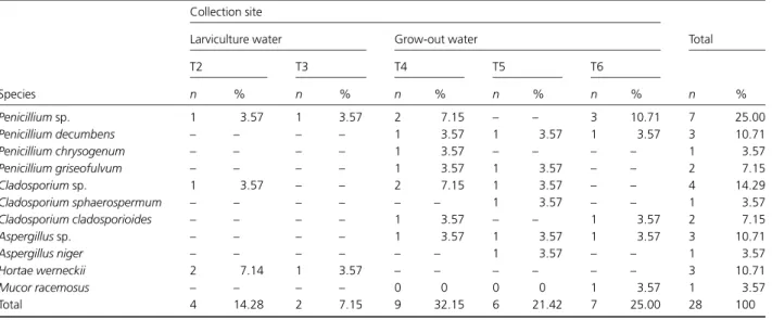
Table 2. Filamentous fungus species isolated from cultivation tanks ofMacrobrachium Amazonicum
Species
Collection site
Total Larviculture water Grow-out water
T2 T3 T4 T5 T6
n % n % n % n % n % n %
Penicilliumsp. 1 3.57 1 3.57 2 7.15 – – 3 10.71 7 25.00
Penicillium decumbens – – – – 1 3.57 1 3.57 1 3.57 3 10.71
Penicillium chrysogenum – – – – 1 3.57 – – – – 1 3.57
Penicillium griseofulvum – – – – 1 3.57 1 3.57 – – 2 7.15
Cladosporiumsp. 1 3.57 – – 2 7.15 1 3.57 – – 4 14.29
Cladosporium sphaerospermum – – – – – – 1 3.57 – – 1 3.57
Cladosporium cladosporioides – – – – 1 3.57 – – 1 3.57 2 7.15
Aspergillussp. – – – – 1 3.57 1 3.57 1 3.57 3 10.71
Aspergillus niger – – – – – – 1 3.57 – – 1 3.57
Hortae werneckii 2 7.14 1 3.57 – – – – – – 3 10.71
Mucor racemosus – – – – 0 0 0 0 1 3.57 1 3.57
Total 4 14.28 2 7.15 9 32.15 6 21.42 7 25.00 28 100
21.43%) and seven (7/28; 25%) isolates were obtained from water samples from grow-out tanks T4, T5 and T6, respec-tively.
Penicillium sp. was the most isolated genus (13/28; 46.42%), followed by Cladosporium sp. (7/28; 25%) and Aspergillussp. (4/28; 14.29%). Other obtained isolates were Hortaea werneckii (3/28; 10.71%) and Mucor sp. (1/28; 3.57%) (Table 2).
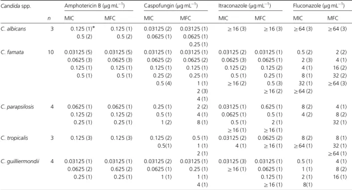
Concerning in vitro antifungal susceptibility tests of Candida spp., the MIC values for all tested isolates are described in Table 3. Briefly, for C. famata (10/24), the MIC for amphotericin B varied from 0.03125 to 0.5mg mL 1, and for caspofungin, itraconazole and flucona-zole, MICs ranged from 0.03125 to 0.5mg mL 1, 0.03125 to
Z16mg mL 1and 0.5 toZ64mg mL 1, respectively. ForC. parapsilosis (4/24), MIC for amphotericin B, caspofungin, itraconazole and fluconazole ranged from 0.0625 to 0.25mg mL 1, 0.25 to 1mg mL 1, 0.03125 to Z16mg mL 1 and 4 to 8mg mL 1, respectively. ForC.guilliermondii(4/24), MICs varied from 0.03125 to 0.25mg mL 1, 0.03125 to 1mg mL 1, 0.03125 to Z16mg mL 1 and 0.5 to 8mg mL 1 for amphotericin B, caspofungin, itraconazole and flucona-zole, respectively. ForC.tropicalis(3/24), the MIC value for amphotericin B was 0.125mg mL 1, while those for caspo-fungin, itraconazole and fluconazole ranged from 0.125 to 0.5mg mL 1, 0.03125 to 4mg mL 1 and 8 toZ64mg mL 1, respectively. Finally, forC.albicans(3/24), MIC values for
amphotericin B and caspofungin varied from 0.125 to 0.5mg mL 1 and 0.03125 to 0.0625mg mL 1, respectively, while those for itraconazole and fluconazole wereZ16 and
Z64mg mL 1, respectively. A positive correlation was ob-served when comparing MICs for amphotericin B and itraconazole (P= 0.0005), amphotericin B and fluconazole (P= 0.0020), and fluconazole and itraconazole (P= 0.0007), but not for caspofungin, when compared with the other tested drugs.
MFC values for all Candida spp. isolates varied from 0.03125 to 0.5mg mL 1, 0.03125 to 8mg mL 1, 0.03125 to
Z16mg mL 1 and 2 to Z64mg mL 1 for amphotericin B, caspofungin, itraconazole and fluconazole, respectively (Ta-ble 3). A positive correlation was also observed when comparing MFCs for amphotericin B and itraconazole (P= 0.0001), amphotericin B and fluconazole (P= 0.0104), and fluconazole and itraconazole (P= 0.0013), but not for caspofungin, when compared with the other tested drugs. When comparing MIC and MFC, a positive correlation was observed for caspofungin (P= 0.0000), fluconazole (P= 0.0000) and itraconazole (P= 0.0000). For amphoter-icin B, MICs were equal to MFCs; for itraconazole, MICs and MFCs were not statistically different (P= 0.1108), and for caspofungin (P= 0.0007) and fluconazole (P= 0.0013), MICs and MFCs were statistically different.
Concerning phospholipase activity, only three (12.5%) isolates presented positive results (Pzo1). The obtained Pz Table 3.MIC and MFC distribution of amphotericin B, caspofungin, itraconazole and fluconazole against 24 isolates ofCandidaspp.
Candidaspp. Amphotericin B (mg mL1) Caspofungin (mg mL1) Itraconazole (mg mL1) Fluconazole (mg mL1)
n MIC MFC MIC MFC MIC MFC MIC MFC
C.albicans 3 0.125 (1) 0.125 (1) 0.03125 (2) 0.03125 (1) Z16 (3) Z16 (3) Z64 (3) Z64 (3) 0.5 (2) 0.5 (2) 0.0625 (1) 0.0625 (1)
0.25 (1)
C.famata 10 0.03125 (5) 0.03125 (5) 0.03125 (1) 0.03125 (1) 0.03125 (2) 0.03125 (1) 0.5 (2) 2 (2) 0.0625 (3) 0.0625 (3) 0.0625 (2) 0.0625 (2) 0.0625 (3) 0.0625 (1) 2 (3) 4 (1) 0.125 (1) 0.125 (1) 0.125 (1) 0.125 (1) 0.125 (2) 0.125 (2) 4 (1) 16 (2)
0.5 (1) 0.5 (1) 0.25 (2) 0.25 (1) 0.5 (1) 0.25 (1) 8 (1) 32 (2)
0.5 (4) 1 (1) Z16 (2) 0.5 (3) 32 (1) Z64 (3)
2 (3) Z16 (2) Z64 (2)
4 (1)
C.parapsilosis 4 0.0625 (1) 0.0625 (1) 0.25 (1) 2 (2) 0.03125 (1) 0.625 (1) 8 (2) 4 (1)
0.125 (2) 0.125 (2) 0.5 (1) 4 (1) 0.0625 (1) 0.5 (1) 4 (2) 8 (2)
0.25 (1) 0.25 (1) 1 (2) 8 (1) 0.5 (1) 2 (1) 32 (1)
Z16 (1) Z16 (1)
C.tropicalis 3 0.125 (3) 0.125 (3) 0.125 (2) 0.5 (1) 0.03125 (2) 0.0625 (2) 8 (2) 8 (1)
0.5(1) 1 (1) 4 (1) Z16 (1) Z64 (1) 32 (1)
2 (1) Z64 (1)
C.guilliermondii 4 0.03125 (1) 0.03125 (1) 0.03125 (2) 0.03125 (1) 0.03125 (3) 0.03125 (1) 0.5 (1) 4 (1) 0.0625 (2) 0.625 (2) 0.0625 (1) 0.25 (1) Z16 (1) 0.0625 (1) 1 (1) 8 (2)
0.25 (1) 0.25 (1) 1 (1) 1 (1) 0.125 (1) 2 (1) 16 (1)
4 (1) Z16 (1) 8(1)
values were 0.64, 0.67 and 0.71 forC.famata,C.tropicalis and C. parapsilosis, respectively. Candida famata was iso-lated from the larviculture water (T1) andC. tropicalisand C. parapsilosis were isolated from the digestive tract of captive adults (T5).
As for protease production, eight isolates presented positive results (8/24; 33.3%), which ranged from 1.0 to 27.0 UmL 1. These isolates were fourC.famata(4/10; 40%; one from T3 water and three from wild-harvested adults), twoC. albicans(2/3; 66.7%; one from wild-harvested adults and one from captive adult from T5), oneC. parapsilosis(1/ 4; 25%; from captive adult from T5) and one C. guillier-mondii(1/4; 25%; from wild-harvested adult).
Discussion
The larval health index was satisfactory for all analyzed tanks during this experiment, with values 41 (Tayamen & Brown, 1999). The water temperature varied from 28 to 301C and
ammonia and nitrite concentrations were maintained at zero throughout the research, within ideal management conditions forM. amazonicum(Moraes-Valenti & Valenti, 2010).
Candidawas the most isolated genus andC. famatathe most frequently isolated species, with 10 isolates (38.46%), followed by C. guilliermondii (15.4%) and C. parapsilosis (15.4%). Other recovered species were C. albicans, C. tropicalis,C.laurentiiandR.mucilaginosa.Candida albicans was recovered from larvae and adult prawns, but not from water, corroborating the generally accepted idea that ecolo-gical niches for this yeast species do not exist in the environment. Medeiroset al., 2008 recovered several Candi-da species from water and sediment samples from two unpolluted lakes, but C. albicans was not recovered, sup-porting this idea.
Candida famatawas the most commonly isolated species from wild-harvested adult prawns and cultivation water (42% and 50%, respectively). From the digestive tract of cultivated prawns,C. tropicalisandC. famatawere the most frequently recovered species (28.6% each). The digestive tract of wild-harvested adultM.amazonicumfrom Catu Lake represented the type of sample where the highest number of species was found, followed by digestive tracts of cultivated animals, cultivation water and larvae. Considering that the intestinal microbiota of aquatic invertebrates resembles that of the environment where they are inserted (Hagler et al., 1995; Kutty & Philip, 2008), this observation was expected, once environments under natural conditions are subjected to several influencing factors, including natural and anthropic ones, harboring a greater diversity of microorganisms (Me-deiroset al., 2008), when compared with environments under controlled conditions, commonly used in prawn farming.
In a study withM.rosenbergiicultivated in Taiwan, the greatest percentage of the isolates (86%) were represented by
Candidaspecies, out of which 70% wereC.sakeand 16% were C. famata (Lu et al., 1998). However, 61% of the animals presented clinical alterations, which may explain the high prevalence ofC.sake, a well-known pathogen of fresh-water prawns. In our study, only healthy prawns were assessed andC.sakewas not recovered, whileC.famatawas the most frequently isolated species, demonstrating the presence of this yeast species in these animals.
In our study, although the number of yeasts recovered from the cultivation water was small,Candidaspp. were the predominantly isolated ones, similar to that observed by Lu et al., 1998 and Lea˜no et al., 2005 in studies with captive giant river prawns and tiger prawns, respectively. The water-sampling methodology used in this research differed from those mentioned by other authors (Luet al., 1998; Lea˜no et al., 2005; Medeiros et al., 2008), which could have accounted for the low recovery rate of yeasts, including the inability to obtainC.albicansfrom cultivation water. How-ever, these authors studied the quality of water from natural sources or ponds, but not from tanks equipped with closed water circulation system, which justifies their need to work with larger volumes of water.
The three predominant genera of filamentous fungi found in cultivation water of this study,Penicillium, Clados-poriumandAspergillus, were also found in an investigation of filamentous fungi in cultivation water of tilapia and tiger prawns by Lea˜noet al., 2005.Penicilliumsp. andAspergillus sp. were the predominant genera found by these authors, butCladosporiumsp. was isolated from only one sample of the tiger prawn cultivation. In contrast, Cladosporium sp. represented 25% of the isolates in our study, which probably reflects the environmental condition under which this research was conducted, considering that the species belong-ing to this genus, along withPenicilliumspp. andAspergillus spp., represent common airborne fungi in indoor and out-door environments (Solomon et al., 2006; Pantoja et al., 2009; Miaoet al., 2010).
In our study, the fungusH.werneckii(3/28) was isolated from water samples from tanks T2 and T3 (salinities of 4000 and 6000 mg L 1, respectively). This fungus inhabits tropical and subtropical regions (De Hoog et al., 2000; Varga & Godoy, 2004) and it is the predominant species in hypersa-line waters, acting as a saprobic microorganism in these environments (Gunde-Cimermanet al., 2000; Mbata, 2008). Hortaea werneckii can inhabit a wide range of salinities, varying from 0 to 25 000 mg L 1, but optimal growth occurs between 3000 and 7500 mg L 1 of NaCl (Petrovic et al., 2002), which includes the salinity of the samples from which this fungus was isolated in our study.
pathogenic fungi for prawns and shrimps belong to the microbiota of cultivation water and are secondary or opportunistic invaders. These microorganisms cause culti-vation problems only when the animals are submitted to inadequate management conditions, which favor the dis-semination of other diseases (Lea˜noet al., 2005). Hence, the animals were kept under adequate management conditions
In this study, all Candida strains were susceptible to amphotericin B, as observed in other researches performed by our group (Britoet al., 2009; Sidrimet al., 2010). Eight out of 24Candida spp. isolates (33.3%) were resistant to itraconazole and/or fluconazole, with particular attention given to the isolates ofC.albicans, which were resistant to both drugs simultaneously. The MFC results obtained in our study were similar to those for clinical isolates of non-albicans Candidaspecies (Tawaraet al., 2000).
Additionally, it is noteworthy that 28.6% (4/14) of the isolates of Candidaspp. obtained from wild-harvested M. amazonicum were resistant to these azole derivatives. The observed resistance phenomenon to this class of drugs arose the curiosity to seek the causes that may be associated with this phenomenon in the environment. Catu Lake is a fresh-water source that has been used for human consumption and supply, animal consumption, agriculture, industries and leisure activities. Considering that industrial wastes and pollutants are reported as causes of mutagenesis in yeasts (Keenan et al., 2007) and that the occurrence of gene mutation (Feng et al., 2010) may result in antifungal resistance, we believe that the resistance phenomenon observed in these Candida strains is related to anthropic activities developed in the studied area, such as pollution with industrial, agricultural and farming wastes.
As mentioned before, the intestinal microbiota of aquatic invertebrates is similar to that of the environment where they are inserted (Hagleret al., 1995; Kutty & Philip, 2008); thus, yeasts isolated from the digestive tract of wild-harvested M. amazonicum may reflect the environmental conditions of Catu Lake. Based on this, crustaceans, includ-ingM.amazonicum, and fishes can be used as sentinels for the occurrence of resistant yeasts in the environment. Several animal species have been reported as sentinels for the occurrence of resistant bacteria in the wild, including predatory fishes (Blackburn et al., 2010) and terrestrian mammals (Routmanet al., 1985; Costaet al., 2008; Mariano et al., 2009). Interestingly, all of these authors reported that the occurrence of resistant bacteria was associated with the close relationship between these wild animals or their environment and human beings and that water was the main vehicle for spreading chemical compounds involved in the development of resistance.
Classically, bacteria have been used as bioindicators of environmental imbalance related to pollution. However, more recently, the use of yeasts has been encouraged,
especially because of the structural and molecular simila-rities between fungal and mammalian cells (Keenan et al., 2007). Several pollutants are capable of altering gene expres-sion or sequence composition (Wegrzyn & Czyz, 2003; Keenan et al., 2007; M¨uller et al., 2007). Considering that azole resistance is basically associated with changes in gene expression or in gene nucleotide sequence, the presence of pollutants in the water of this lake, including azole deriva-tives commonly used in agricultural practices, may be responsible for the observed resistance. Based on these observations, we propose the use of M. amazonicum and other aquatic species as sentinels for the occurrence of resistant yeasts in the environment and the use of the isolated yeast as bioindicators for the presence of pollutants in natural water sources.
Additionally, the risk of yeast infections occurring in hu-mans should be considered when manipulating these animals or the water they inhabit, considering that potentially patho-genic species were isolated from these sources. Three species should be highlighted, C. albicans, C. parapsilosis and C. tropicalis, for being the most commonly involved in human candidiasis in Latin America (Palacio et al., 2009). Besides, special attention must be given toC.albicans, considering that animals can represent a source of infection for humans (Edelmannet al., 2005) and that the recovered isolates of this species were all azole resistant, which is not common among human strains (Kanafani & Perfect, 2008). Thus, studies on genetic diversity of this Candida species could help to elucidate whether or not the recovered isolates are commonly distributed among humans.
There are several researches concerning phospholipase activity of fungi isolated from humans (Fotedar & Al-Hedaithy, 2005; Zenget al., 2008). However, researches with yeasts isolated from animals are scarce (Sidrimet al., 2010) and no reports have been found for yeasts from prawns or shrimps. For many years, it was believed that only C. albicanswas able to produce phospholipase. However, now it is known that other species ofCandidaalso produce this enzyme, usually in smaller amounts (Ghannoum, 2000), as demonstrated by our research. Paradoxically, the isolates of C.albicansobtained in this study did not present phospho-lipase activity, while 14.29% (3/21) of the non-albicans Candidaspecies were positive for this enzyme.
Some studies have evaluated the protease activity of Candida species; however, in those researches, the strains were obtained from humans with candidiasis (Kantarcioglu & Yucel, 2002; Mohan & Ballal, 2008; Ombrellaet al., 2008). In our study, the production of protease byCandida spp. strains isolated from wild-harvested and captive crustaceans was demonstrated for the first time.
isolation of azole-resistantCandidaspp. from wild-harvested animals, showing the role of these animals as sentinels for the occurrence of resistant yeasts in the natural environment.
Acknowledgements
The authors thank the financial support of FUNCAP (Cear´a State Research Funding; Proc. 071.02.00/09), CAPES/PNPD (Proc. 23038.027637/2009-68) and CNPq (National Counsel for Technological and Scientific Development; Brazil, Proc. 473881/2008-0).
References
Blackburn JK, Mitchell MA, Blackburn MCH, Curtis A & Thompson BA (2010) Evidence of antibiotic resistance in free-swimming top-level marine predatory fishes.J Zoo Wildlife Med41: 7–16.
Brilhante RS, Castelo-Branco DSCM, Soares GD, Astete-Medrano DJ, Monteiro AJ, Cordeiro RA, Sidrim JJ & Rocha MF (2010) Characterization of the gastrointestinal yeast microbiota of cockatiels (Nymphicus hollandicus): a potential hazard to human health.J Med Microbiol59: 718–723.
Brito EHS, Fontenelle ROS, Brilhante RSN, Cordeiro RA, Monteiro AJ, Sidrim JJC & Rocha MFG (2009) The anatomical distribution and antimicrobial susceptibility of yeast species isolated from healthy dogs.Vet J182: 320–326.
Cenci E, Francisci D, Belfiori B, Pierucci S, Baldelli F, Bistoni F & Vecchiarelli A (2008) Tipranavir exhibits different efects on opportunistic pathogenic fungi.J Infect56: 58–64.
Charney J & Tomarelli RM (1947) A colorimetric method for the determination of the proteolytic of duodenal juice.J Biol Chem
23: 501–505.
Costa D, Poeta P, S´aenz Y, Vinu´e L, Coelho AC, Matos M, Rojo-Bezares B, Rodrigues J & Torres C (2008) Mechanisms of antibiotic resistance inEscherichia coliisolates recovered from wild animals.Microb Drug Resist14: 71–77.
Coyle SD, Alston DE & Sampaio CMS (2010) Nursery systems and management.Freshwater Prawns: Biology and Farming
(New MB, Valenti WC, Tidwell JH, D’abramo LR & Kutty MN, eds), pp. 108–126. Wiley-Blackwell, Oxford.
De Hoog GS, Guarro J, Gen´e J & Figueiras MJ (eds) (2000)Atlas of Clinical Fungi. Centraalbureau voor Schimmslcultures, The Nederlands.
De Hoog GS, Guarro J & Gen´e J (eds) (2002)Atlas of Clinical Fungi. Centraalbureau voor Schinmelculture/Universitat Rovira i Virgilli, Delf.
Edelmann A, Kr¨uger M & Schmid J (2005) Genetic relationship between human and animal isolates ofCandida albicans.J Clin Microbiol43: 6164–6166.
Feng L, Wan Z, Wang X, Li R & Liu W (2010) Relationship between antifungal resistance of fluconazole resistantCandida albicansand mutations inERG11gene.Chin Med J123:
544–548.
Fotedar R & Al-Hedaithy SSA (2005) Comparison of
phospholipase and proteinase activity inCandida albicansand
C.dubliniensis.Mycoses48: 62–67.
Gatesoupe FJ (2007) Live yeasts in the gut: natural occurrence, dietary introduction, and their effects on fish health and development.Aquaculture267: 20–30.
Ghannoum MA (2000) Potential role of phospholipases in virulence and fungal pathogenesis.Clin Microb Rev13:
122–143.
Gunde-Cimerman N, Zalar P, De Hoog S & Plemenitas A (2000) Hypersaline waters in salterns – natural ecological niches for halophilic black yeasts.FEMS Microbiol Ecol32: 235–240.
Hagler AN, Mendonc¸a-Hagler LC, Rosa CA & Morais PP (1995) Yeast as an example of microbial diversity in brazilian ecosystems.Oecologia brasiliensis1: 225–244.
Holthuis LB (1952) A general revision of the Palemonidae (Crustacea, Decapoda, Natantia) of the Americas.Allan Hancock Foundation12: 1–396.
Johnson SK & Bueno SLS (2000) Health management.Freshwater Prawn Culture: The Farming of Macrobrachium Rosenbergii,
Vol. 1(New MB & Valenti WC, eds), pp. 18–40. Blackwell Science, London.
Kanafani ZA & Perfect JR (2008) Resistance to antifungal agents: mechanisms and clinical impact.Clin Infect Dis46: 120–128.
Kantarcioglu AS & Yucel A (2002) Phospholipase and protease activities in clinical Candida isolates with reference to the sources of strains.Mycoses45: 160–165.
Keenan PO, Knight AW, Billinton N, Cahill PA, Dalrymple IM, Hawkyard CJ, Stratton-Campbellc D & Walmsleya RM (2007) Clear and present danger? The use of a yeast biosensor to monitor changes in the toxicity of industrial effluents subjected to oxidative colour removal treatments.J Environ Monitor9: 1394–1401.
Kutty SN & Philip R (2008) Marine yeasts – a review.Yeast25:
465–483.
Lea˜no EM, Lio-Po GD, Nadong LA, Tirado AC, Sadaba RB & Guanzon NG Jr (2005) Mycoflora of the ‘green water’ culture system of tiger shrimpPenaeus monodonFabricius.Aquac Res
36: 1581–1587.
Lu CC, Tang KFJ & Chen SN (1998) Identification and genetic characterization of yeasts isolated from freshwater prawns,
Macrobrachium rosenbergiide Man, in Taiwan.J Fish Dis21:
185–192.
Mariano V, McCrindle CME, Cenci-Goga B & Picard JA (2009) Case–control study to determine whether river water can spread tetracycline resistance to unexposed impala (Aepyceros melampus) in Kruger National Park (South Africa).Appl Environ Microb75: 113–118.
Martin JW & Davis GE (2001) An update classification of the recent Crustacea.Nat Hist Mus Los Angeles39: 1–124.
Mbata TI (2008) Isolation of fungi in hyper saline Dead Sea water.
Sudanese J Public Health3: 170–172.
yeasts from tropical freshwater environments in Southeastern Brazil.Water Res42: 3921–3929.
Miao Z, Chai T, Qi C, Cai Y, Liu J, Yuan W & Yao M (2010) Composition and variability of airborne fungi in an enclosed rabbit house in China.Aerobiologia26: 135–140.
Mohan V & Ballal M (2008) Proteinase and phospholipase activity as virulence factors inCandidaspecies isolated from blood.Rev Iberoam Micol25: 208–210.
Moraes-Valenti P & Valenti WC (2010) Culture of the Amazon river prawnMacrobrachium amazonicum.Freshwater Prawns: Biology and Farming(New MB, Valenti WC, Tidwell JH, D’abramo LR & Kutty MN, eds), pp. 485–501.
Wiley-Blackwell, Oxford.
M¨uller FMC, Staudigel A, Salvenmoser S, Tredup A, Miltenberger R & Herrmann JV (2007) Cross-resistance to medical and agricultural azole drugs in yeasts from the oropharynx of human immunodeficiency virus patients and from
environmental bavarian vine grapes.Antimicrob Agents Ch51:
3014–3016.
National Committee for Clinical Laboratory Standards (2002) Reference method for broth dilution antifungal susceptibility testing of yeasts: approved standard. NCCLS, document M27-A2.
Ombrella AM, Racca L & Ramos L (2008) Actividades proteinasa y fosfolipasa de aislamientos deCandida albicansprovenientes de secreciones vaginales con distintos valores de pH.Rev Iberoam Micol25: 12–16.
Palacio A, Villar J & Alhambra A (2009) Epidemiolog´ıa de las candidiasis invasoras em poblaci ´on pedi´atrica y adulta.Rev Iberoam Micol26: 2–7.
Pantoja LDM, Moreira-Filho RE, Brito EHS, Araga˜o TB, Brilhante RSN, Cordeiro RA, Rocha MFG, Monteiro AJ, Quinet YP & Sidrim JJC (2009) Ants (Hymenoptera: Formicidae) as carriers of fungi in hospital environments: an emphasis on the generaTapinomaandPheidole.J Med Entomol
46: 895–899.
Petrovic U, Gunde-Cimerman N & Plemenitas A (2002) Cellular responses to environmental salinity in the halophilic black yeastHortaea werneckii.Mol Microbiol45: 665–672.
Pfaller MA, Boyken L, Hollis RJ, Messer SA, Tendolkar S & Diekema DJ (2006)In vitrosusceptibilities ofCandidaspp. To
caspofungin: four years of global surveillance.J Clin Microbiol
44: 760–763.
Price MF, Wilkinson ID & Gentry LO (1982) Plate method for detection of phospholipase activity ofCandida albicans.
Sabouraudia22: 201–207.
Routman E, Miller RD, Phillips-Conroy J & Hartl DL (1985) Antibiotic resistance and population structure inEscherichia colifrom free-ranging African yellow baboons.Appl Environ Microb50: 749–754.
Sidrim JJC, Castelo-Branco DSCM, Brilhante RSN, Soares GDP, Cordeiro RA, Monteiro AJ & Rocha MFG (2010) Candida species isolated from the gastrointestinal tract of cockatiels (Nymphicus hollandicus):in vitroantifungal susceptibility profile and phospholipase activity.Vet Microbiol145:
324–328.
Solomon GM, Hjelmroos-Koski M, Rotkin-Ellman M & Hammond K (2006) Airborne mold and endotoxin concentrations in New Orleans, Louisiana, after flooding, October through November 2005.Environ Health Persp114:
1381–1386.
Tawara S, Ikeda F, Maki Ket al. (2000)In vitroactivities of a new lipopeptide antifungal agent, FK463, against a variety of clinically important fungi.Antimicrob Agents Ch44:
57–62.
Tayamen M & Brown JH (1999) A condition index for evaluating larval quality ofMacrobrachium rosenbergii(De Man, 1879).
Aquac Res30: 917–922.
Valenti WC, Mallasen M & Silva CA (1998) Larvicultura em sistema fechado dinaˆmico.Carcinicultura de ´agua doce: Tecnologia para Produc¸a˜o de Camaro˜es,Vol. 1(Valenti WC, ed), pp. 115–143. FAPESP/IBAMA, S a˜ o Paulo, Brazil.
Varga VES & Godoy P (2004)Tinea Nigra.Micologia m´edica a` luz de autores contemporaˆneos,Vol. 1(Sidrim JJC & Rocha MFG, eds), pp. 124–128. Guanabara Koogan, Rio de Janeiro. Wegrzyn G & Czyz A (2003) Detection of mutagenic pollution of
natural environment using microbiological assays.J Appl Microbiol95: 1175–1181.