Digestive Surgery Division, São Paulo University Medical School – São Paulo/ SP, Brazil.
Department of Gastroenterology, São Paulo University Medical School – São Paulo/SP, Brazil.
Institute of Biomedical Sciences, Department of Microbiology, São Paulo University Medical School – São Paulo/SP, Brazil.
E-mail: [email protected]
Received for publication on July 20, 2006. Accepted for publication on October 10, 2006.
CLINICAL SCIENCES
DIGESTIVE TRACT MICROBIOTA IN HEALTHY
VOLUNTEERS
Bruno Zilberstein, Alina G Quintanilha, Manoel A A Santos, Denis Pajecki, Eduardo G Moura, Paulo Roberto Arruda Alves, Fauze Maluf Filho, João Ary Ubriaco de Souza, Joaquim Gama-Rodrigues
Zilberstein B, Quintanilha AG, Santos MAA, Pajecki D, Moura EG, Alves PRA, Maluf Filho F, Souza JAU de, Gama-Rodrigue J. Digestive tract microbiota in healthy volunteers. Clinics. 2007:62(1):47-54.
PURPOSE:The aim of this study was to standardize the methods of sample collection of mucus from the digestive tract and to determine the microbiota in healthy volunteers from Brazil, collecting samples from the mouth, esophagus, stomach, duodenum, jejunum, ileum, colon, and rectum.
METHODS: Microbiota of selected healthy volunteers from the oral cavity (n=10), the esophagus (n=10), the upper digestive tract (n=20), and the lower digestive tract (n=24) were evaluated through distinct collection methods. Collection methods took into account the different sites, using basic scraping and swabbing techniques, stimulated saliva from the oral cavity, irrigation-aspiration with sterile catheters especially designed for the esophagus, a probe especially designed for upper digestive tract, and a special catheter for the lower digestive tract.
RESULTS: (i) Mixed microbiota were identified in the oral cavity, predominantly Gram-positive aerobic and anaerobic cocci; (ii) transitional flora mainly in the esophagus; (iii) Veillonella sp, Lactobacillussp, and Clostridium sp in the stomach and duodenum;
(iv) in the jejunum and upper ileum, we observed Bacteroides sp, Proteus sp, and Staphylococcus sp, in addition to Veillonella sp;
(v) in the colon, the presence of “nonpathogenic” anaerobic bacteria Veillonella sp (average 105 UFC) indicates the existence of a
low oxidation-reduction potential environment, which suggests the possibility of adoption of these bacteria as biological markers of total digestive tract health.
CONCLUSIONS: The collection methods were efficient in obtaining adequate samples from each segment of the total digestive tract to reveal the normal microbiota. These procedures are safe and easily reproducible for microbiological studies.
KEYWORDS: Bacteria Anaerobic. Bacteria Aerobic. Fungi. Colony count microbial. Gastrointestinal tract.
INTRODUCTION
Interest in the microbiologic composition of the normal digestive tract began in the 19th century with Pasteur, who believed that a symbiotic relationship between man and bacteria was essential to life. However, at the beginning
of the 20th century, several authors including Metchnikoff
et al1 believed that microbiota competed with the host
or-ganism for nutrients. The development of experimental models to study the relationship between microbiota and the human organism in totally germ-free laboratory animals demonstrated the role of the normal microflora in vitamin synthesis, metabolism of nitrogen compounds and lipids, and especially their participation as a barrier against the invasion of pathogenic microorganisms.2-4 In 1964,
Donaldson5 standardized the quantitative analysis of
bac-teria in the feces of healthy volunteers, and in 1966, Kalser6
have been some improvements in cultivation techniques and the use of genetic probes for identification of the microor-ganism.7
Over the last 20 years, there has been an increased in-terest in the study of the microbiota and their role in dis-eases of the digestive system including neoplasias,8,9
intes-tinal inflammatory disease,10,11 diarrhea associated with
an-tibiotic use,12-14 and bacterial translocation.15,16 However,
one of the difficulties in performing conclusive repeatable studies has always been the lack of a standard collection method that can be used to establish a uniform control group of healthy individuals against which the different dis-eases could be compared.
This shortcoming becomes even more important when there is the intention to study the eventual modification of the normal microbiota caused by or as a consequence of digestive diseases.11,17
The aim of this study was to standardize collecting sam-ple methods from the different segments of the digestive tract in healthy volunteers in order to better characterize their microbiota.
PATIENTS AND METHODS
A multidisciplinary prospective investigation was de-veloped involving the Digestive Surgery Division and the Gastrointestinal Endoscopy Unit of the São Paulo Univer-sity Medical School, and the Institute of Biomedical Sci-ences of the University of São Paulo.
The study was approved by the Ethic Committees and informed consent was obtained from all volunteers.
The different segments of the digestive tract were stud-ied in separate groups of healthy volunteers. Sample col-lection methods were specifically developed for each seg-ment. Healthy individuals were selected based on the fol-lowing exclusion criteria: use of antibiotics and/or anti-in-flammatory drugs in the preceding 90 days, dental pros-theses, smoking, alcoholism, diabetes, presence of malig-nant tumors, scleroderma, previous surgical procedures on the digestive tract, or any other problem that could affect the digestive tract, the esophagus, stomach, intestines, or specifically the mouth flora; a dental caries index (CPO – D) greater than 2.618was also was also an exclusion
crite-rion.
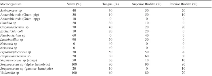
Standardized collection method from the mouth. Ten healthy individuals were prospectively studied, including 5 men and 5 women, with a mean age of 33.3 years. Sam-ples for microbiological study were collected from saliva, the back of the tongue, and supragingival and subgingival bacterial biofilm. A sterile Petri dish was used for saliva collection after stimulation by masticatory mouth
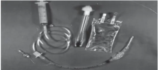
move-ments. From the back of the tongue, a sterile cotton swab was used with a front-to-back movement. The supragingival bacterial biofilm was collected from the vestibular face of the first lower left molar and from the subgingival vestibular face of the contralateral first molar with a periodontal (Gracey) curette (Figure 1). The collected materials were placed in a test tube containing phosphate-buffered saline for immediate transport.
Standardized collection method from the esophagus. Ten individuals were prospectively studied, including 6 women and 4 men, with a mean age of 43.4 years. Volteers were selected among those who were going to un-dergo an upper digestive tract endoscopy. After local mouth anesthetic with 10% lidocaine spray and intravenous se-dation with diazepam (3-5 mg), a nº14(Fr) Levine catheter was introduced with extreme care into the mouth through an nº7.5 or 29 Fr orotracheal tube. The catheter was pushed forward to touch the esophagus lumen, where 10 mL of saline was injected using a 20 mL syringe connected to its proximal end; aspiration was then performed. The collected material was transported in a phosphate buffer saline solu-tion (1 mL/9 mL) to the microbiology laboratory within 1 hour (Figure 2). Upper digestive endoscopy was then im-mediately performed to identify any abnormality of the upper digestive tract.
Standardized collection method from the stomach, duodenum, jejunum, and proximal ileum. This was per-formed in 20 other volunteers: 8 men and 12 women with a mean age of 42 years. All individuals presented normal upper digestive tract endoscopies. A flexible silicone sam-pling probe was used with a small (3 mm) distal opening and with a lead weight bound in its extremity.19-23 This Figure 1 - Saliva; back of the tongue; sub-gingival biofilm
probe was introduced by mouth and swallowed until it reached the stomach, being located by radioscopy. The first sample was collected, aspirating 1 mL of mucus with a 20 mL syringe connected to its proximal end. The probe was allowed to migrate by peristaltic movement to the more distal segments and was periodically monitored by radioscopy (Figure 3). The same probe was used for col-lection of material in each intestinal segment. The evalua-tion of the exact site from which the mucus was obtained was determined by the length of the probe introduced and its location through radioscopy. To avoid contamination and eventual false results, the probe was washed out several times, and the contents were discarded before the actual collection of mucus. A series of 1-mL samples was taken from the duodenum, proximal jejunum, distal jejunum, and proximal ileum. The samples were placed into the test tubes with 9 mL of phosphate buffer saline solution and taken to the microbiology laboratory within the first hour.
Standardized collection method from distal ileum, co-lon, and rectum. This method was performed in another 24 volunteers, 10 men and 14 women, with a mean age of 53 years, through colonoscopy after previous colon preparation with a 10% mannitol solution until the stool was liquid, clear, and free of residues.24 Colonoscopy was performed at least
5 hours after the colonic preparation to avoid interference in the determination of the microbiologic contents of the co-lonic mucus.25 Colonoscopy was performed after sedation
with diazepam (to 10 mg) and meperidine (to 100 mg). The colonoscope was introduced into the distal ileum, checking the integrity of the lower digestive tract (distal ileum; cecum; ascending, transverse, descending, and sigmoid colon; and rectum). A proper catheter, especially developed for this pur-pose, was used to pass through the working channel of the colonoscope to collect the samples of mucus from the dif-ferent segments of the lower digestive tract. This catheter, obtained from a polyethylene probe, was especially designed,
being 2.20 m long and 8 Fr in diameter; it was internally coated with silicone (Figure 4). The distal end of the cath-eter was covered by a protective membrane; this was rup-tured at the time of collection by inflation with air. Sample volume was always 0.1 mL, determined by step marks on the distal end of the catheter5,2,25-27 that could be visualized
by the colonoscope. A fresh sterile catheter was used at each site of the lower digestive tract for collecting the samples. After collection, the samples were taken to the microbio-logical laboratory in a sterile Eppendorf tube containing 0.9 mL of VMGA III (viability-maintaining transport medium).28
Culture media
The collected materials from all segments were placed in selective culture media for aerobic and anaerobic mi-croorganisms and yeasts.29 The following culture media
were used: Sabouraud agar *, MacConkey agar**-blood, Enterococcus-selective agar**, reinforced Clostridium me-dium** ; Phenylethylalcohol agar*, Veillonella medium*, Brain-Heart infusion (BHI) agar* + vitamin K + hemin +
streptomycin, Chapman-Stone medium**, Bacteroides
fragilis bile-esculin agar medium (BBE) *, Bifidobacterium medium,Propionibacterium medium*, BHI* + extract of yeast, Blood-trypticase soy agar (TSA) * + vitamin K + hemin (*DIFCO. St. Louis, MO, USA; **MERCK DIAGNOSTICA, RJ, Brazil).
Statistical analysis
Descriptive analysis of findings from the mouth, esophagus, and upper digestive tract was done. The chi square (X2) test was used to verify the difference in
distri-bution of each bacterial species in the various regions of the lower digestive tract. The expected frequency of each bacterial species in different regions of the lower digestive tract was calculated using nonparametric tests.30
The results of the concentration of microorganisms were expressed in units of colony formation/mL (ucf) expressed in logarithm base 10 (log10). Significance was assigned to aP value of < 0.05.
Figure 3 - Agar-blood; Bile esculin (BBE); Tioglicolato; Flexible silicone sampling probe with distal orifice and lead weight bound to extremity
RESULTS
Mouth microbiota. Table 1 shows the frequency and locations of microorganisms found in the mouth. Mixed microbiota were identified here, with as predominance of Gram-positive aerobic and anaerobic cocci
Esophagus microbiota. Table 2 shows the frequency and average concentration of microorganisms found in the esophagus. It can be seen that in this segment the flora is transitional.
Stomach, duodenum, jejunum, and proximal ileum microbiota. Table 3 shows the mean concentrations and prevalence of the different microorganisms found in each segment of the upper digestive tract.Veillonella sp,
Lacto-bacillus sp, and Clostridium sp are predominant in the
stomach and duodenum; while Bacteroides sp, Proteus sp, andStaphylococcus sp, in addition to Veillonella sp are pre-dominant in the jejunum and upper ileum;
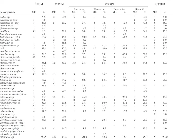
Distal ileum, colon, and rectum microbiota. Table 4 shows the different microorganisms found in each segment. The most distinctive feature here is the presence of “nonpatho-genic” anaerobic bacteria Veillonella sp (average 105 UFC)
Table 5 shows the association between microorganisms and sites of the lower digestive tract.
DISCUSSION
The complex interactions between the different micro-organisms or between them and healthy hosts or their in-teraction in digestive diseases are poorly understood. The study of the microbiota in humans starts with the need for standardizing sample collection methods from the differ-ent segmdiffer-ents of the digestive tract, including availability of different technical resources and ethical issues. This base is necessary when we intend to compare different studies.
This investigation describes the sample collection meth-ods from different segments of the digestive tract. This methodology is characterized by the following character-istics: (i) it is innocuous for patients; (ii) it allows mate-rial to be collected from restricted sites, avoiding contami-nation; (iii) it uses available materials with accessible prices, and (iv) it is easily reproducible.
Material sampling from the mouth18,31,32and esophagus33
was done by modifying previously validated methods al-ready described in the literature. For the stomach, duode-num, jejunum (proximal and distal), and proximal ileum, similar methods were used.19-22 Finally, samples from the
distal ileum, colon, and rectum were collected using a new method developed by the authors.28
The oral cavity presented very diverse microbiota due to the different anatomical sites and constant exposure to the external environment. The method of collecting saliva without using chewing gum or paraffin tablets gave better results than in previous studies,34-38suggesting that the
tech-nique used in this study is more sensitive and causes fewer alterations to indigenous flora. Results from the back of the tongue were similar to those of other studies.35-38
Periodontal Gracey curettes were preffered for sampling from the supra- and subgingival biofilm. Our results were Table 1 - Frequency and locations of microorganisms in the oral cavity
Microorganism Saliva (%) Tongue (%) Superior Biofilm (%) Inferior Biofilm (%)
Actinomycessp 40 30 30 20
Anaerobic rods (Gram- pig) 30 10 50 10
Anaerobic rods (Gram- npg) 10 0 0 0
Candidasp 20 30 10 0
Corynebacteriumsp 70 40 20 20
Escherichia coli 10 20 20 0
Fusobacteriumsp 60 0 40 0
Lactobacillussp 90 50 30 0
Neisseriasp 0 40 0 0
Neisseriasp 0 40 0 0
Peptostreptococcus sp 70 50 50 20
Propionibacterium sp 20 30 60 30
Staphylococcus sp (coag-) 50 30 10 10
Streptococcus sp (alpha- hemolytic) 100 90 90 80
Streptococcus sp (gamma- hemolytic) 20 10 0 10
Veillonella sp 100 60 80 70
sp = species; pig = pigmented; npg = nonpigmented; coag- = coagulase negative
Table 2 - Frequency and concentration of microorganisms in the esophagus
Frequency (%) Concentration (ufc/mL)
Streptococcus sp 40 101
Staphylococcussp 20 102
Corynebacterium sp 10 102
Lactobacillus sp 10 102
Peptococcus sp 10 101
similar to those reported in the literature,32,38,39 validating
this method.
From a microbiological point of view, the esophagus has always been considered to be a saliva secretion pas-sageway with transitory microbiota similar to the mouth and of little interest in research. However, when alimen-tary stasis occurs, as in neoplasia or achalasia,40,41 there is
an excessive increase in bacterial growth, with the risk of serious infections after surgery or endoscopy.42,43
Sample collection for studying the microbiota of the esophagus cannot be performed with catheters that pass through the mouth because of the risk of contamination during their insertion. The use of sterile catheters that pass through the working channel of an endoscope is expensive and does not permit aspiration of sufficient thick material from an esophagus with stasis.
Gagliardi et al33 developed a similar collection method
to that used in our study. However, by taking the samples
before endoscopic examination, we avoided possible con-tamination during the introduction or removal of the en-doscopic device. This small technical modification enabled us to obtain fewer positive cultures (40% vs 66%) with similar strains of bacteria, with predominantly Gram posi-tive aerobes and without Gram negaposi-tive aerobes, as de-scribed by Gagliardi in 13% of cases.
The samples obtained from the stomach for microbio-logical study presented no problems because of the ease of access, and because gastric pH is responsible for the de-struction of most swallowed bacteria, all of which facili-tates the isolation of the appropriate microorganisms.19
In the stomach, there was a predominance of Lactoba-cillussp,Veillonella sp, and Clostridiumsp, which are all resistant to the acid environment, demonstrating potential residual microflora that could develop down to the lower regions of the digestive tract.
Other species such as Escherichia coli, Peptococcus sp,
Table 3 - Mean concentration and prevalence of microorganism in the upper digestive tract
Site STOMACH DUODENUM JEJUNUM ILEUM
Proximal Distal Proximal
Microorganisms MC % MC % MC % MC % MC %
Bacillussp 4 9.1
Bacteroides sp 2 9.1 3 9.1
Bacteroides sp (npg) 2 16 2 9.1 3.5 36.4 3 45.5 4.5 40.0
Bifidobacterium sp 3 9.1 3 9.1
Candidasp 2 9.1 3 18.2 1 10.0
Clostridium ramosum Clostridiumsp
Clostridiumsp (gel-) 4 33.3 2 27.3 2.5 18.2 5 27.3 3.5 20.0
Clostridiumsp (gel+)
Corynebacteriumsp 3 25 3 18.2 2 9.1 5.5 18.2 3 30.0
Escherichia coli 5.5 16 2.5 18.2 5.5 18.2 6 36.4 3 30.0
Enterobacter sp 7 8.3 5 27.3 7 27.3 7 50.0
Enterococcus faecalis 4 9.1
Enterococcus sp 7 8.3 2 27.3 4.5 18.2 6 18.2 5 20.0
Eubacterium lentum Eubacteriumsp
Fusobacterium fusiformes 3 8.3 1 9.1
Fusobacteriumsp 2 8.3 4 9.1 2 18.2 2 18.2 3 10.0
Klebsiella pneumoniae
Klebsiella sp 3.5 16 2 45.5 3.5 18.2 7 36.4 9 10.0
Lactobacillus acidophillus
Lactobacillussp 4 41.6 2 27.3 3 45.5 4 27.3 4.5 20.0
Leptotrichia sp 1 9.1
Peptococcus sp 3.5 16 2 9.1 3 9.1 3 27.3 4 30.0
Peptostreptococcus sp 5 8.1 3 10.0
Propionibacterium sp 3 8.1 2.5 18.2 9.1 2 36.4
Proteus sp 5 8.1 2 45.5 4 45.5 6 20.0
Pseudomonassp
Rodothorula sp 4 8.1 1 9.1 4 18.2 4.5 20.0
Sarcinia lútea
Staphylococcussp 2 8.1 1 9.1 18.2 2 45.5 3 50.0
Staphylococcussp (coag-) 3 27.3
Streptococcus sp 4 2.5 2 27.3 4 9.1 5 10.0
(alpha hemolytic viridans group)
Torulopsis sp 4 9.1
Veillonella sp 4 41.6 3 45.5 4 54.5 4 63.6 4 50.0
Klebisiellasp,Bacteroides sp (npg) start an ascending curve of growth or “re-population” of the lower sites of the di-gestive tract, being dormant in the stomach because of the acidic environment.
Sample collection from small intestine segments is tech-nically more difficult because of difficulties relating to ac-cess and to the standardization of specific locations. Nev-ertheless, it is of major interest because of the range of dis-eases associated with alterations in its microbiota.44,45
Shiner46 developed a stainless steel capsule that only
opens at collection time to aspirate the contents of the je-junum; this avoids contamination during its passage, but it is very complex to use. Kalser et al6 developed a double
lumen polyvinyl catheter with a mercury weight on the distal end for taking samples starting 75 cm from Treitz’s ligament to near the distal ileum and cecal valve. However, there was no mechanism to protect from contamination. Their results were very similar to those of Shiner. 46
This method was tested in our midst by Machado et al,19
who collected samples of the jejunum liquid in patients suf-fering from Chagasic achalasia. It was also used by Quintanilha et al20,21 for qualitative evaluation of microbiota
alterations in the proximal jejunum of patients with Chagasic megacolon, both before and after surgery.
A similar method has been used by different authors in the study of bacterial translocation in critical patients. Belov et al47 evaluated sepsis mediators (TNF and IL-1)
using jejunal aspiration in septic shock patients. Pardo et al16demonstrated a reduction in bacterial overgrowth and
translocation in cirrhotic patients using cisapride. Bernhardt et al48 studied Candida sp colonization in digestive tracts
of critical patients under long-term antibiotic treatment. In this study, the method of collecting mucus from the upper digestive tract was used to evaluate healthy volun-teers with the aim of establishing a normal pattern that could serve as a control in studies of microbiotic changes in disease states. Even though the sample group was small, we obtained similar results to those described by Kalser6
and Shiner.46
It must be said that sample collection from the jejunum and proximal ileum is an uncomfortable procedure for the patient. Therefore, recruiting healthy volunteers is not easy; this makes it difficult for researchers to recruit control groups for their studies.
The study of lower digestive tract microbiota is a ma-jor challenge because of the high concentration and vari-ety of microorganisms pertaining to the indigenous and transitory microbiota.49 In early trials in which researchers
attempted to study the microbiology of dregs,4,5they were
unable to differentiate indigenous from transitory microbiota or determine the different levels of bacteria in
the different segments of the lower digestive tract. Studies that have employed sample collection during laparotomy50
have not respected the physiological conditions of the pa-tient, because the quantitative study was prejudiced due to dilution of mucus during collection.
The need for more precise and reliable methods moti-vated us to develop a special catheter to collect adequate volume of undiluted mucus from specific regions of the lower digestive tract during colonoscopy using an aseptic technique. This is an efficient method as shown by the large variety of aerobic, anaerobic, microaerophile, and faculta-tive microorganisms, and yeasts obtained. The results show a stable increase in anaerobic microorganisms (Veillonella sp,Peptococcus sp, and Fusobacterium sp) with the capac-ity to digest amino acids but with little abilcapac-ity to ferment carbohydrates. These and other similar microorganisms comprise a distinct metabolism group in the large intes-tine.4
There was also the constant presence of other bacteria with average concentrations of 105 such as Clostridium sp
(gel-) Corynebacterium sp, E. coli, Enterobacter sp, Klebisiella sp, Lactobacillus sp, Propionibacterium sp, Pro-teus sp, and Veillonella sp; there were others at lower con-centrations.
Bacterial types were characterized colonizing restricted areas of the lower digestive tract with average concentra-tions of 105 (Table 5); the following could be interpreted
as “biological markers” of health: Fusobacterium sp in the rectum; Peptococcus sp in the sigmoid; and Enterococcos sp in the transverse colon. Bacteroides sp tend to reside in the more proximal regions, gradually decreasing in preva-lence in the sigmoid and rectum.
CONCLUSION
The presented sampling collection methods are safe and efficient for obtaining suitable samples of mucus for quali-tative and quantiquali-tative microbiologic studies, revealing an environment of low oxidation-reduction (redox) potential.
These results open the possibility for many other stud-ies in this area, using low-risk and highly reliable method-ology, to define the role of microbiota in the other gastrointestinal diseases as well as for standardization of future prophylactic treatment50,51 in gastroenterology.
ACKNOWLEDGMENTS
The authors thank Ulysses Ribeiro Junior for his help-ful suggestions.
RESUMO
Zilberstein B, Quintanilha AG, Santos MAA, Pajecki D, Moura EG, Alves PRA, Maluf Filho F, Souza JAU de, Gama-Rodrigue J. Microbiota no trato digestivo em volun-tários saudáveis. Clinics. 2007:62(1):47-54.
OBJETIVO: Padronizar os métodos de coleta do muco do trato digestivo e determinar a microbiota, em voluntários saudáveis no Brasil, coletando amostras da boca, esôfago, estômago, duodeno, jejunos e íleo, cólons e reto.
MÉTODOS: A microbiota de voluntários saudáveis foi ava-liada através de diferentes métodos de coleta: cavidade oral (n=10 voluntários), do esôfago (n=10), do trato digestivo alto (n=20) e do trato digestivo baixo (n=24). Métodos de cole-ta foram adocole-tados em cada sítio restrito, usando derramar saliva, técnica de esfregar a mucosa e saliva estimulada da cavidade oral, irrigação-aspiração, cateteres específicos de-signados para o esôfago, sonda especial para o trato digesti-vo alto e cateteres especiais para o trato digestidigesti-vo baixo. RESULTADOS: Identificados: (i) na cavidade oral, microbiota mista, predominando cocos aeróbios e
anaeróbios Gram positivos; (ii) no esôfago, flora
transitó-ria; (iii) no estômago e duodeno, Veillonella sp,
Lactobacillus sp and Clostridium sp; (iv) no jejuno e íleo proximal,Bacteróides sp, Proteus sp and Staphilococcus sp, além da Veillonella sp ; (v) no colon, foi revelada a pre-sença “não patogênica” da bactéria anaeróbica Veillonella sp numa concentração média de 105 unidades formadoras
de colônia, indicando um meio de baixo potencial de oxi-do-redução e a possibilidade de se conceituar esta bacté-ria como um marcador biológico do trato digestivo total em sadios.
CONCLUSÃO: Estes métodos de coleta foram conside-rados eficientes para obtenção adequada de amostra em cada segmento do trato digestivo total para caracterizar a microbiota normal. Estes procedimentos são seguros e fa-cilmente reprodutível para estudo microbiológico.
UNITERMOS:Bactérias Anaeróbias. Bactérias Aeróbias. Fungos. Contagem de Colônia Microbiana. Trato Gastrointestinal.
REFERENCES
1. Metchnikoff E. Sur la Flore du corps humain. Manchester: Lit hilosSoc; 1901. v.45, p. 1-38, apud.
2. Mackrowiak PA. The normal microbial flora. N Engl J Med. 1982;307:83-93.
3. Drasar BS, Shiner M, McLeod GM. Studies on the intestinal flora. I. the bacterial flora of the gastrointestinal tract in healthy and achlorhydric persons. Gastroenterology. 1969;56:71-9.
4. Freter R. Interactions between mechanisms controlling the Intestinal microflora. Am J Clin Nutr. 1974;27:1409-16.
5. Macfarlane GT, Macfarlane S. Human colonic microbiota: ecology, physiology and metabolic potential of intestinal bacteria. Scand J Gastroenterol. 1997;222 (Suppl):3-9.
6. Donaldson Jr RM. Normal bacterial population of the intestine and their relation to intestinal function. N Engl J Med. 1964;270:938-45. 7. Kalser MH, Cohen R, Arteaga I, Yawn E, Mayoral L, Hofeert W, et al.
Normal viral and bacterial flora of the human small and large intestine. N Engl J Med. 1966;274;558-63.
8. Turene CY, Witwicki E, Hoban DJ, Karlowsky JA, Kabani AM. Rapid identification of bacteria from positive blood cultures by fluorescence-based PCR-single-strand conformation polymorphism analysis of the 16S rRNA gene. J Clin Microbiol. 2000;38:513-20.
9. Marchesini JB, Wiederkehr.JC, Zeni Neto C, Repka JC, Brenner S, Malafaia O, et al. Intragastric microflora study in patients with gastroduodenal disease. ABCD Arq Bras Cir Dig. 1990;5:63-6.
10. Pajecki D, Zilberstein B, Santos MAA, Quintanilha AG, Cecconello I, Gama-Rodrigues J. Microbiota do megaesôfago e carcinogênese. Arq Gastroenteorol. 2003;40:16-9.
11. Hugot JP, Alberti C, Berrebi D, Bingen E, Cezard J. Crohn’s disease: the cold chain hypothesis. Lancet. 2003;362:2012-5.
12. Quintanilha AG, Almeida MG, Zilberstein B, Teixeira MG, Azevedo MAA, Kiss DR, et al. Intestinal microbiota in ulcerative colitis patients with surgical indication – What is its role? Hepatogastroenterology. 2003;50(suppl.1):162.
13. Colledge CL, Carson CF, O’Neill GL, Bowman RA, Riley TV. Ciprofloxacin and Clostridium difficile-associated diarrhoea. J Antimicrob Chemother. 1992;30:141-7.
14. Gerding DN. Clindamycin, cephalosporins, fluoroquinolone and Clostridium difficile-associated diarrhea: is an antimicrobial resistance problem. Clin Infect Dis. 2004;38:646-8.
15. Thibault A, Miller MA, Gaese C. Risk factors for the development of Clostridium difficile-associated diarrhea during a hospital outbreak. Infect Control Hosp Epidemiol. 1991;12:345-8.
16. MacFiej J, Boyle C, Mitchell CJ, Buckley PM, Johnstone D, Sudworth P. Gut origin of sepsis: a prospective study investigating associations between bacterial translocation, gastric microflora, and septic morbidity. Gut 1999;42:223-8.
18. Pajecki D, Zilberstein B, Santos MAA, Quintanilha AG, Cecconello I, Gama-Rodrigues J. Microbiota esofágica: fatores que interfere no seu determinismo. ABCD Arq Bras Cir Dig. 2002;15:54-7.
19. Ainamo J, Barmes D, Beagrie G, Cutress T, Martin J, Sardo-Infirri J. Development of the World Health Organization (WHO) community periodontal index of treatment needs (CPITN). Int Dent J. 1982;32:281-91.
20. Machado WM, Moraes-Filho JP, Santos MAA, Betarello A. Jejunal flora of patients with megaesophagus secondary to Chagas disease. Trans R Soc Trop Med Hyg. 1989;83:199-201.
21. Quintanilha AG. Microbiota do jejuno proximal em pacientes com megacolon chagásico: identificação qualitativa e quantitativa antes e apos tratamento cirúrgico [tese]. São Paulo: Faculdade de Medicina, Universidade de São Paulo; 1993.
22. Quintanilha AG, Santos MAA, Ávila-Campos MJ, Saad WA, Pinotti HW, Zilberstein B. Chagasic megacolon and proximal jejunum microbiota. Scand J Gastroenterol. 2000;35:623-6.
23. Quintanilha AG, Zilberstein B, Pinto Jr PE, Pinotti HW. Microbiota no jejuno proximal em pacientes com megacolon chagásico: identificação qualitativa e quantitativa antes e após tratamento cirúrgico. Rev Bras Nutr Clin. 1997;12:145.
24. Quintanilha AG, Zilberstein B, Saad WA, Pinotti HW, Habr Gama A. Microbiota no jejuno proximal em pacientes com megacolon chagásico. Braz J Infect Dis. 2001;5(suppl.2):S137.
25. Alves PRA, Souza AHS, Habr-Gama A, Gama-Rodrigues J, Pinotti HW. Express mannitol: a safe and fast bowel preparation for colonoscopy used on 3400 consecutive patients. ABCD Arq BrasCir Dig. 1991;6:20-3.
26. Arabi Y, Dimock F, Burdon DW, Alexander-Williams J, Keighley MRB Influence of bowel preparation and antimicrobials on colonic microflora. Br J Surg, 1978;65:555-8.
27. Quintanilha AG, Zilberstein B, Santos MAA, Pajecki D, Moura EGH, Gama-Rodrigues J. Investigação de microbiota do trato digestivo baixo em voluntários hígidos. Braz J Infect Dis. 2001;5(suppl.2):S111. 28. Quintanilha AG, Zilberstein B, Santos MAA, Pajecki D, Moura EG,
Martins B, et al. Sample method of determination of the lower gastrointestinal tract microbiota. Hepatogastroenterology. 2003;50 (suppl.1):161.
29. Möller AJR. Microbiological examination of root canals and periapical tissues of human teeth. Odontologisk. 1966;74:380.
30. Krieg NR, Holt JG. Bergey‘s manual of systematic bacteriology. London: Williams & Wilkins; 1992.
31. Siegel S. Estatística não paramétrica: para as ciências do comportamento. São Paulo: Mcgraw-Hill do Brasil; 1975. 75p.
32. Socransky SS. Predominant cultivable micro-organisms inhabiting periodontal pockets. Br Dent J. 1968;124:560-4.
33. Weiger R, Von Ohle C, Decker E, Axmann-Krcmar D, Netuschil L. Vital microorganisms in early supragingival dental plaque and in stimulated human saliva. J Periodont Res. 1997;32:233-40.
34. Gagliardi D, Makiharas S, Corsi PR, Toledo Viana A, Wiczer MVF, Nakakubo S, et al. Microbial flora of the normal esophagus. Dis Esophagus. 1998;11:248-50.
35. Marcotte H, Lavoie MC. Oral microbial ecology and the role of salivary immunoglobulin A. Microbiol Mol Biol Vet. 1998;62:71-109. 36. Almstähl A, WikstrQm M. Oral microflora in subjects with reduced
salivary secretion. J Dent Res. 1999;78:1410-6.
37. Igarashi K, Lee IK, Schachtele CF. Comparison of in vivo human dental plaque pH changes within artificial fissures and at interproximal sites. Caries Res. 1989;23:417-22.
38. Klein H, Palmer CE. Studies on dental caries: sex differences in dental caries experience of elementary school children. Publ Health. 1938;53:165-90.
39. Papapanou PN, Sellen A, Wennstrom JL, Dahlen G. Analysis of the subgingival microflora in randomly selected subjects. Oral Microbiol Immunol. 1993;8:24-9.
40. Percival RS, Challacombe SJ, Marsh PD. Age-related microbiological changes in the salivary and plaque microflora of healthy adults. J Med Microbiol. 1991;35:5-11.
41. Pajecki D, Zilberstein B, Santos MAA, Ubriaco J, Quintanilha AG, Cecconello I, et al. Megaesophagus microbiota: a qualitative and quantitative analysis. J Gastrointest Surg. 2002;6:723-9.
42. Lau WF, Wong J, Lam KH, Ong GB. Oesophageal microbial flora in carcinoma of the esophagus. Aust N Z J Surg. 1981;51:522-55. 43. Nair LA, Reynolds.JC, Parkman HP, Ouyang A, Strom BL, Rosato EF,
et al. Complications during pneumatic dilatation for achalasia or diffuse esophageal spasm: analysis of risk factors, early clinical characteristics and outcome. Dig Dis Sci. 1993;38:1893-904.
44. Pinotti HW, Felix VN, Zilberstein B, Cecconello I. Surgical complications of Chagas’ disease: megaesophagus, achalasia of the pylorus and cholelithiasis. World J Surg. 1991:15:198-204.
45. Shindo K, Machida M, Miyakawa K, Fukumura MA. Syndrome of cirrhosis, achlorhydria, small intestinal bacterial overgrowth and fat malabsorption. Am J Gastroenterol. 1993;88:2084-91.
46. Shinner M. Capsule for obtaining sterile samples of gastrointestinal fluids. Lancet. 1963;9:532.
47. Belov L, Meher-Homji V, Putaswamy V, Miller R. Western blot analysis of bile or intestinal fluid from patients with septic shock or systemic inflammatory response syndrome, using antibodies to TNFa and IL -IB. Immunol Cell Biol. 1999;77: 34-40.
48. Bernhardigestive tract H, Knoke M. Mycological aspects of gastrointestinal microflora. Scand J Gastroenterol. 1997;222 (Suppl):102-6.
49. Savage DC. Microbial ecology of the gastrointestinal tract. Annu Rev Microbiol. 1977;31:107-33.
50. Bentley DW, Nichols RL, Condon RE, Gorbach SL. The microflora of the human ileum and intra-abdominal colon: results of direct needle aspiration at surgery and evaluation of technique. J Lab Clin Med. 1972;79:421-9.
CLINICS 2007;62(1):47-54
Page 52, 1º Column, Line 22
Replace: Quintanilha et al 20,21 for Quintanilha et al 21-22
Page 52, Tables 4 e 5
Table 4 - Mean Concentration and Prevalence (%) of Microorganisms in each Site of the LDT
ÍLEUM CECUM COLON RECTUM
Ascending Transverse Descending Sigmoid
Microorganisms MC % MC % MC % MC % MC % MC % MC %
Bacillus sp 1 9.5 2 4.2 5 4.2 3 4.2 1 4.3 3 5.0
Bacteroide sp (pig.) 5 4.8 5 4.3 1 5.0
Bacteroides sp (npg) 3 47.6 5 29.2 4 37.5 5 12.5 5 12.5 5 47.8 5 65.0
Bacteroides sp 2 4.8 2 4.2 3 5.0
Bifidobacterium sp 2 9.5 3 4.2 4 4.2 4 8.3 4 8.7 3 5.0
Candida sp 3.5 9.5 2 20.8 3 20.8 2 29.2 4 16.7 3 34.8 3 35.0
Clostridium rammosum 1 4.8 3 4.2
Clostridium sp (gel -) 4 66.7 4 45.8 5 50.0 4.5 58.3 5 54.2 5 69.6 7 60.0
Clostridium sp (gel+) 2 4.2 4 4.2 3 5.0
Clostridium sp 1 4.8 4 45.8 7 4.2 5 5.0
Corynebacterium sp 3 57.1 3 54.2 3.5 50.0 4 41.7 4 45.8 5 60.9 5 65.0
E.coli 5 47.6 5 37.5 5 45.8 4.5 50.0 5 37.5 5 69.6 7 80.0
Enterobacter cloacae 4 4.2 1 4.2
Enterobacter sp 4 28.6 4 20.8 7 37.5 4 29.2 5 45.8 5 52.2 7 35.0
Enterococcus faecalis 4.5 9.5 3 4.2 4 4.2 3 4.2 1 4.2 4 8.7
Enterococcus faecium 5 4.2
Enterococcus sp 4 38.1 2.5 33.3 3.5 33.3 3 58.3 5 58.3 5 34.8 5 60.0
Eubacterium lentum 4 4.8
Eubacterium sp 3.5 9.5
Fusobacterium fusiformes 3 4.8
Fusobacterium sp 3.5 19.0 2.5 25.0 3 20.8 4 16.7 4 8.3 3 21.7 4 55.0
Klebsiella pneumoniae 5 4.2
Klebsiella sp 5 76.2 4 54.2 6 62.5 5 54.2 5 41.7 7 69.6 7 65.0
Lactobacillus acidophillus 4 4.8 3 4.2
Lactobacillus sp 4 33.3 2 29.2 2.5 33.3 3 37.5 3 25.0 2 43.5 4 70.0
Leptotrichia sp 1 4.2 1 4.3
Peptococcus anaerobius 2 4.8 4 4.2 2 4.2 1 4.3
Peptococcus assachalyticus 2 4.8
Peptococcus sp 2 28.6 2.5 33.3 3 25.0 3 33.3 3 25.0 3 56.5 3 35.0
Peptostreptococcus sp 3 9.5 2 8.3 1 4.2 5 13.0
Propionibacterium sp 3 52.4 3 20.8 4 33.3 3 50.0 3 29.2 5 26.1 5 30.0
Proteus sp 3.5 19.0 4 12.5 5 33.3 5 37.5 5 25.0 7 34.8 7 30.0
Pseudomonas sp 3 9.5 1 4.2
Rodothorula sp 4 14.3 2 20.8 1 4.2 1 12.5 3 16.7 1 4.3 3.5 20.0
Selenomonas sp 2 5.0
Staphylococcus sp 5 4.8 2 4.2
Staphylococcus sp coag - 2 33.3 2 20.8 1.5 8.3 2 20.8 2 8.3 3 21.7 3 45.0
Staphylococcus sp (coag+ 3 5.0
Streptococcus/ gama hem 2 8.3
Streptococcussp/alfa 4 14.3 4 16.7 2 8.3 3.5 8.3 2 13.0 3 5.0
hemolítico grupo Viridans
Veillonella sp (Gel - ) 3 8.3
Veillonella sp 4 90.5 2.5 83.3 4 70.8 4 62.5 5 75.0 5 95.7 5 90.0
MC = Mean concentration (UFC / Log10), % = prevalence
Table 5 - Association between microorganisms/sites of the lower digestive tract
Microbiota Lower Digestive Tract site
Klebisiella sp, Clostridium sp (gel-), Veillonella sp All sites
Enterobacter sp Sigmoid*
Cândida sp Sigmoid and Rectum*