CLINICAL SCIENCE
Statin therapy blunts inflammatory activation and
improves prognosis and left ventricular performance
assessed by Tissue Doppler Imaging in subjects with
chronic ischemic heart failure: results from the
Daunia Heart Failure Registry
Michele Correale, Natale Daniele Brunetti, Antonio Totaro, Deodata Montrone, Anna Rita Russo, Anna Maria Fanigliulo, Riccardo Ieva, Matteo Di Biase
Department of Cardiology, University of Foggia, Italy.
BACKGROUND:A limited number of studies have used Tissue Doppler Imaging (TDI) to evaluate the effect of statin therapy on left ventricular dysfunction in patients with chronic heart failure. In this work, we aimed to determine whether statin administration influenced prognosis, inflammatory activation and myocardial performance evaluated by Tissue Doppler Imaging in subjects enrolled in the Daunia Heart Failure Registry, a local registry of patients with chronic heart failure.
METHODS:This study retrospectively analyzed 353 consecutive outpatients with chronic heart failure (mean follow-up 384 days), based on whether statin therapy was used. In all patients, several Tissue Doppler Imaging parameters were measured; circulating levels of interleukin (IL)-6, IL-10 and C-reactive protein were also assayed.
RESULTS:Statin administration in 128 subjects with ischemic heart disease was associated with a lower incidence of adverse events (rehospitalization for HF 15% vs. 46%, p,0.001; ventricular arrhythmias 5% vs. 21%, p,0.01; cardiac death 1% vs. 8%, p,0.05), lower circulating levels of IL-6 (p,0.05) and IL-10 (p,0.01), lower rates of chronic heart failure (p,0.001) and better Tissue Doppler Imaging performance (E/E’ ratio 12.82¡5.42 vs. 19.85¡9.14, p,0.001; ET: 260.62¡44.16 vs. 227.11¡37.58 ms, p,0.05; TP: 176.79¡49.93 vs. 136.7¡37.78 ms, p,0.05 and St: 352.35¡43.17 vs. 310.67¡66.46¡37.78 ms, p,0.05).
CONCLUSIONS:Chronic ischemic heart failure outpatients undergoing statin treatment had fewer readmissions for adverse events, blunted inflammatory activation and improved left ventricular performance assessed by Tissue Doppler Imaging.
KEYWORDS: Chronic Heart Failure; Statins; Echocardiography; Tissue Doppler Imaging; Inflammation.
Correale M, Brunetti ND, Totaro A, Montrone D, Russo AR, Fanigliulo AM, et al. Statin therapy blunts inflammatory activation and improves prognosis and left ventricular performance assessed by Tissue Doppler Imaging in subjects with chronic ischemic heart failure: results from the Daunia Heart Failure Registry. Clinics. 2011;66(5):777-784.
Received for publication onNovember 23, 2010;First review completed onDecember 27, 2010;Accepted for publication onFebruary 10, 2011 E-mail: [email protected]
Tel.: 0881733652
INTRODUCTION
Chronic heart failure (CHF) is almost always character-ized by impaired systolic and diastolic function and increased inflammatory activation. In addition, the inflam-matory activation depends on the type of initial insult sustained by the myocardium. The increased production of
pro-inflammatory cytokines, including TNF-alpha, interleu-kin (IL)-6, IL-1, and IL-18, jeopardizes the surrounding tissue through the propagation of the inflammatory response and by directly affecting the cardiac myocyte structure and function. Cardiac myocyte hypertrophy, contractile dysfunction, cardiac myocyte apoptosis, and extracellular matrix remodeling contribute enormously to the development and progression of CHF.1
Left ventricular (LV) performance may be assessed by several methods. Tissue Doppler Imaging (TDI), a newly developed echocardiographic tool, quantitatively assesses LV systolic and diastolic function. TDI can be used to measure systolic time (ST) and ejection time (ST and ET) intervals in a noninvasive, geometrically independent,
easily applicable fashion.2Few researchers, however, have
evaluated these intervals in CHF patients.3,4
Observational studies,5,6,7 prospective studies,7-8 and post-hoc analyses9-10 of randomized clinical trials have suggested that statins could be beneficial in patients with CHF, although the mechanisms in CHF patients are still not completely known. Small prospective clinical studies using atorvastatin and simvastatin for systolic heart failure (HF) have documented an improved LV systolic function and decreased inflammatory biomarker levels after statin therapy.11
A limited number of studies have evaluated the effect of statin therapy on LV dysfunction in patients with CHF, particularly using TDI. We therefore aimed to determine whether statin administration would influence prognosis, myocardial performance evaluated by TDI and inflamma-tory activation in subjects with CHF enrolled in the Daunia Heart Failure Registry.
METHODS
Between January 1, 2008 and June 1, 2010, a total of 353 consecutive patients with CHF were enrolled in the Daunia
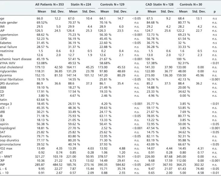
Heart Failure Registry; their clinical characteristics are given inTable I. Each patient’s medical history, heart rate, systolic blood pressure, body mass index, NYHA class, and medications were recorded. All patients underwent con-ventional 2D and TDI echocardiography in an ambulatory setting and under resting conditions. Clinical follow-up was performed every 6 months, for a mean of 384¡254 days of follow-up. Clinical follow-up was anticipated in cases of worsening decompensated heart failure. Patients were retrospectively analyzed according to the presence of statin therapy (N = 224, 63.6% of the study population) and the presence of coronary heart disease (158 patients with a history of previous myocardial infarction, known coronary artery disease, prior percutaneous coronary interventions [PCIs] and coronary artery bypass grafting [CABG]). Of 158 ischemic subjects, 128 were treated with statins. The incidence of major adverse cardiac events (e.g., cardiac death, readmission for HF and ventricular arrhythmias) was evaluated by direct clinical examination or by direct interrogation of the patient’s relatives. Cardiac death was considered in cases of sudden death or death associated with documented myocardial infarction, congestive HF or malignant ventricular arrhythmias.
Table I -Clinical characteristics.
All Patients N = 353 Statin N = 224 Controls N = 129 CAD Statin N = 128 Controls N = 30
Mean Std. Dev. Mean Std. Dev. Mean Std. Dev. p Mean Std. Dev. Mean Std. Dev. p
age 66.0 12.2 67.0 10.4 64.1 14.7 ,0.05 67.5 9.2 68.4 13.1 n.s
male gender 69.52% 70.37 % 70.16 % n.s 84.68 % 80.77 % n.s.
BMI 29.5 5.0 29.9 4.4 28.9 6.0 n.s. 29.2 4.2 28.2 4.2 n.s.
SAP 126.5 24.5 126.4 25.3 126.3 23.5 n.s. 124.7 25.6 122.2 22.7 n.s.
hypertension 68.62 % 75.23 % 56.30 % ,0.001 72.73 % 69.23 % n.s
COPD 52.23 % 56.19 % 45.45 % n.s. 56.78 % 61.54 % n.s.
diabetes 31.45 % 36.19 % 22.60 % ,0.05 36.75 % 32 % n.s
renal failure 28.57 % 31.37 % 22.88 % n.s 36.28 % 33.33 % n.s.
creatinine 1.5 0.6 0.3 0.5 0.2 0.4 n.s. 1.5 0.6 1.6 0.5 n.s
Hb 12.6 2.0 12.7 1.9 12.6 2.1 n.s. 13.0 2.0 12.7 2.3 n.s.
ischemic heart disease 45.19 % 57.41 % 21.67 % ,0.001 100 % 100 % n.s.
NYHA III/IV 53.08% 49.53 % 58.68% n.s. 57.38% 92.31% ,0.01
total cholesterol 179.13 42.50 164.11 45.25 173.00 45.53 n.s. 141.33 37.90 133.00 0.00 n.s. triglycerides 118.31 36.85 131.29 23.78 97.80 48.69 n.s. 122.00 36.77 48.00 0.00 n.s glycemia 152.15 81.53 147.14 101.12 147.20 80.29 n.s. 215.00 136.30 159.50 45.96 n.s.
atrial fibrillation 19.19 % 14.55 % 25.81 % ,0.05 10.74 % 42.13 % ,0.001
QRS duration 86.9 36.6 88.6 37.3 86.1 35.4 n.s. 85.2 37.5 88.4 36.2 n.s.
LBBB 19.10 % 18.27 % 21.49 % n.s. 14.88 % 20.00 % n.s.
ICD 17.91 % 17.54 % 19.33 % n.s. 23.33 % 34.62 % n.s.
CRT 3.80 % 4.67 % 2.46 % n.s. 4.96 % 0.00 % n.s.
statin 63.64 %
omega 3 18.45 % 26.51 % 4.20 % ,0.001 35.77 % 3.85 % ,0.01
ACE – I 45.35 % 48.36 % 39.63 % n.s. 59.17 % 53.85 % n.s.
ARB 30.21 % 33.49 % 24.17 % n.s. 21.67 % 7.69 % n.s.
BB 71.18 % 75.93 % 63.11 % ,0.05 78.05 % 80.77 % n.s.
CCB 18.13 % 21.05 % 13.33 % n.s. 13.22 % 3.85 % n.s.
aspirin 56.38 % 59.62 % 50.41 % n.s. 72.95 % 50.00 % ,0.05
clopidogrel 18.81 % 27.70 % 2.52 % ,0.001 47.54 % 3.85 % ,0.001
OAT 25.82 % 25.82 % 25.62 % n.s. 14.75 % 34.62 % ,0.05
diuretics 79.71 % 83.80 % 72.95 % ,0.05 82.11 % 92.31 % n.s.
loop diuretics 77.17 % 80.20 % 71.30 % n.s. 83.19 % 95.88 % n.s.
spironolactone 39.52 % 40.74 % 37.93 % n.s. 43.09 % 66.67 % ,0.05
VO2 max 13.49 4.35 13.39 4.03 13.92 4.88 n.s. 14.07 4.44 14.45 4.31 n.s.
AT 0.92 0.74 0.85 0.28 1.06 1.20 n.s. 0.91 0.28 0.80 0.17 n.s.
6 – MWT 321.27 103.19 221.00 50.95 378.57 76.91 ,0.01 226.00 87.68 345.00 0.00 n.s.
CRP 10.36 21.22 6.73 13.02 14.49 29.41 n.s. 9.68 17.59 112.00 0.00 ,0.001
BNP 285.87 484.52 185.76 273.16 390.35 584.82 ,0.05 240.17 367.93 364.25 302.32 n.s. IL – 6 9.95 22.27 7.87 15.44 15.71 35.74 n.s. 9.47 21.07 61.43 74.40 ,0.05
All patients provided informed consent. This study was approved by the Ethics Committee of University of Foggia according the ethical standards for experiments in human subjects established by the Declaration of Helsinki.
Inflammatory markers. Circulating levels of 6 and IL-10 were evaluated in all patients using a commercially available solid-phase, enzyme-labeled, chemiluminescent sequential immunometric assay (Immulite 1000 system, Siemens). The components of this system were a matched pair of beads: one coated with a monoclonal murine anti-IL-6 antibody and one coated with the equivalent anti-IL-10 antibody. Biochemical levels were measured using optimal concentrations of standards and antibodies according to the manufacturer’s instructions. Incubation cycles were either twice for 30 minutes (IL-6) or once for 60 minutes.
The concentration of CRP was determined using a particle-enhanced turbidimetric immunoassay with an assay range of 0.2 to 12 mg/dl (The Dimension Flex Immunoassay System, Dade Behring, Inc., IL, USA).
Echocardiography. Conventional echocardiography was used to assess LV dimensions and ejection fraction (EF), peak velocities of transmitral early (E) and late diastolic (A) LV (left ventricle) filling, the ratio of transmitral early to late LV filling velocity (E/A ratio), and E-deceleration time (EDT). TDI measurements recorded at the mitral annulus in the apical four-chamber view included systolic velocity (S’), early (E’) and late (A’) diastolic velocities, and the ratio of early to late diastolic velocity (E’/A’). The transmitral to mitral annular early diastolic velocity ratio (E/E’) was also calculated (Figure 1).
The time to regional peak systolic velocity (TP: the beginning of the QRS complex was used as the reference point), St (the end of the A’ wave to the end of the S wave) and ET (the start to the end of the S wave) were also measured by TDI (Figure 1).
Transthoracic echocardiography was performed using iE33 (Philips Medical Systems, Andover, MA, USA). All echocardiographic studies were performed and interpreted by experienced physicians. The LV dimensions and LVEF were calculated using recommendations from the combined ASE/ESC guidelines. The LVEF was calculated according to Simpson’s rule. Pulsed Doppler mitral inflow velocities were obtained by placing a 1- to 2-mm sample volume between the tips of the mitral leaflets in the apical four-chamber view. The Doppler beam was aligned parallel to the direction of flow.
TDI was performed using apical views for the long-axis motion of the ventricles, as previously described.12 Two-dimensional echocardiography with TDI color imaging was performed using an S5-1 Sector Array Transducer with PureWave Crystal Technology (5 to 1 MHz). Two-dimen-sional echocardiography with TDI color imaging views was optimized for pulse repetition frequency, color saturation, sector size, and depth and was allowed the highest possible frame rate. At least 3 consecutive beats were stored, and the images were analyzed offline with the aid of a customized software package (QLAB quantification software, Philips). The peak myocardial velocity during the ejection phase (S’) and the time to peak Sm (TP) were measured with reference to the QRS complex.
STATISTICAL ANALYSES
Continuous variables are expressed as mean¡standard
deviation; categorical variables are presented as percen-tages. Mean values were compared using Student’s t-test for variables with a normal distribution or using the Mann-Whitney U non-parametric test for non-normally distribu-ted variables. Percentages were compared using a x2 test. Event-free survival was shown with Kaplan-Meier curves
and compared using the log-rank test. Univariate results were adjusted in a multivariate analysis for age, gender, LV ejection fraction (LVEF) and other significant factors according to a Cox regression analysis. A p,0.05 was considered to be statistically significant.
RESULTS
353 patients were enrolled in the registry; 224 (63%) were treated with statins. A total of 77.3% (N = 172) were treated with atorvastatin (mean daily dose 30.2¡16.3 mg), 2.3% (N = 4) with pravastatin (mean daily dose 26.7¡10.3 mg),
15.7% (N = 33) with simvastatin (mean daily dose
19.7¡6.3 mg), 4.2% (N = 14) with rosuvastatin (mean daily
dose 10.6¡3.9 mg), and 0.5% (N = 1) with fluvastatin. In the
subgroup of patients with ischemic heart disease (N = 158, 45%), 128 were treated with statins: 79% (N = 101) were treated with atorvastatin (mean daily dose 35.3¡18.1 mg),
2% (N = 2) with pravastatin (mean daily dose 30.0¡14.1 mg),
16.9% (N = 22) with simvastatin (mean daily dose
19.5¡8.0 mg), and 2.4% (N = 3) with rosuvastatin (mean
daily dose 13.3¡5.8 mg).
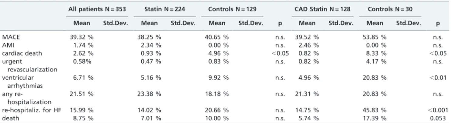
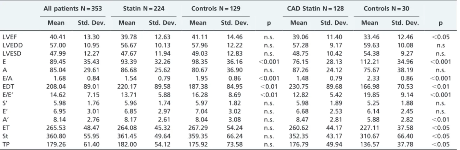
In a retrospective, non-randomized analysis, therapy with statins was associated with a lower incidence of cardiac death (1% vs. 5%, p,0.05) (Table II). The association remained statistically significant even after correction in a multivariable analysis for age, gender, LVEF, cholesterol levels, and therapy with ACE inhibitors and beta blockers. No correla-tion was seen between statin dose and outcome at follow-up. Patients prescribed statins exhibited lower values of E/A (1.53¡0.79 vs. 1.95¡0.86, p,0.001) and E/E’ (13.71¡5.88
vs. 16.28¡8.69, p,0.01), as well as higher values of EDT (220.17¡89.58 vs. 187.38¡84.95 ms, p,0.01) (Table III).
In a subgroup analysis of 150 patients with ischemic HF, those receiving therapy with statins showed a lower incidence of readmissions for worsening HF (15% vs. 46%, p,0.001), ventricular arrhythmias (5% vs. 21%, p,0.01), and cardiac death (1% vs. 8%, p,0.05) (Table II); lower circulating levels of inflammatory markers (IL-6: 9.47¡21.06
vs. 61.43¡74.39 pg/ml, p,0.05; IL-10: 0.65¡1.99 vs. 5.0¡4.58 pg/ml, p,0.01; and CRP: 9.68¡17.59 vs. 112 mg/ dl, p,0.001); a lower E/E’ ratio (12.82¡5.42 vs. 19.85¡9.14,
p,0.001); longer systolic time intervals (ET: 260.62¡44.16
vs. 227.11¡37.58 ms, p,0.05 and St 352.35¡43.17 vs.
310.67¡66.46¡37.78 ms, p,0.05); a higher value of TP (176.79¡49.93 vs. 136.7¡37.78 ms, p,0.05); a lower E/A ratio (1.48¡0.79 vs. 2.33¡0.85, p,0.001); and a higher EDT
(230.75¡89.67 vs. 166.98¡70.52 ms, p,0.01) (Table III).
Episodes of decompensated HF requiring rehospitaliza-tion were related to E (odds ratio [OR] 1.33, 95% confidence interval [CI] 1.11-1.59, p,0.01); A (OR 0.81, 95% CI 0.65-0.99, p,0.05); E/A (OR 1.37, 95% CI 1.16-1.63, p,0.001); EDT (OR 0.72, 95% CI 0.60-0.87, p,0.001); E/E’ (OR 1.46, 95% CI 1.23-1.74, p,0.001); S (OR 0.78, 95% CI 0.65-0.94, p,0.01); E’ (OR 0.85, 95% CI 0.71-1.01, p = 0.07); and A’ (OR 0.82, 95% CI 0.67-1.01, p = 0.06). Using multivariable analysis correlation, only E/E’ (p,0.01) remained significant.
Kaplan-Meier survival analysis showed a poorer prognosis in subjects not receiving statin therapy (log-rank p,0.01), with higher rates of rehospitalization for decompensated HF (Figure 2). Using multivariate Cox regression analysis, higher readmission rates in subjects not receiving statins were independent of age, gender, LVEF, NYHA class, cholesterol levels, presence of atrial fibrillation, or concomitant therapy with ACE inhibitors, beta blockers,v3 fatty acids, clopido-grel, aspirin or spironolactone (p,0.01) (Figure 3).
DISCUSSION
Several observational studies of HF cohorts have linked statin therapy to an improved survival rate.10-15However, the beneficial mechanisms have not been completely established. Small, prospective clinical studies on atorvas-tatin and simvasatorvas-tatin in systolic HF have documented an improved ventricular systolic function and decreased levels of inflammatory biomarkers after statin therapy.29,13 Previous evidence has shown that, in patients with prior myocardial infarction, statin therapy initiated prior to hospital discharge significantly reduced subsequent hospi-talizations for HF;14 furthermore, the initiation and
main-tenance of treatment with statins has been associated with better survival in patients with LV systolic dysfunction.15In a recent report, Sankaranarayanan et al.16 showed that mortality in patients with ischemic CHF treated with statins was significantly lower than in controls. Univariate analysis also showed fewer HF readmissions (7% vs. 32%) and HF deaths (4% vs. 13%), with effects independent of cholesterol levels, age, sex, drugs, revascularization, and implantable cardioverter-defibrillator or cardiac resynchronization ther-apy using multivariable analysis.
Our results corroborate the above evidence; therapy with statins was associated with a lower incidence of cardiac death, and the association remained significant even after correction in a multivariable analysis for age, gender, LVEF and therapy with ACE inhibitors and beta blockers. Furthermore, ischemic HF patients receiving therapy with
Table II -Clinical outcomes.
All patients N = 353 Statin N = 224 Controls N = 129 CAD Statin N = 128 Controls N = 30
Mean Std.Dev. Mean Std.Dev. Mean Std.Dev. p Mean Std.Dev. Mean Std.Dev. p
MACE 39.32 % 38.25 % 40.65 % n.s. 39.52 % 53.85 % n.s.
AMI 1.74 % 2.34 % 0.00 % n.s. 2.46 % 0.00 % n.s.
cardiac death 2.62 % 0.93 % 4.96 % ,0.05 0.82 % 8.33 % ,0.05
urgent
revascularization
0.58% 0.47 % 0.83 % n.s. 0.82 % 4.17 % n.s.
ventricular arrhythmias
6.71 % 5.16 % 9.92 % n.s. 4.96 % 20.83 % ,0.01
any re-hospitalization
21.51 % 23.38 % 18.18 % n.s. 21.31 % 20.83 % n.s.
re-hospitaliz. for HF 15.99 % 14.02 % 20.66 % n.s. 14.75 % 45.83 % ,0.001
statins showed a lower incidence of readmissions for worsening HF. In a multivariate Cox regression analysis, higher readmission rates in subjects not receiving statins were independent of age, gender, LVEF, and concomitant therapy with ACE inhibitors and beta blockers.
In previous research, statins have appeared to provide similar benefits to patients with non-ischemic and ischemic cardiomyopathy;17 our results, however, suggest a wider benefit of statin therapy in patients with coronary heart disease. In fact, those receiving therapy with statins, in addition to a lower incidence of readmissions for worsening HF, also demonstrated a lower incidence of cardiac deaths (1% vs. 8%) and ventricular arrhythmias (5% vs. 21%).
Previous studies have shown that non-antiarrhythmic drugs, such as those acting on the renin-angiotensin-aldosterone system, fish oil, and statins, can reduce the likelihood of future ventricular tachycardia/ventricular fibrillation in patients with coronary artery disease or congestive HF.18In our study, among patients with ischemic
HF, those receiving therapy with statins showed a lower incidence of ventricular arrhythmias than controls.
The mechanisms by which statins exert their positive effects are not completely known. In our study, statin administration was related to a better LV performance as measured by TDI. There is evidence that abnormal para-meters identified by TDI can identify subjects at risk for
Table III -Echocardiographic characteristics.
All patients N = 353 Statin N = 224 Controls N = 129 CAD Statin N = 128 Controls N = 30
Mean Std. Dev. Mean Std. Dev. Mean Std. Dev. p Mean Std. Dev. Mean Std. Dev. p
LVEF 40.41 13.30 39.78 12.63 41.11 14.46 n.s. 39.06 11.40 33.46 12.46 ,0.05
LVEDD 57.00 10.95 56.67 10.13 57.96 12.22 n.s. 57.28 9.17 59.63 10.08 n.s
LVESD 47.99 12.27 47.67 11.94 49.03 12.83 n.s. 48.75 10.42 54.38 9.27 n.s. E 89.45 35.43 93.39 32.26 98.35 36.16 ,0.001 76.15 28.13 112.21 34.96 ,0.001
A 85.04 29.61 86.68 25.62 80.67 36.90 n.s. 87.26 24.12 75.67 38.19 n.s.
E/A 1.68 0.84 1.54 0.79 1.95 0.86 ,0.001 1.48 0.79 2.33 0.86 ,0.001
EDT 208.04 89.01 220.17 89.58 187.38 84.95 ,0.01 230.75 89.68 166.98 70.53 ,0.01
E/E’ 14.62 7.15 13.71 5.88 16.28 8.69 ,0.01 12.82 5.42 19.85 9.14 ,0.001
S’ 5.98 1.76 5.96 1.74 5.97 1.82 n.s. 5.98 1.89 5.25 1.88 n.s.
E’ 6.95 3.01 6.85 2.97 7.04 3.02 n.s. 6.68 2.53 6.14 2.45 n.s.
A’ 8.14 2.76 8.17 2.61 8.04 3.08 n.s. 8.47 2.81 5.88 2.82 ,0.01
ET 265.53 48.47 264.08 45.32 267.29 54.24 n.s. 260.62 44.17 227.11 37.58 ,0.05 St 360.80 55.95 361.45 49.64 359.35 66.24 n.s. 352.35 43.17 310.67 66.40 ,0.05 TP 179.26 61.40 182.00 54.12 175.92 73.58 n.s. 176.79 49.94 136.57 37.78 ,0.05
adverse events in major cardiac diseases, such as HF. In particular, patients with reduced S’ or E’ values of,3 cm/s have an extremely poor prognosis. During HF and after myocardial infarction, non-invasive assessment of LV diastolic pressure by transmitral to mitral annular early diastolic velocity ratio (E/E’) is a strong predictor of prognosis, especially when E/E’ is$15. Several researchers have assessed the effect of statin therapy on LV function by TDI and showed that compared with baseline, S’ and E’ increased significantly after atorvastatin therapy in patients with coronary artery disease.19 In our study, HF patients administrated statins had lower values of E/A and E/E’ and higher values of EDT. Furthermore, we saw lower values of the E/E’ ratio and higher EDT, suggesting a minor grade of diastolic dysfunction, in ischemic CHF patients receiving therapy with statins. In addition, TDI can be used to measure ST and ET intervals in a noninvasive, geome-trically independent, easily applicable fashion.14-19 Few authors have evaluated these intervals in CHF patients; Reant P et al.8 showed that ST intervals could be used to
detect alterations in LV systolic function. Chen HM et al. determined the cutoff values for ET in predicting high N-terminal pro-brain natriuretic peptide (NT-proBNP) levels.9 To our knowledge, there have been no previous studies evaluating the effect of statin therapy on ST intervals detected by TDI. In the present work, we showed higher values of ET and ST in ischemic CHF patients receiving therapy with statins. Therefore, lower values of ST intervals, as for ET, in patients not receiving statin therapy also suggest a higher grade of LV diastolic dysfunction, as previously demonstrated by Jarnert et al.20In statin therapy
patients, we were also able to show a smaller grade of diastolic dysfunction than in controls by conventional Doppler (E/A and EDT), TDI (E/E’) and longer systolic intervals (ST and ET). In addition to lipid-lowering and anti-atherosclerotic effects, statins have demonstrated other non-lipid related or pleiotropic effects that could be beneficial in HF. Statins may inhibit or reverse myocardial remodeling,21-22 inhibit inflammation in HF, improve endothelial function,11,23,24 and restore autonomic nervous system balance.25Furthermore,
statin treatment may lower inflammatory activation in several cardiac diseases. ‘‘Increased circulating and intra-cardiac levels of pro-inflammatory cytokines have been associated with CHF. Following an initial insult, the increased produc-tion of pro-inflammatory cytokines, including TNF-alpha, IL-6, IL-1, and IL-18, jeopardizes the surrounding tissue through propagation of the inflammatory response and direct effects on the cardiac myocyte structure and function. Cardiac myocyte hypertrophy, contractile dysfunction, cardiac myo-cyte apoptosis, and extracellular matrix remodeling contri-bute enormously to the development and progression of CHF’’.1 Previous authors have shown that atorvastatin treatment may induce a significant decrease in the activity of matrix metalloproteinase-9, high-sensitivity CRP, TNF-alpha, IL-6, and malondialdehyde, and a significant increase in endothelial superoxide dismutase activity compared with the placebo. In CHF patients, atorvastatin therapy has been associated with a decrease in inflammation (as measured by IL-6 and CRP levels) and extracellular matrix remodeling, improving both endothelial function and exercise capacity.26
Atorvastatin treatment significantly decreased the concentra-tion of IL-6 in patients with dilated cardiomyopathy after two months of therapy.27,28Recently, Amir et al.29showed that
elevated circulating IL-10 levels in systolic HF patients did not have a protective counterbalance effect on mortality. In addition, Wang et al.30demonstrated that plasma levels of
IL-10 were higher in HF patients than controls. In contrast, other studies31have shown a better event-free survival rate with higher IL-10 levels in hospitalized CHF patients. In a retrospective analysis, the authors found a significant interaction between hs-CRP and the effect of rosuvastatin for most end points whereby rosuvastatin treatment was associated with better outcomes in patients with hs-CRP $2.0 mg/L.32In addition, Liu et al.33have shown that twelve weeks of treatment with atorvastatin (10 mg/day) signifi-cantly decreased serum CRP.
Our findings appear to confirm these previous results; in ischemic CHF patients receiving statin therapy, circulating levels of the inflammatory markers IL-6, IL-10 and CRP were significantly lower, thus supporting an additional hypothesis for the mechanism of the better outcomes seen in HF patients. It still remains not completely clear whether statin administration improves LV performance (evaluated in our study by TDI) through reducing inflammatory activation featuring LV dysfunction or blunted inflamma-tory activation associated with statin therapy may some-times positively affect LV performance.
LIMITATIONS
These are preliminary data from a non-randomized observational registry with a limited number of patients. Statin administration depended on clinician judgment. Further, randomized trials are needed to confirm these results.
CONCLUSIONS
Ischemic outpatients with CHF undergoing statin treat-ment showed fewer readmissions, a blunted inflammatory activation and a better LV performance as assessed by TDI.
REFERENCES
1. Hedayat M, Mahmoudi MJ, Rose NR, Rezaei N. Proinflammatory cytokines in heart failure: double-edged swords. Heart Fail Rev. 2010;15:543-62, doi: 10.1007/s10741-010-9168-4.
2. Pai RG, Gill KS. Amplitudes, durations and timings of apically directed left ventricular myocardial velocities: I. Their normal pattern and coupling to ventricular filling and ejection. J Am Soc Echocardiogr. 1998;11:105-11, doi: 10.1016/S0894-7317(98)70067-7.
3. Reant P, Dijos M, Donal E, Mignot A, Ritter P, Bordachar P, et al. Systolic time intervals as simple echocardiographic parameters of left ventricular systolic performance: correlation with ejection fraction and longitudinal two-dimensional strain. Eur J Echocardiogr. 2010;11:834-44, doi: 10.1093/ ejechocard/jeq084.
4. Cheng HM, Chuang SY, Hsu PF, Chou P, Chen CH. Systolic time intervals revisited: correlations with N-terminal pro-brain natriuretic peptide in a community population. Heart Vessels. 2005;20:256-63, doi: 10.1007/s00380-005-0844-6.
5. Dilaveris P, Giannopoulos G, Riga M, Synetos A, Stefanadis C. Beneficial effects of statins on endothelial dysfunction and vascular stiffness. Curr Vasc Pharmacol. 2007;5:227–37, doi: 10.2174/157016107781024091. 6. Levy WC. Observational studies of statins in systolic heart failure. Heart
Fail Clin. 2008;4:201–8, doi: 10.1016/j.hfc.2008.01.006.
7. Node K, Fujita M, Kitakaze M, Hori M, Liao JK. Short-term statin therapy improves cardiac function and symptoms in patients with idiopathic dilated cardiomyopathy. Circulation. 2003;108:839–43, doi: 10.1161/01. CIR.0000084539.58092.DE.
8. Bielecka-Dabrowa A, Goch JH, Mikhailidis DP, Rysz J, Maciejewski M, Banach M. The influence of atorvastatin on parameters of inflammation and function of the left ventricle in patients with dilated cardiomyo-pathy. Med Sci Monit. 2009;15:MS12-23.
9. Krum H, Latini R, Maggioni AP, Anand I, Masson S, Carretta E, et al. Statins and symptomatic chronic systolic heart failure: a post-hoc analysis of 5010 patients enrolled in Val-HeFT. Int J Cardiol 2007;119:48–53, doi: 10.1016/j.ijcard.2006.07.106.
10. Khush KK, Waters DD, Bittner V, Deedwania PC, Kastelein JJ, Lewis SJ, et al. Effect of high-dose atorvastatin on hospitalizations for heart failure: subgroup analysis of the Treating to New Targets (TNT) study. Circulation. 2007;115:576–83, doi: 10.1161/CIRCULATIONAHA.106. 625574.
11. Horwich TB, MacLellan WR. Atorvastatin and statins in the treatment of heart failure. Expert Opin Pharmacother. 2007;8:3061-8, doi: 10.1517/ 14656566.8.17.3061.
12. Yu CM, Chau E, Sanderson JE, Fan K, Tang MO, Fung WH, et al. Tissue Doppler echocardiographic evidence of reverse remodeling and improved synchronicity by simultaneously delaying regional contraction after biventricular pacing therapy in heart failure. Circulation. 2002;105:438-45, doi: 10.1161/hc0402.102623.
13. Kishi T, Yamada A, Okamatsu S, Sunagawa K. Atorvastatin might improve ventricular electrostability and decelerate the deterioration of renal function in patients with heart failure and diabetes mellitus. J Cardiol. 2009;53:341-8, doi: 10.1016/j.jjcc.2008.12.002.
14. Aronson D, Mutlak D, Lessick J, Kapeliovich M, Dabbah S, Markiewicz W, et al. Relation of statin therapy to risk of heart failure after acute myocardial infarction. Am J Cardiol. 2008;102:1706-10, doi: 10.1016/j. amjcard.2008.07.057.
15. Huan Loh P, Windram JD, Tin L, Reddy P, Velavan P, Rigby AS, et al. The effects of initiation or continuation of statin therapy on cholesterol level and all-cause mortality after the diagnosis of left ventricular systolic dysfunction. Am Heart J. 2007;153:537-44, doi: 10.1016/j.ahj.2007.01.029. 16. Sankaranarayanan R, Maini S, James MA, Burtchaell S, Chatterjee AK. Do statins improve heart failure outcome in post-myocardial infarction patients with moderate to severe left ventricular dysfunction? Congest Heart Fail. 2010;16:181-6, doi: 10.1111/j.1751-7133.2010.00165.x. 17. Dickinson MG, Ip JH, Olshansky B, Hellkamp AS, Anderson J, Poole JE,
et al. Statin use was associated with reduced mortality in both ischemic and nonischemic cardiomyopathy and in patients with implantable defibrillators: mortality data and mechanistic insights from the Sudden Cardiac Death in Heart Failure Trial (SCD-HeFT). Am Heart J. 2007;153:573-8, doi: 10.1016/j.ahj.2007.02.002.
18. Das MK, Zipes DP. Antiarrhythmic and nonantiarrhythmic drugs for sudden cardiac death prevention. J Cardiovasc Pharmacol. 2010;55:438-49.
19. Qie L, Meng X, Wang Y, Feng M, Zhong M, Li L. Assessment of regional systolic and diastolic functions affected by atorvastatin in coronary artery disease using tissue Doppler imaging. Clin Cardiol. 2008;31:551-5, doi: 10.1002/clc.20287.
20. Jarnert C, Mejhert M, Ring M, Persson H, Edner M. Doppler tissue imaging in congestive heart failure patients due to diastolic or systolic dysfunction: a comparison with Doppler echocardiography and the atrio-ventricular plane displacement technique. Eur J Heart Fail. 2000;2:151-60, doi: 10.1016/S1388-9842(00)00075-1.
21. Wassmann S, Laufs U, Baumer AT, Mu¨ller K, Konkol C, Sauer H, et al. Inhibition of geranylgeranylation reduces angiotensin II-mediated free radical production in vascular smooth muscle cells: involvement of angiotensin AT1 receptor expression and Rac1 GTPase. Mol Pharmacol. 2001;59:646–54.
23. Hernandez-Perera O, Perez-Sala D, Navarro-Antolin J Sa´nchez-Pascuala R, Herna´ndez G, Dı´az C, Lamas S. Effects of the 3-hydroxy-3-methylglutaryl-CoA reductase inhibitors, atorvastatin and simvastatin, on the expression of endothelin-1 and endothelial nitric oxide synthase in vascular endothelial cells. J Clin Invest. 1998;101:2711–9, doi: 10.1172/ JCI1500.
24. Saijonmaa O, Nyman T, Stewen P, Fyhrquist F. Atorvastatin completely inhibits VEGF-induced ACE upregulation in human endothelial cells. Am J Physiol Heart Circ Physiol, 2004;286:H2096–102, doi: 10.1152/ ajpheart.00894.2003.
25. Horwich TB, MacLellan WR. Atorvastatin and statins in the treatment of heart failure. Expert Opin Pharmacother. 2007;8:3061-8, doi: 10.1517/ 14656566.8.17.3061.
26. Castro PF, Miranda R, Verdejo HE, Greig D, Gabrielli LA, Alcaino H, et al. Pleiotropic effects of atorvastatin in heart failure: role in oxidative stress, inflammation, endothelial function, and exercise capacity. J Heart Lung Transplant. 2008;27:435-41, doi: 10.1016/j.healun.2008.01.012. 27. Bielecka-Dabrowa A, Goch JH, Mikhailidis DP, Rysz J, Maciejewski M,
Banach M. The influence of atorvastatin on parameters of inflammation and function of the left ventricle in patients with dilated cardiomyo-pathy. Med Sci Monit. 2009;15:MS12-23.
28. Stumpf C, Petzi S, Seybold K, Wasmeier G, Arnold M, Raaz D, et al. Atorvastatin enhances interleukin-10 levels and improves cardiac
function in rats after acute myocardial infarction. Clin Sci. 2009;116:45-52, doi: 10.1042/CS20080042.
29. Amir O, Rogowski O, David M, Lahat N, Wolff R, Lewis BS. Circulating interleukin-10: association with higher mortality in systolic heart failure patients with elevated tumor necrosis factor-alpha. Isr Med Assoc J. 2010;12:158-62.
30. Wang Y, Zhou Y, Meng L, Lu X, Ou N, Li X. Inflammatory mediators in Chinese patients with congestive heart failure. J Clin Pharmacol. 2009;49:591-9, doi: 10.1177/0091270009333265.
31. Parissis JT, Farmakis D, Nikolaou M, Birmpa D, Bistola V, Paraskevaidis I, et al. Plasma B-type natriuretic peptide and anti-inflammatory cytokine interleukin-10 levels predict adverse clinical outcome in chronic heart failure patients with depressive symptoms: a 1-year follow-up study. Eur J Heart Fail. 2009;11:967-72, doi: 10.1093/eurjhf/hfp125.
32. McMurray JJ, Kjekshus J, Gullestad L, Dunselman P, Hjalmarson A, Wedel H, et al. Effects of statin therapy according to plasma high-sensitivity C-reactive protein concentration in the Controlled Rosuvastatin Multinational Trial in Heart Failure (CORONA): a retrospective analysis. Circulation. 2009;120:2188-96, doi: 10.1161/CIRCULATIONAHA.109.849117. 33. Liu M, Wang F, Wang Y, Jin R. Atorvastatin improves endothelial