CLINICAL SCIENCE
Longitudinal reference ranges for fetal ultrasound
biometry in twin pregnancies
Adolfo Wenjaw Liao, Maria de Lourdes Brizot, Helenice Ju´lio Kang, Renata Almeida Assunc¸a˜o, Marcelo Zugaib
Hospital das Clı´nicas, Instituto Central, Faculdade de Medicina da Universidade de Sa˜o Paulo, Department of Obstetrics and Gynecology.
OBJECTIVE:The purpose of this study was to establish longitudinal reference ranges for fetal ultrasound biometry measurements and growth parameters in twin pregnancies.
METHOD: A total of 200 uncomplicated twin pregnancies before 21 weeks of gestation were recruited for this prospective, longitudinal study. Women who abandoned follow-up, pregnancies with unknown outcomes or pregnancies with complications were excluded. Ultrasound scans were performed every three weeks, and biparietal and occipitofrontal diameters, head and abdominal circumferences, and femur diaphysis length measurements were obtained for each fetus at each visit. Estimated fetal weight, biparietal/occipitofrontal diameter, head circumference/abdominal circumference, and femur diaphysis length/abdominal circumference ratios were also calculated. Multilevel regression analysis was performed on normalized data.
RESULTS:A total of 807 ultrasound examinations were performed in 125 twin pregnancies between 14 and 38 weeks of gestation (6.5¡1.4 scans/pregnancy). Regression analysis demonstrated significant correlations for all variables with gestational age, namely log of the biparietal diameter (r = 0.98), log of the occipitofrontal diameter (r = 0.98), log of the head circumference (r = 0.99), log of the abdominal circumference (r = 0.98), square root of the femur length (r = 0.99), log of the estimated fetal weight (r = 0.99), biparietal/occipitofrontal ratio (r = -0.11), head/abdomen circumference ratio (r = -0.56), and log of the femur length/abdominal circumference ratio (r = 0.61). Values corresponding to the 10th, 50th, and 90th percentiles for estimated fetal weight at 28, 32, and 36 weeks, respectively, were as follows: 937, 1,096, 1,284 g; 1,462, 1,720, 2,025 g; and 2,020, 2,399, 2,849 g.
CONCLUSION: In twin pregnancies, fetal ultrasound biometry measurements and growth parameters show a significant correlation with gestational age.
KEYWORDS: Twins; Pregnancy; Ultrasonography; Biometry; Reference Values.
Liao AW, Brizot ML, Kang HJ, Assunc¸a˜o RA, Zugaib M. Longitudinal reference ranges for fetal ultrasound biometry in twin pregnancies. Clinics. 2012;67(5):451-455.
Received for publication onDecember 26, 2011;First review completed onJanuary 5, 2012;Accepted for publication onJanuary 19, 2012 E-mail: [email protected]
Tel.: 55 11 2661 6209
INTRODUCTION
The mean birth weight adjusted for gestational age in twin pregnancies is lower compared to singletons at the end of the second trimester (1). Nevertheless, neonatal mortality appears to be similar in both groups (2). Therefore, it is unclear whether smaller growth in twins should be interpreted as normal or pathological (3,4).
Fetal size and weight can be evaluated prenatally by ultrasound. However, most centers still use singleton reference charts in the assessment of twin pregnancies, which leads to frequent diagnosis of fetal growth restriction
(5). Under these circumstances, subsequent management usually includes serial growth scans and fetal well-being tests. These measures will inevitably increase parental anxiety and financial costs. Moreover, false-positive results may eventually lead to mismanagement of the pregnancy and unnecessary iatrogenic deliveries.
The aim of this study was to establish longitudinal reference ranges for ultrasound fetal biometry and growth parameters in uncomplicated, twin pregnancies from our study population.
MATERIALS AND METHODS
This was a prospective study conducted at the Twin’s Clinic, Hospital das Clı´nicas da Faculdade de Medicina da Universidade de Sa˜o Paulo, Brazil, Department of Obstetrics and Gynecology, between May 2007 and June 2010. The study protocol was approved by the hospital’s ethics committee (418/04).
Copyrightß2012CLINICS– This is an Open Access article distributed under
the terms of the Creative Commons Attribution Non-Commercial License (http:// creativecommons.org/licenses/by-nc/3.0/) which permits unrestricted non-commercial use, distribution, and reproduction in any medium, provided the original work is properly cited.
Participants
Women at less than 21 weeks of gestation and with an uncomplicated, naturally conceived, diamniotic, twin preg-nancy were included in the study. Those who abandoned follow-up, pregnancies with unknown outcomes or preg-nancies that were complicated by oligohydramnios, increased umbilical artery pulsatility index (above the 95th percentile), pre-eclampsia, gestational diabetes, placenta previa, twin-to-twin transfusion syndrome, fetal structural malformation, chromosomal abnormalities, or fetal death were excluded.
Pregnancy outcome information was obtained from hospital notes and delivery records or by direct phone contact with the patients. Chorionicity was confirmed by histological examination of the placenta after delivery.
Gestational age was calculated from the first day of the last menstrual period (LMP) and confirmed either by an ultrasound crown-rump length measurement during the first trimester or by an estimate based on multiple ultrasound parameters (biparietal diameter [BPD], head circumference [HC], abdominal circumference [AC] and femur length [FL]) of the larger fetus during the second trimester. When the first day of the LMP was uncertain or unknown, or when there was a discrepancy between gestational age based on the LMP and ultrasound dates, gestational age was determined based on the earliest ultrasound findings.
Ultrasonography protocol
At the first evaluation, each twin was defined as ‘‘1’’ or ‘‘2’’ according to the relative position of its amniotic sac and the uterine internal cervical os. This definition was used throughout all subsequent examinations, which were performed every three weeks. Scans were carried out transabdominally with a 3.5-MHz curvilinear transducer and a Corevision SSA-350A (Toshiba, Japan), Envisor (Philips, Netherlands), or Voluson (General Electric, Austria) ultrasound machine.
At each visit, BPD, occipitofrontal diameter (OFD), HC, AC, and FL measurements were obtained for each fetus according to standard techniques (Figure 1) (6). Fetal weight was estimated according to the mathematical formula proposed by Hadlock et al. (7), and BPD/OFD, HC/AC, and FL/AC ratios were calculated for each fetus.
Statistical analysis
All data were prospectively recorded in a computer fetal database system and exported to a Microsoft Excel spreadsheet. Statistical analysis was performed with MLwiN version 2.19 (Centre for Multilevel Modelling, University of Bristol, United Kingdom).
The data were tested for normal distributions using the Kolmogorov-Smirnov test, and variables that were not normally distributed underwent transformation. Multilevel regression analysis was performed to examine the associa-tions between each parameter and gestational age and to construct reference curves for the full gestation period. In the multilevel analysis, the first level was the variance between measurements obtained from the same fetus, the second was the variance between fetuses within the same pregnancy, and the third was the variance between different pregnancies. Values corresponding to the 5th, 10th, 50th, 90th, and 95th percentiles at each gestational week were
deter-mined for each fetal growth parameter.
RESULTS
Participants and pregnancy outcomes
A total of 200 women with twin pregnancies were recruited for the study, and 75 (37.5%) were excluded for the following reasons: three abandoned follow-up, seven had an unknown pregnancy outcome, and 65 developed clinical or obstetrical complications. The final study group included 125 women with normal pregnancy outcomes.
The mean gestational age at delivery was 35.5¡2.9 weeks,
and the mean birth weight was 2,266¡546 g. Regarding
chorionicity, 103 pregnancies (82.4%) were dichorionic, 16 (12.8%) were monochorionic diamniotic and chorionicity was not determined in six (4.8%) cases.
Ultrasound measurements
A total of 807 ultrasound examinations were performed between 14 and 38 weeks of gestation (6.5¡1.4 scans/ pregnancy). Gestational age at the first scan was 17.9¡2.0 weeks, and the mean interval between ultrasound examina-tions was 3.0¡0.6 weeks. All examinations were performed by
a group of eight experienced physicians, and measurements of both fetuses were successfully obtained in all examinations.
To normalize the distribution, the square roots of femur length measurements were calculated, and the biparietal and occipitofrontal diameters, head and abdominal circum-ferences, estimated fetal weight, and FL/AC ratio were log-transformed.
All of the ultrasound parameters showed significant correlations with gestational age. Table 1 presents poly-nomial regression equations for each parameter according to gestational age. Gestational age-specific reference values for the 5th, 10th, 50th, 90th, and 95thpercentiles are presented in Tables 2-6.
DISCUSSION
In this study, longitudinal reference ranges for traditional ultrasound fetal growth parameters in uncomplicated, twin pregnancies were established from our study population.
Although twin ultrasound reference ranges have been published based on large sets of cross-sectional data (8), studies based on longitudinal data are more appropriate to evaluate fetal growth (9-11). From a strictly statistical perspective, ranges derived from cross-sectional data should be considered to indicate size curves, which are suitable for single observations, rather than growth curves. In a longitudinal study, serial measurements are obtained from the same fetus at different gestational ages. However, previous longitudinal studies have applied simple poly-nomial regression analysis. On the other hand, we based our statistical analysis on multilevel modeling (12), which takes into account variance in measurements obtained from the same fetus at different occasions, variance related to fetuses
within the same pregnancy, and variance related to different pregnancies.
Moreover, due to sample size limitations, the results presented in some previous studies are limited to mean values (10,11) or clinically inadequate percentiles (13). The present study defines values for different percentiles throughout the second and third trimesters of pregnancy, which will be useful in the clinical setting. In most clinical situations, values at the 5th- 10thand 90th- 95thpercentiles are used to define the limits of normality.
The present data were collected prospectively and evaluated by only a small number of experienced physi-cians. These study aspects guaranteed strict adherence to a single, standardized fetal biometry technique and are
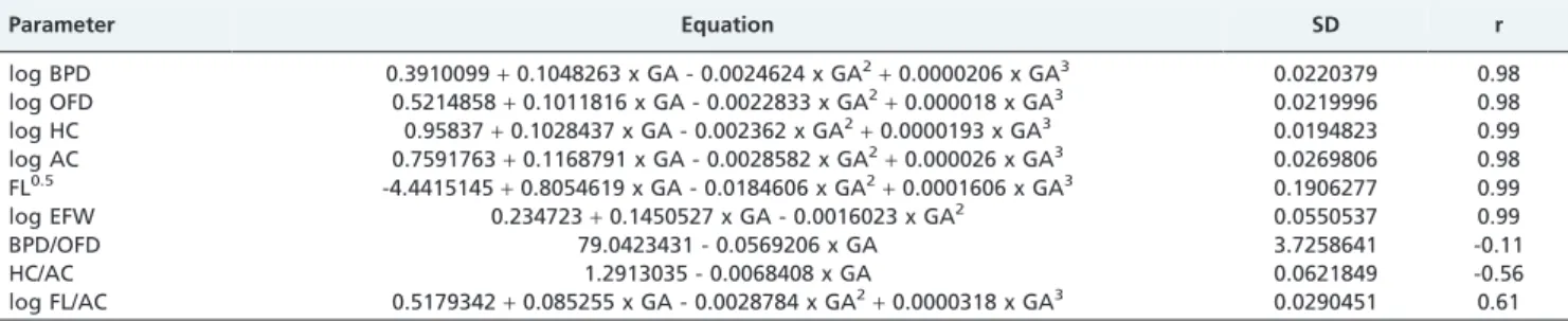
Table 1 -Polynomial regression formulas for ultrasound fetal biometry parameters according to gestational age in 125 uncomplicated, twin pregnancies.
Parameter Equation SD r
log BPD 0.3910099+0.1048263 x GA - 0.0024624 x GA2
+0.0000206 x GA3 0.0220379 0.98
log OFD 0.5214858+0.1011816 x GA - 0.0022833 x GA2+0.000018 x GA3 0.0219996 0.98 log HC 0.95837+0.1028437 x GA - 0.002362 x GA2
+0.0000193 x GA3 0.0194823 0.99
log AC 0.7591763+0.1168791 x GA - 0.0028582 x GA2+0.000026 x GA3 0.0269806 0.98
FL0.5 -4.4415145
+0.8054619 x GA - 0.0184606 x GA2
+0.0001606 x GA3 0.1906277 0.99
log EFW 0.234723+0.1450527 x GA - 0.0016023 x GA2 0.0550537 0.99
BPD/OFD 79.0423431 - 0.0569206 x GA 3.7258641 -0.11
HC/AC 1.2913035 - 0.0068408 x GA 0.0621849 -0.56
log FL/AC 0.5179342+0.085255 x GA - 0.0028784 x GA2+0.0000318 x GA3 0.0290451 0.61
AC: abdominal circumference; BPD: biparietal diameter; EFW: estimated fetal weight; FL: femur length; HC: head circumference; OFD: occipitofrontal diameter; SD: standard deviation.
p,0.001 for all equations.
Table 2 -Longitudinal reference ranges for biparietal diameter based on 807 ultrasound examinations
performed on 250 fetuses from 125 uncomplicated, twin pregnancies.
Gestational age
(weeks) Biparietal diameter (mm)
P5 P10 P50 P90 P95
14 24.5 25.1 27.1 29.2 29.9
15 27.4 28.0 30.1 32.4 33.1
16 30.3 31.0 33.3 35.8 36.5
17 33.4 34.0 36.5 39.2 40.0
18 36.4 37.2 39.8 42.7 43.5
19 39.6 40.3 43.2 46.2 47.1
20 42.7 43.5 46.5 49.7 50.6
21 45.8 46.7 49.8 53.1 54.1
22 48.9 49.8 53.1 56.6 57.6
23 51.9 52.8 56.2 59.9 61.0
24 54.8 55.8 59.3 63.1 64.2
25 57.6 58.6 62.3 66.3 67.4
26 60.3 61.3 65.2 69.2 70.5
27 62.8 63.9 67.9 72.1 73.4
28 65.2 66.3 70.4 74.8 76.1
29 67.4 68.6 72.9 77.4 78.7
30 69.5 70.7 75.1 79.8 81.2
31 71.4 72.7 77.3 82.1 83.5
32 73.2 74.5 79.2 84.3 85.8
33 74.9 76.2 81.1 86.3 87.8
34 76.4 77.8 82.8 88.2 89.8
35 77.8 79.2 84.5 90.1 91.7
36 79.1 80.6 86.0 91.8 93.5
37 80.3 81.9 87.5 93.5 95.3
38 81.5 83.1 88.9 95.2 97.0
P: percentile.
Table 3 -Longitudinal reference ranges for head circumference based on 807 ultrasound examinations performed on 250 fetuses from 125 uncomplicated, twin pregnancies.
Gestational age
(weeks) Head circumference (mm)
P5 P10 P50 P90 P95
14 88.9 90.7 97.3 104.4 106.5
15 99.2 101.2 108.3 116.0 118.2
16 109.9 112.0 119.7 128.0 130.4
17 121.0 123.2 131.5 140.3 142.9
18 132.3 134.7 143.5 152.9 155.6
19 143.8 146.3 155.6 165.5 168.5
20 155.3 158.0 167.8 178.3 181.3
21 166.8 169.7 180.0 190.9 194.1
22 178.3 181.2 192.0 203.4 206.7
23 189.5 192.5 203.8 215.6 219.1
24 200.4 203.6 215.2 227.6 231.2
25 211.0 214.3 226.3 239.1 242.8
26 221.1 224.6 237.0 250.2 254.1
27 230.8 234.4 247.2 260.8 264.8
28 240.0 243.7 257.0 271.0 275.1
29 248.7 252.5 266.1 280.5 284.8
30 256.8 260.7 274.8 289.6 294.0
31 264.4 268.4 282.9 298.2 302.6
32 271.5 275.6 290.5 306.2 310.8
33 278.0 282.2 297.6 313.8 318.5
34 284.0 288.4 304.2 320.9 325.8
35 289.6 294.1 310.4 327.6 332.7
36 294.8 299.4 316.3 334.1 339.3
37 299.7 304.4 321.8 340.2 345.6
38 304.2 309.2 327.1 346.2 351.8
reflected in the success rate of obtaining adequate ultra-sound images from both fetuses in all scans.
Accurate gestational age determination is crucial to correctly evaluate and classify fetal growth. In this regard, some previous studies did not report how gestational age was determined (10,11,14), and others were based solely on clinical parameters such as LMP and/or neonatal clinical examination (9). In the present study, gestational age was confirmed by ultrasound, and antenatal follow-up of all participants was conducted at our Twin’s clinic according to previously established protocols. Only data from uncompli-cated pregnancies were included in the statistical analysis.
Chorionicity was not a selection criterion for the present study. In fact, in a monochorionic pregnancy, a single placenta is shared by both fetuses, and fetal growth in such pregnancies may be slightly different from that of dichor-ionic twins. However, only approximately 12% of our cases were monochorionic twins.
Twin pregnancies resulting from assisted reproduction techniques are predominant in several clinical settings. However, due to the social and economic characteristics of our population, the reference ranges presented here are applicable to naturally conceived twins only.
A large dataset from the United States has shown that the mean birth weight in twin pregnancies is lower than that of singletons. Deviation begins at approximately 28 weeks and increases progressively throughout the pregnancy. For instance, at 38 weeks of gestation, the 50th percentile for twins is equivalent to a singleton’s 10thpercentile (1). Despite
lower mean birth weights, perinatal outcomes in twin pregnancies are comparable to those in pregnancies with
Table 4 -Longitudinal reference ranges for abdominal circumference based on 807 ultrasound examinations performed on 250 fetuses from 125 uncomplicated, twin pregnancies.
Gestational age
(weeks) Abdominal circumference (mm) P5 P10 P50 P90 P95
14 72.0 73.8 80.7 88.1 90.4
15 81.0 83.0 90.6 98.8 101.3
16 90.4 92.6 100.9 110.0 112.7
17 100.1 102.6 111.6 121.5 124.5
18 110.2 112.8 122.6 133.3 136.5
19 120.4 123.2 133.8 145.3 148.7
20 130.7 133.7 145.0 157.3 161.0
21 141.0 144.2 156.3 169.4 173.3
22 151.2 154.7 167.5 181.4 185.5
23 161.4 165.0 178.5 193.2 197.6
24 171.3 175.2 189.4 204.8 209.4
25 181.0 185.1 200.0 216.1 220.9
26 190.5 194.7 210.3 227.2 232.2
27 199.7 204.0 220.3 237.9 243.1
28 208.5 213.1 230.0 248.3 253.7
29 217.0 221.8 239.4 258.3 264.0
30 225.3 230.2 248.4 268.1 273.9
31 233.2 238.3 257.2 277.6 283.6
32 240.9 246.2 265.8 286.8 293.1
33 248.5 253.9 274.1 296.0 302.5
34 255.9 261.5 282.4 305.0 311.7
35 263.2 269.0 290.7 314.0 321.0
36 270.5 276.6 299.0 323.2 330.4
37 278.0 284.3 307.5 332.6 340.1
38 285.7 292.1 316.2 342.3 350.1
P: percentile.
Table 5 -Longitudinal reference ranges for femur diaphysis length based on 807 ultrasound examinations performed on 250 fetuses from 125 uncomplicated, twin pregnancies.
Gestational age
(weeks) Femur length (mm)
P5 P10 P50 P90 P95
14 11.0 11.5 13.4 15.4 16.0
15 13.6 14.2 16.2 18.4 19.1
16 16.4 17.0 19.2 21.5 22.2
17 19.2 19.8 22.1 24.6 25.3
18 22.0 22.7 25.1 27.7 28.4
19 24.8 25.5 28.1 30.8 31.5
20 27.6 28.4 31.0 33.8 34.6
21 30.4 31.1 33.9 36.7 37.5
22 33.1 33.8 36.6 39.6 40.4
23 35.6 36.4 39.3 42.4 43.2
24 38.1 39.0 41.9 45.0 45.9
25 40.6 41.4 44.4 47.6 48.5
26 42.9 43.7 46.8 50.1 51.0
27 45.0 45.9 49.1 52.4 53.4
28 47.1 48.0 51.3 54.7 55.7
29 49.1 50.1 53.4 56.9 57.9
30 51.0 52.0 55.4 59.0 60.0
31 52.8 53.8 57.3 61.0 62.0
32 54.6 55.6 59.2 62.9 64.0
33 56.2 57.2 60.9 64.8 65.9
34 57.8 58.9 62.7 66.6 67.7
35 59.3 60.4 64.3 68.4 69.6
36 60.8 62.0 66.0 70.1 71.3
37 62.3 63.5 67.6 71.9 73.1
38 63.8 65.0 69.2 73.7 74.9
P: percentile.
Table 6 -Longitudinal reference ranges for estimated fetal weight based on 807 ultrasound examinations performed on 250 fetuses from 125 uncomplicated, twin pregnancies.
Gestational age
(weeks) Estimated fetal weight (g) P5 P10 P50 P90 P95
14 72 76 89 106 111
15 91 95 112 133 139
16 113 119 140 165 173
17 140 147 173 203 213
18 173 181 212 249 261
19 211 220 258 303 317
20 255 267 313 366 383
21 307 321 375 439 459
22 366 382 447 523 547
23 433 452 529 618 646
24 508 531 621 726 758
25 593 619 724 846 884
26 685 716 837 979 1023
27 787 822 962 1125 1176
28 896 937 1096 1284 1342
29 1012 1059 1241 1454 1521
30 1135 1188 1394 1635 1711
31 1264 1323 1554 1826 1912
32 1396 1462 1720 2025 2121
33 1530 1603 1890 2229 2336
34 1664 1745 2062 2437 2555
35 1796 1884 2232 2645 2775
36 1924 2020 2399 2849 2992
37 2045 2149 2559 3048 3203
38 2157 2269 2710 3237 3405
singletons born at the same gestational age (2). Furthermore, some studies have shown a survival advantage despite lower birth weights in multiple pregnancies (15,16).
In multiple pregnancies, the uterine fundal height measurement does not allow an adequate evaluation of growth for each fetus. Therefore, fetal growth assessment is essentially based on serial ultrasound scans. However, most centers still use singleton reference ranges in the assessment of twins’ fetal growth. This will inevitably result in frequent misdiagnosis of fetal growth restriction (5) and consequent misclassification of risk. Moreover, under these circum-stances, repeat follow-up scans to monitor fetal growth and well-being surveillance are usually scheduled and may potentially lead to parental anxiety and increased risk of mismanagement due to additional false-positive results.
It is therefore plausible that the smaller fetal size characteristic of twin pregnancies constitutes a physiologi-cal phenomena; the use of appropriate reference charts for normal fetal growth in twin pregnancies should be advocated to help reduce false-positive diagnoses and unnecessary interventions.
AUTHOR CONTRIBUTIONS
Liao AW was responsible for the data collection, statistical analysis and the draft of manuscript. Brizot ML was responsible for the data collection and the draft of manuscript. Kang HJ and Assunc¸a˜o RA were responsible for the data collection. Zugaib M was responsible for the manuscript revision and discussion
REFERENCES
1. Alexander GR, Kogan M, Martin J, Papiernik E. What are the fetal growth patterns of singletons, twins, and triplets in the United States? Clin Obstet Gynecol. 1998;41(1):114-25.
2. Kilpatrick SJ, Jackson R, Croughan-Minihane MS. Perinatal mortality in twins and singletons matched for gestational age at delivery at.or = 30
weeks. Am J Obstet Gynecol. 1996;174(1 Pt 1):66-71, http://dx.doi.org/ 10.1016/S0002-9378(96)70375-7.
3. Blickstein I. Is it normal for multiples to be smaller than singletons? Best Pract Res Clin Obstet Gynaecol. 2004;18(4):613-23, http://dx.doi.org/ 10.1016/j.bpobgyn.2004.04.008.
4. Joseph KS, Fahey J, Platt RW, Liston RM, Lee SK, Sauve R, et al. An outcome-based approach for the creation of fetal growth standards: do singletons and twins need separate standards? Am J Epidemiol. 2009;169(5):616-24.
5. Sebire NJ, Nicolaides KH. Screening for fetal abnormalities in multiple pregnancies. Baillieres Clin Obstet Gynaecol. 1998;12(1):19-36, http:// dx.doi.org/10.1016/S0950-3552(98)80037-0.
6. Snijders RJ, Nicolaides KH. Fetal biometry at 14-40 weeks’ gestation. Ultrasound Obstet Gynecol. 1994;4(1):34-48, http://dx.doi.org/10.1046/ j.1469-0705.1994.04010034.x.
7. Hadlock FP, Harrist RB, Carpenter RJ, Deter RL, Park SK. Sonographic estimation of fetal weight. The value of femur length in addition to head and abdomen measurements. Radiology. 1984;150(2):535-40.
8. Ong S, Lim MN, Fitzmaurice A, Campbell D, Smith AP, Smith N. The creation of twin centile curves for size. Bjog. 2002;109(7):753-8. 9. Grumbach K, Coleman BG, Arger PH, Mintz MC, Gabbe SV, Mennuti
MT. Twin and singleton growth patterns compared using US. Radiology. 1986;158(1):237-41.
10. Reece EA, Yarkoni S, Abdalla M, Gabrielli S, Holford T, O’Connor TZ, et al. A prospective longitudinal study of growth in twin gestations compared with growth in singleton pregnancies. I. The fetal head. J Ultrasound Med. 1991;10(8):439-43.
11. Reece EA, Yarkoni S, Abdalla M, Gabrielli S, Holford T, O’Connor TZ, et al. A prospective longitudinal study of growth in twin gestations compared with growth in singleton pregnancies. II. The fetal limbs. J Ultrasound Med. 1991;10(8):445-50.
12. Royston P, Altman DG. Design and analysis of longitudinal studies of fetal size. Ultrasound Obstet Gynecol. 1995;6(5):307-12, http:// dx.doi.org/10.1046/j.1469-0705.1995.06050307.x.
13. Socol ML, Tamura RK, Sabbagha RE, Chen T, Vaisrub N. Diminished biparietal diameter and abdominal circumference growth in twins. Obstet Gynecol. 1984;64(2):235-8.
14. Yarkoni S, Reece EA, Holford T, O’Connor TZ, Hobbins JC. Estimated fetal weight in the evaluation of growth in twin gestations: a prospective longitudinal study. Obstet Gynecol. 1987;69(4):636-9.
15. McCarthy BJ, Sachs BP, Layde PM, Burton A, Terry JS, Rochat R. The epidemiology of neonatal death in twins. Am J Obstet Gynecol. 1981;141(3):252-6.