SELEX Aptamer Used as a Probe to Detect
Circulating Tumor Cells in Peripheral Blood of
Pancreatic Cancer Patients
Jinqiang Zhang1, Shaohua Li2, Fang Liu1, Lanping Zhou1, Ningsheng Shao2, Xiaohang Zhao1*
1State Key Laboratory of Molecular Oncology, Cancer Institute and Hospital, Chinese Academy of Medical Sciences and Peking Union Medical College, Beijing, China,2Department of Biochemistry and Molecular Biology, Beijing Institute of Basic Medical Sciences, Beijing, China
Abstract
Many studies have shown that the quantity and dynamics of circulating tumor cells (CTCs) in peripheral blood of patients afflicted with solid tumours have great relevance in therapeu-tic efficacy and prognosis. Different methods based on various strategies have been devel-oped to isolate and identify CTCs, but their efficacy needs to be improved because of the rarity and complexity of CTCs. This study was designed to examine the possibility of using a SELEX aptamer (BC-15) as a probe to identify rare CTCs out of background nucleated cells. Aptamer BC-15 was selected from a random oligonucleotide library screened against human breast cancer tissue. Fluorescence staining showed that BC-15 had a high affinity for nuclei of human cancer cell lines of various origins as well as CTCs isolated from pancre-atic cancer patients, whereas its binding capacity for non-tumor breast epithelial cells and leukocytes was almost undetectable. BC-15+/CD45-cells in cancer patient blood were also
found to be cytokeratins 18-positive and aneuploid by immunofluorescence staining and fluorescent in situ hybridization, respectively. Finally, the aptamer method was compared with the well-established anti-cytokeratin method using 15 pancreatic cancer patient blood samples, and enumeration indicated no difference between these two methods. Our study establishes a novel way to identify CTCs by using a synthetic aptamer probe. This new ap-proach is comparable with the anti-cytokeratin-based CTC identification method.
Introduction
Circulating tumor cells (CTCs) have been found in the peripheral blood of patients afflicted with all major solid carcinomas[1] and their quantity and fluctuation in cancer patient blood correlated with tumor development, therapeutic efficacy, tumor recurrence, and long-term prognosis[2–4]. In addition, CTC detection also provides a possible way to monitor patient re-sponse to certain anti-cancer therapies[5,6]. Many techniques have been developed to identify CTCs in patients with different types of cancer[7]. The predominant strategy exploits the OPEN ACCESS
Citation:Zhang J, Li S, Liu F, Zhou L, Shao N, Zhao X (2015) SELEX Aptamer Used as a Probe to Detect Circulating Tumor Cells in Peripheral Blood of Pancreatic Cancer Patients. PLoS ONE 10(3): e0121920. doi:10.1371/journal.pone.0121920
Academic Editor:Natasha Kyprianou, University of Kentucky College of Medicine, UNITED STATES
Received:November 7, 2014
Accepted:February 5, 2015
Published:March 23, 2015
Copyright:© 2015 Zhang et al. This is an open access article distributed under the terms of the
Creative Commons Attribution License, which permits unrestricted use, distribution, and reproduction in any medium, provided the original author and source are credited.
Data Availability Statement:All relevant data are within the paper.
Funding:This work was supported by grants of the High-tech R & D Program (No. 2012AA020206 and 2012AA02A503), State Key Projects for Basic Research (2011CB910703) and NSFC (30700992, 81372591, 81321091) of China. The funders had no role in study design, data collection and analysis, decision to publish, or preparation of the manuscript.
Competing Interests:The authors have declared
epithelial origin of CTCs to capture and identify them using antibodies that target epithelial markers such as epithelial cellular adhesion molecule (EpCAM) and cytokeratin (CK)[2]. However, some CTCs may lose their epithelial characteristics because of the epithelia-mesen-chymal transition (EMT) and thus cannot be detected by these antibodies[8]. Furthermore, these methods have difficulty discriminating malignant cells from benign cells that express epi-thelial markers. The development of novel and more effective approaches for CTCs detection is urgently needed.
Aptamers represent a group of single-stranded nucleic acid fragments that were screened against specific targets from a random synthetic nucleic acid library by the method of systemat-ic evolution of ligands by exponential enrsystemat-ichment (SELEX)[9,10]. One can even get aptamers, which specifically binding to a molecule without knowing its characteristics. Aptamers can bind to targets with high affinity via specific structural regions that are induced by sequence-dependent folds[11]. This technique has been used for many purposes, including protein inhib-itor design, molecular detection, and therapeutic drug and antibody replacement[7]. In previ-ous study[12], we have identified a tumor specific aptamer BC-15 using a newin situtissue slide-based SELEX strategy. Aptamer BC-15 has also been proved to bind to multiple cancer cells of various origins with high specificity. Through streptavidin magnetic beads mediated af-finity purification assay followed by mass spectrometry identification and western blot confir-mation, the target of BC-15 was characterized to be heterogeneous nuclear ribonucleoprotein A1 (hnRNP A1). The hnRNP family proteins play important roles in biogenesis and transport of messenger RNAs. Up-regulation of hnRNPs usually precedes morphological differentiation and is considered a good biomarker in the early stages of cancer development[13]. Enhanced amounts of hnRNP A1 has been reported in many cancer tissues including breast, and small cell lung, ovarian, colorectal carcinoma, and pancreatic cancer with a location that is mainly nuclear [13–16]. High levels of hnRNP A1 expression was also proved in pancreatic tumor cell lines, whereas in normal primary pancreatic cells hnRNP expression was undetectable. These results strongly suggest that hnRNP could be a good candidate for diagnosis of pancreatic can-cer[13]. Therefore, aptamer BC-15 could also be used as diagnosis biomarker for multiple types of cancer, including pancreatic cancer due to its high specific affinity to hnRNA A1. Here, we reported the feasibility of using an aptamer BC-15 as a probe to identify CTCs in the peripheral blood of patients with pancreatic cancer.
Material and Methods
Blood specimen collection
This study was approved by the ethics review committees of Cancer Hospital of Chinese Acad-emy of Medical Sciences, and informed written consents were obtained from all the pancreatic cancer patients and healthy donors. A total of 30 blood samples were collected, including 15 samples taken from pancreatic cancer patients and 15 samples from healthy donors. For each patient or healthy donor, peripheral blood (7.5ml) was drawn from the median cubital vein into acid citrate dextrose vacutainer tubes (BD Diagnostics, Franklin Lakes, NJ) after discard-ing the first 3 ml of blood to avoid epithelial cell contamination durdiscard-ing venipuncture. All sam-ples were maintained at room temperature and processed within 12 h after collection. To avoid bias, samples were blindly processed by different persons.
SELEX procedure
rounds of screening, BC-15 was selected for its high affinity for tumor tissue specifically, which was also verified using cell lines. The entire SELEX screening process has been described in our
previous study[12]. The aptamer probe BC-15 (5’
-GCAATGGTACGGTACTTCCTGTGGC-GAGGTAGGTGGGGTGTGTGTGTATCCAAAAGTGCACGCTACTTTGCTAA-3’) was
synthesized and labelled with fluorescein isothiocyanate (FITC) on the 5’end (Life Technolo-gies, Grand Island, NY, USA), and a mixture of random sequences labelled the same way acted as a control probe.
Cell culture
Human cancer PL-45 (ATCC CRL-2558, pancreatic adenocarcinoma), MCF-7 (ATCC HTB22, breast adenocarcinoma), A549 (ATCC CCL185, lung carcinoma), MDA-MB-231 (ATCC HTB-26, metastatic breast adenocarcinoma), HT-29 (ATCC HTB-38, colorectal ade-nocarcinoma) and MCF10A (ATCC CRL10317, breast fibrocystic disease) cell lines were pur-chased from American Type Culture Collection (Rockville, MD, USA). All cells except MCF10A were cultured in Dulbecco’s Modified Eagle’s Medium (DMEM) supplemented with 10% fetal bovine serum (Gibco, Grand Island, NY, USA). MCF10A was cultured in DMEM supplemented with 20% fetal bovine serum, 20ng/ml human epidermal growth factor (EGF), 10μg/ml insulin, and 0.5μg/ml hydrocortisone. All cells were incubated in a humidified
atmo-sphere with 5% CO2at 37°C. For fluorescence staining, overnight cultured cells were fixed in
methanol at−20°C for 2 h and washed with phosphate-buffered saline (PBS, pH 7.4), and then
cells were stained with aptamer as described in the following section.
Enrichment and identification of CTCs
The CTCs enrichment method was similar to the previously published methods[17]. Briefly, 7.5 ml of peripheral blood collected in a vacutainer tube was washed with PBS once, and then red blood cells were lysed with lysis buffer (150 mM NH4Cl, 10mM NaHCO3, and 0.1mM
EDTA). The reaction mixture was spun down at 300 ×gfor 5 min at room temperature. The resulting cell pellet was resuspended in PBS and subsequently incubated with 0.1 ml of anti-CD45- antibody coated magnetic beads (Miltenyi Biotec, Bergisch Gladbach, Germany) for 30 min at room temperature, followed by separation using a magnetic stand (Promega, Madison, WI, USA). Supernatants were transferred into a centrifuge tube followed by spinning at 500 ×gfor 3 min at room temperature. Each cell pellet was resuspended in PBS and dropped equally onto two slides (Thermo Fisher Scientific, Waltham, MA, USA), air dried, and then subjected to staining.
CTCs were identification by BC-15 aptamer or anti-CK staining. For BC-15, slides were fixed with methanol for 30 min at -20°C followed by penetration with 0.1% (w/v) Tween 20 in PBS at room temperature for 15 min. After blocking with buffer (0.1mg/ml tRNA, 0.1mg/ml salmon sperm DNA, 1% BSA and 0.02% (w/v) Tween-20 in PBS) at 37°C for 20 min, slides were incubated with 10ug/ml FITC-labelled BC-15 in blocking buffer at 37°C for 1 h. Anti-CK CTC identification was conducted as described in many studies [2,7,18]. Briefly, slides were fixed with paraformaldehyde for 40 min followed by 0.1% (w/v) Triton X-100 penetration and 2% BSA blocking, and then incubated with FITC-labelled anti-CK (Miltenyi Biotec; 1:1000 di-lution in PBS), which recognises CKs 8, 18, and 19, at room temperature for 1 h. Alexa 594 la-belled anti-CD45 (Miltenyi Biotec; 1:1000 dilution in PBS) was used to co-staining
considered as CTCs: cell size>4μm in diameter, intact with round-to-oval morphology with a DAPI-stained nucleus, and positive for CK or BC-15 staining and negative for CD45[18].
Aneuploid chromosome analysis using fluorescence
in situ
hybridisation
(FISH)
Chromosome enumeration probes (CEP) 8 (Vysis, Des Plaines, IL, USA) was denatured in hy-bridization buffer (0.15 M sodium chloride, 0.015 M sodium citrate, 70% formamide) at 73°C for 5min. The probe was then incubated with a slide in a humidified chamber at 42°C overnight followed by washing with 0.3% (w/v) NP-40 in 0.4x saline-sodium citrate (SSC; 1x SSC con-taining 150 mM NaCl and 15 mM Na3Citrate) at 73°C for 2 min and with 2x SSC/0.1% NP-40
at room temperature for 30 s. After briefly drying, slides were analyzed after mounting with DAPI mounting medium (Vector Labs) under fluorescence microscope.
Statistical analysis
Difference between these two identification methods was evaluated by SPSS 19 (IBM, Armonk, NY, USA) software with Wilcoxon Signed Ranks test and the correlation between BC-15 and anti-cytokeratin results was assessed by the nonparametric Spearman’s rho value. AP-value of<0.05 was consider as the statistical significance.
Results
BC-15 binds to the nucleus of human tumor cells specifically
Our previous study shows that BC-15 can bind to breast cancer cells and tissues with high af-finity, whereas its binding capacity for non-tumorigenic cells and normal breast tissues is much lower; in addition to fluorescence staining, flow cytometric confirmed that BC-15 has a high affinity for tumor cells [12]. To validate the binding of BC-15 to tumor cells other than breast cancer cells, a set of human cancer cells was stained with BC-15 (Fig. 1). BC-15 signals accumulated mainly in nuclei of cancer cells but were much lower in immortalised breast epi-thelial MCF10A cells. No positive signals were observed in any cells when staining with a ran-dom control aptamer probe. This observations suggested that BC-15 exclusively binds to nuclei of cancer cells.
BC-15
+cells in patient peripheral blood are also CK-positive
BC-15 has the similar efficacy as anti-CK8/18/19 for identifying CTCs
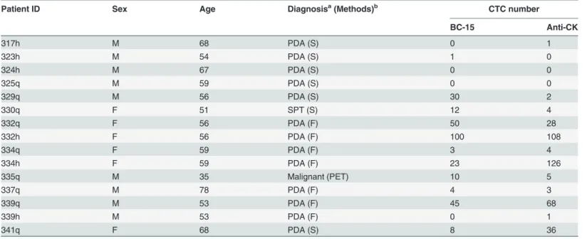
Anti-CK-based immunostaining is an established method to detect CTCs in many systems and shows relative satisfactory consistency and repeatability in various kinds of cancers types[19]. The malignant characteristics of CK+cells have also been proven by FISH with various CEP probes. To compare the CTC identification capacity of BC-15 with that of anti-CK methods, 15 peripheral blood samples from pancreatic cancer patients (Table 1) and 15 from healthy do-nors were collected. The cells enriched from these samples were divided equally and stained side by side using these two methods. Neither method detected positive cells in the samples from healthy donors. Of the 15 patients samples, 12 (80.0%) had positive cells according to the anti-CK method, whereas the positive rate was 73.3% (11/15) using BC-15 staining. The aver-age number of positive cells detected by anti-CK or the BC-15 staining was 25.7 and 19.1 per sample respectively. No statistic difference was found between the CTC enumeration results of the two methods by Wilcoxon Signed Ranks test (P= 0.699) and there was significant correla-tion between the results of these two different CTC deteccorrela-tion methods by Spearman's rank cor-relation test (Spearman’s rho = 0.810,P<0.01).
Fig 1. BC-15 binds to various kinds of human cancer cells.Cells were stained with FITC-labelled BC-15 aptamer (Upper row) or FITC-labelled random control aptamer (Middle row, green). All the cells were counter stained with 0.25% Evans blue for 10 min to reveal whole-cell morphology (red). Lower row show CK immunofluorescence staining of different types of cancer cells. These cultured cells were trypsinized, dropped onto the coated glass slides, followed by staining with Alexa 594 labelled anti-CK18 antibody, and counter stained with DAPI. PL45, pancreatic adenocarcinoma cell; MCF-7, breast cancer cell; A549, lung carcinoma cell; MDA-MB-231, breast cancer cell; HT-29, colon adenocarcinoma cell; MCF10A, non-tumourigenic immortalised mammary epithelial cells. Scale bar, 20μm.
Discussion
As a surrogate of primary or metastatic tumors, CTCs are showing increasing importance both in cancer research and clinical practice, and CTC detection has gained intensified interest with-in the cancer research community. In the present study, we successfully established a CTC de-tection system combining a leukocyte depletion enrichment strategy and BC-15 aptamer identification. This assertion is supported by the following observations: (1) BC-15 recognised various kinds of tumor cells specifically, and non-tumorigenic epithelial cells and leukocytes were negative for BC-15 staining; (2) BC-15 had the same staining pattern in BC-15+/CD45
-Fig 2. CTCs identified in pancreatic cancer patient peripheral blood by BC-15.(A) BC-15+(green) and CK18+(red) signals coincided in both PL-45 cells (Upper row) and CTCs from pancreatic cancer patients (Lower row). (B) Morphology of dispersed CTCs from pancreatic cancer patients identified by immunofluorescence staining. CTCs were demonstrated as CD45–cells with BC-15+nuclei (green) and the arrows in the bottom images of panel A indicat
the white blood cells (CD45+, red). (C) Clustered CTCs recognized by BC-15 in blood samples from pancreatic cancer patients. White arrows indicate leukocytes (CD45+). (D) Immunofluorescence of a BC-15+cells (noted by yellow arrow) was quenched and the same slide was subjected to FISH using a CEP8 probe. BC-15+cell had a chromosome 8 aberration (7 copies), whereas the background leukocyte was diploid for chromosome 8 (white arrow). The cell indicated by green arrow was not classified as CTC because of its positivity for both BC-15 and CD45. Scale bars, 10μm.
cells enriched from pancreatic cancer patient blood as in cancer cells and tumor tissues; (3) BC-15+cells in cancer patients blood were CK+[20]; (4) BC-15+/CD45-cells in blood from can-cer patients showed aneuploidy, a cytogenetic abnormality typical of malignant cells; and (5) the CTC identification efficacy of BC-15 was equivalent to that of anti-CK methods.
Because of their heterogeneic characteristics and the mesenchymal-epithelial transition in circulation, CTCs cannot be fully enumerated by antibodies that target epithelial markers (e.g. anti- CK)[21]. However, aptamers could be developed to recognise subtle characteristics and/ or low-immunogenic molecules of malignant cells. Our preliminary results presented here demonstrate that it is feasible to use an aptamer as a CTC detection probe and also provide per-spectives that would make CTC detection more customised and personalised. Although our pilot results show no improvement on efficacy of CTC identification compared with antibody-based methods, the aptamer probe holds great promise for CTCs detection (both isolation and identification) owing to its capacity to bind to various types of molecular targets, high affinity, well defined synthesis/modification process, stability and uniformity. Furthermore, based on the well-developed cell/tissue-SELEX technique, it is possible to generate particular aptamers with high stability by using tumor cells or tumor tissues from a given patient as selecting tar-gets[22]. These personalised aptamers can partly overcome the weakness raised by uniform an-tibodies[23] and can be used as the guidance molecular of targeting therapy or used to monitor therapeutic response and anticipate long term prognosis [22].
Acknowledgments
The authors wish to thank Nathan Ungerleider who assisted in the English language editing of this manuscript.
Table 1. CTCs detection in patient samples using BC-15 aptamer versus anti-CK.
Patient ID Sex Age Diagnosisa(Methods)b CTC number
BC-15 Anti-CK
317h M 68 PDA (S) 0 1
323h M 54 PDA (S) 1 0
324h M 67 PDA (S) 0 0
325q M 59 PDA (S) 0 0
329q M 56 PDA (S) 30 2
330q F 51 SPT (S) 12 4
332q F 56 PDA (F) 50 28
332h F 56 PDA (F) 100 108
334q F 59 PDA (F) 3 4
334h F 59 PDA (F) 23 126
335q M 35 Malignant (PET) 10 5
337q M 78 PDA (F) 4 3
339q M 53 PDA (F) 45 68
339h M 53 PDA (F) 0 1
341q F 68 PDA (S) 8 36
a: PDA, pancreatic ductal adenocarcinoma; SPT, solid pseudopapillary tumor of the pancreas; PET, pancreatic endocrine tumor. b: S, surgical biopsy; F,fine needle aspiration; PET, positron emission tomography.
Author Contributions
Conceived and designed the experiments: XZ JZ NS. Performed the experiments: JZ SL FL LZ. Analyzed the data: JZ SL NS XZ. Contributed reagents/materials/analysis tools: NS XZ. Wrote the paper: JZ XZ.
References
1. Allard WJ, Matera J, Miller MC, Repollet M, Connelly MC, Rao C, et al. Tumor cells circulate in the pe-ripheral blood of all major carcinomas but not in healthy subjects or patients with nonmalignant dis-eases. Clin Cancer Res. 2004; 10(20):6897–6904. PMID:15501967
2. Cristofanilli M, Budd GT, Ellis MJ, Stopeck A, Matera J, Miller MC, et al. Circulating tumor cells, disease progression, and survival in metastatic breast cancer. N Engl J Med. 2004; 351(8):781–791. PMID:
15317891
3. Moreno JG, Miller MC, Gross S, Allard WJ, Gomella LG, Terstappen LW. Circulating tumor cells predict survival in patients with metastatic prostate cancer. Urology. 2005; 65(4):713–718. PMID:15833514
4. Zippelius A, Pantel K. RT-PCR-based detection of occult disseminated tumor cells in peripheral blood and bone marrow of patients with solid tumors. An overview. Ann N Y Acad Sci. 2000; 906:110–1123. PMID:10818606
5. Fehm T, Sagalowsky A, Clifford E, Beitsch P, Saboorian H, Euhus D, et al. Cytogenetic evidence that circulating epithelial cells in patients with carcinoma are malignant. Clin Cancer Res. 2002; 8(7):2073– 2084. PMID:12114406
6. Meng S, Tripathy D, Shete S, Ashfaq R, Saboorian H, Haley B, et al. uPAR and HER-2 gene status in individual breast cancer cells from blood and tissues. Proc Natl Acad Sci U S A. 2006; 103(46):17361– 17365. PMID:17079488
7. Greene BT, Hughes AD, King MR. Circulating tumor cells: the substrate of personalized medicine? Front Oncol. 2012; 2:69. doi:10.3389/fonc.2012.00069PMID:22783545
8. Yu M, Bardia A, Wittner BS, Stott SL, Smas ME, Ting DT, et al. Circulating breast tumor cells exhibit dy-namic changes in epithelial and mesenchymal composition. Science. 2013; 339(6119):580–584. doi:
10.1126/science.1228522PMID:23372014
9. Tuerk C, Gold L. Systematic evolution of ligands by exponential enrichment: RNA ligands to bacterio-phage T4 DNA polymerase. Science. 1990; 249(4968):505–510. PMID:2200121
10. Ye M, Hu J, Peng M, Liu J, Liu J, Liu H, et al. Generating Aptamers by Cell-SELEX for Applications in Molecular Medicine. Int J Mol Sci. 2012; 13(3):3341–3353. doi:10.3390/ijms13033341PMID:
22489154
11. Bridonneau P, Chang YF, Buvoli AV, O'Connell D, Parma D. Site-directed selection of oligonucleotide antagonists by competitive elution. Antisense Nucleic Acid Drug Dev. 1999; 9(1):1–11. PMID:
10192284
12. Li S, Xu H, Ding H, Huang Y, Cao X, Yang G, et al. Identification of an aptamer targeting hnRNP A1 by tissue slide-based SELEX. J Pathol. 2009; 218(3):327–336. doi:10.1002/path.2543PMID:19291713 13. Yan-Sanders Y, Hammons GJ, Lyn-Cook BD. Increased expression of heterogeneous nuclear ribonu-cleoprotein A2/B1 (hnRNP) in pancreatic tissue from smokers and pancreatic tumor cells. Cancer Lett. 2002; 183(2):215–220. PMID:12065097
14. Patry C, Bouchard L, Labrecque P, Gendron D, Lemieux B, Toutant J, et al. Small interfering RNA-me-diated reduction in heterogeneous nuclear ribonucleoparticule A1/A2 proteins induces apoptosis in human cancer cells but not in normal mortal cell lines. Cancer Res. 2003; 63(22):7679–7688. PMID:
14633690
15. Zerbe LK, Pino I, Pio R, Cosper PF, Dwyer-Nield LD, Meyer AM, et al. Relative amounts of antagonistic splicing factors, hnRNP A1 and ASF/SF2, change during neoplastic lung growth: implications for pre-mRNA processing. Mol Carcinog. 2004; 41(4):187–196. PMID:15390079
16. Ushigome M, Ubagai T, Fukuda H, Tsuchiya N, Sugimura T, Takatsuka J, et al. Up-regulation of hnRNP A1 gene in sporadic human colorectal cancers. Int J Oncol. 2005; 26(3):635–640. PMID:
15703818
17. Molnar B, Ladanyi A, Tanko L, Sreter L, Tulassay Z. Circulating tumor cell clusters in the peripheral blood of colorectal cancer patients. Clin Cancer Res. 2001; 7(12):4080–4085. PMID:11751505 18. Wu C, Hao H, Li L, Zhou X, Guo Z, Zhang L, et al. Preliminary investigation of the clinical significance of
19. Balic M, Lin H, Williams A, Datar RH, Cote RJ. Progress in circulating tumor cell capture and analysis: implications for cancer management. Expert Rev Mol Diagn. 2012; 12(3):303–312. doi:10.1586/erm. 12.12PMID:22468820
20. Pantel K, Brakenhoff RH, Brandt B. Detection, clinical relevance and specific biological properties of disseminating tumour cells. Nat Rev Cancer. 2008; 8(5):329–340. doi:10.1038/nrc2375PMID:
18404148
21. Ksiazkiewicz M, Markiewicz A, Zaczek AJ. Epithelial-mesenchymal transition: a hallmark in metastasis formation linking circulating tumor cells and cancer stem cells. Pathobiology. 2012; 79(4):195–208. doi:10.1159/000337106PMID:22488297
22. Song KM, Lee S, Ban C. Aptamers and their biological applications. Sensors (Basel). 2012; 12 (1):612–631. doi:10.3390/s120100612PMID:22368488
23. Kunii T, Ogura S, Mie M, Kobatake E. Selection of DNA aptamers recognizing small cell lung cancer using living cell-SELEX. Analyst. 2011; 136(7):1310–1312. doi:10.1039/c0an00962hPMID: