INTRODUCTION
S
urgical incision, dissection, coagulation and va-porization with electrocautery are widely used and recognized as a major advance in surgical technique. However, these techniques intentional-ly destroy tissue, creating vapors, popularintentional-ly known as cautery or surgical smoke (SS)1. This smoke, withcharacteristic odor and made up of particles with micro and / or submicron size, diffuses in the en-vironment and is inhaled by professional medical staff present in operating rooms. It is produced when the heat reaches the cells, ruptures their membranes and vaporizes its constituents, dispers-ing them and generatdispers-ing other substances durdispers-ing tissue combustion2.
In vitro experiments have demonstrated the smoke constituents from the use of cautery on sub-cutaneous and prostate tissues, in breast lifting pro-cedures, laparotomy and TURP3,4. It is known today
that many of these components are toxic, mutagen-ic, such like the cigarette smoke, the smoke
generat-ed by a gram of tissue destroygenerat-ed equals the one of six cigarettes without filter5.
The constituents present in greater quanti-ties in the smoke of subcutaneous tissue are hydro-carbons and nitrogen compounds, the hydrogen cy-anide, formaldehyde, and benzene being the most toxic5. The number, proportion, the amount and
nature of the substances present in the smoke de-pend on the tissue, on its condition and on the area under treatment with electrocautery, on the duration of the procedure, on the electric power and on the technique used (incision, coagulation, vaporization or dissection)6.
Although there is a reasonable number of studies that analyze these constituents, the size and shape of these particles in the smoke, the interfer-ence in the surgical field visualization7 and the use of
smoke suction8, those analyzes were performed only
on the subcutaneous tissue. However, electrocautery is widely used in other tissues such as muscle and liver, producing a lot of smoke. Thus, this study aims to comparatively demonstrate which compounds are
Analysis of electrocautery generated smoke by
chromatographic-mass spectrometry
Análise, mediante cromatografia/espectrometria de massas, da fumaça gerada
por eletrocautério
JeFFerson kalil1; FranCisCo B. t. pessine2; Carlos h. v. Fidelis2; FaBio h. Menezes3; paulo Cesar rodrigues palMa, tCBC-sp3.
A B S T R A C T
Objective: to analyze the chemical components of the smoke from electrocautery from coagulating muscle and liver tissues of pigs.
Methods: we collected smoke produced by electrocautery applied to porcine tissue in previously evacuated bottles, with qualitative and quantitative analysis of the compounds present through the hyphenated technique gas chromatography / mass spectrometry. Results: there was a majority of decanal aldehyde in the fumes from the subcutaneous, muscle and liver tissues. Fumes of subcutaneous and muscular tissues also showed the presence of hexanal and phenol. In the fumes of subcutaneous and liver tissues we also found toluene and limonene and, finally, nonanal smoke was present in the muscle and liver tissues. Conclusion: there is increasing evidence showing that smoke from electrocautery used in subcutaneous, muscle and liver tissue is harmful to human health. Thus, there is need to reduce exposure to it or wear masks with filters capable of retaining these particles.
Keywords: Smoke. Subcutaneous Tissue. Mass Spectrometry. Chromatography, Gas. Aldehydes.
present in the smoke from three electrocauterized tissue, subcutaneous, muscle and liver, from pigs.
METHODS
The tissue used for the research was from a pig of the Large White breed, which is closest to human tissue5. The animal had its used approved
for teaching and research by the Ethics Committee on Animal Use of the Biology Institute of the Univer-sidade Estadual de Campinas.
The collection was performed at the Ex-perimental Surgery Center of the Universidade Estadual de Campinas, with fresh tissues, using a monopolar electrocautery with 30w power, long enough to produce smoke.
The samples were collected in four vials, previously evacuated and hermetically sealed. One of the vials was used to collect air in the operating room prior to cautery use, serving as a control. In the three other vials we collected smoke from the
cautery use, in pure coagulation mode, at the site of its production in the subcutaneous, muscle and liver tissues.
These previously evacuated vials are made from Pyrex glass, provided with a telon high vacuum tap with a tap screw cap containing silicone septa for the introduction of the needle containing the gas absorbing iber, and then introduced into the gas chromatograph using helium as the carrier gas. Before the introduction of the samples we performed the chromatogram / mass spectra of the reference (only the iber) to verify that the peaks relating to the chromatograph eluted samples were not due to the reference. The chromatogram / mass spectra were compared with the chromatogram / mass spectra of the samples library existing in the equipment to iden-tify the substances responsible for chromatographic peaks present in the samples collected.
The equipment used in the analysis was the gas chromatograph (Agilent 7890A model) coupled to the mass spectrometer (Agilent, 5975C model).
Table 1. Compounds present in the ambient air sample.
Substance % (area) Elution time (min) Quality
carbon dioxide 12.35 1.464 4 ethylene oxide 12.35 1.464 3
acetonitrile 12.71 1.719 7
ethylamine 12.71 1.719 5
trimethylphosphine oxide 30.75 2.956 9 dimetilsilanodiol 30.75 2.956 9 2 chlorine 2 nitro propane 30.75 2.956 4 hexametilciclotrisiloxano 13.48 5.925 91
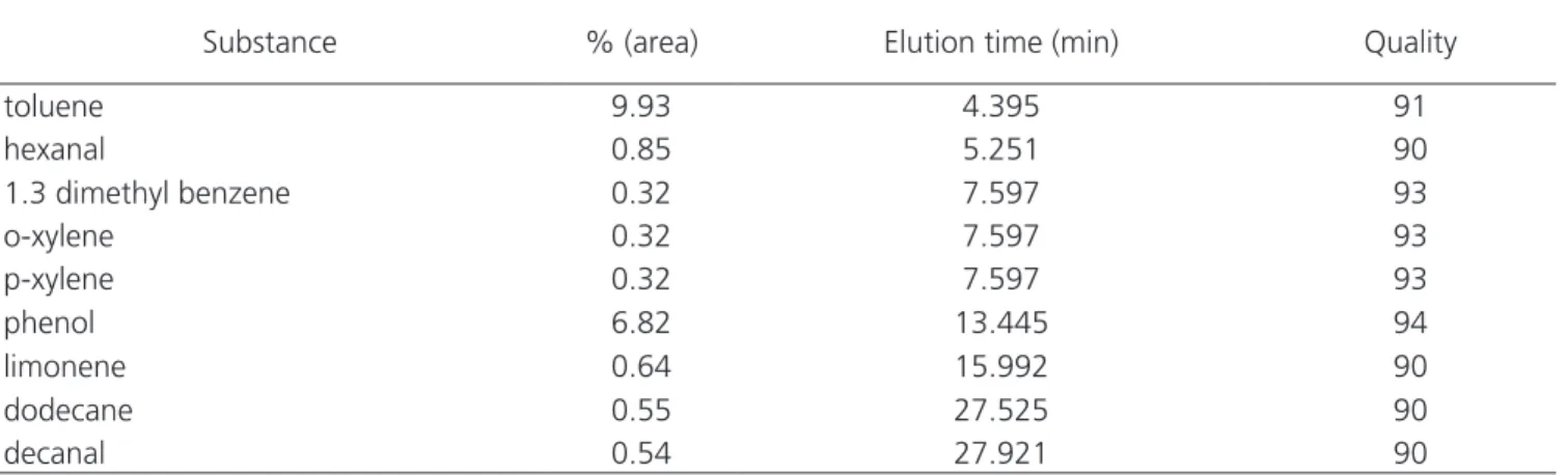
Table 2. Compounds present in subcutaneous tissue sample.
Substance % (area) Elution time (min) Quality
toluene 9.93 4.395 91
hexanal 0.85 5.251 90
1.3 dimethyl benzene 0.32 7.597 93
o-xylene 0.32 7.597 93
p-xylene 0.32 7.597 93
phenol 6.82 13.445 94
limonene 0.64 15.992 90
dodecane 0.55 27.525 90
The technique for sampling was Solid Phase Micro Extraction (SPME) using a needle with SUPELCO, gas-absorbing triple fiber: 50/30mm DVB/CAR/PDMS (polydimethylsiloxane), heated at 100oC for 40
min-utes to release the adsorbed compounds.
We tabulated and presented data in a qual-itative way, with no statistical study.
RESULTS
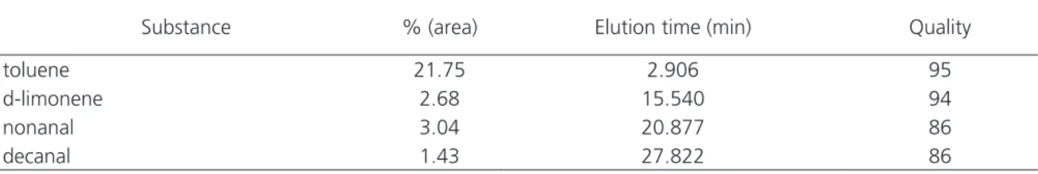
The results of each sample components analysis are shown in Tables 1, 2, 3 and 4, being, respectively, the ambient air control sample, subcu-taneous, muscle, and liver tissues. Tables indicate chemicals, the percentage area of the chromato-graphic peak for each compound, its elution time (in minutes), and quality. This last parameter refers to the degree of similarity between the detected sub-stance and the existing compounds in the mass spec-trometer database.
We found decanal in all three tissues; com-mon substances in the smokes from subcutaneous and muscle tissues were hexanal and phenol; com-mon compounds in the smokes from subcutaneous and liver tissues were toluene and limonene; and the common compound in the smokes from muscle and liver tissues was nonanal.
DISCUSSION
The aromatic hydrocarbon toluene
has been widely found in subcutaneous tissue
smoke
3. However, there was no evidence in the
literature of its presence in the smoke from liver
tissue. Aldehydes have also been widely cited
in the literature as present in subcutaneous
tis-sue smoke and, as shown in this study, are not
restricted to it, being also present in the smoke
from muscle and liver, in the forms of hexanal,
nonanal and decanal. The presence of
d-limo-nene has not been reported in other studies in
subcutaneous tissue smoke.
Wenig
et al
.
9evaluated cautery smoke
exposure in rats and noticed that they were
stunned during the exposure period, returning
to normal after an exposure-free period.
Fur-thermore, when analyzing the rats’ lungs, they
observed vessels hypertrophy, cellular
conges-tion and emphysematous changes. They
sup-ported the idea that these changes were from
exposure to benzene, formaldehyde and
acrole-in, substances present in the subcutaneous
tis-sue, muscle and liver smoke.
The presence of volatile organic
com-pounds within the smoke, as mentioned by
Table 3. Compounds present in the muscle sample.Substance % (area) Elution time (min) Quality
hexanal 2.97 5.238 91
tetrachloroethylene 0.77 5.501 97
heptanal 2.57 9.064 95
phenol 2.48 13.479 95
octanal 6.10 14.581 90
nonanal 13.17 21.092 91
decanal 17.84 27.900 91
Table 4. Compounds present in the liver sample.
Substance % (area) Elution time (min) Quality
toluene 21.75 2.906 95
d-limonene 2.68 15.540 94
nonanal 3.04 20.877 86
Moot
et al
.
10, although in low concentrations,
can chronically inflict the same health hazards
of passive smoking. Furthermore, two
com-pounds identified by this group, hydrogen
cy-anide and butadiene, are implicated as
cardio-toxic and carcinogenic, respectively. They also
showed that benzene, butadiene and decene
are carcinogenic substances
10.
El Ghawabi
et al
.
11and Chandra
et al
.
12showed that chronic exposure to low
concentra-tions of hydrocarbons – hexanal, heptanal,
oc-tanal, nonanal and decanal – cause headache,
weakness, touch and smell changes,
lacrima-tion, salivalacrima-tion, abdominal colic pain and
ner-vous instability. Moreover, Blanc
et al
.
13showed
that hydrocarbons can lead to deficiency of
vi-tamin B12 and folate and increase in thyroid
stimulating hormone (TSH), leading to goiter.
Laugesen
et al
.
14, in a review study, stated that
in cigarette smoke, the butadiene amounted
to 45% of the cancer risk, hydrocarbons
corre-sponded to 89% risk of cardiovascular disease,
and acrolein (aldehyde, like the others found in
all three tissues) corresponded to 97% risk of
lung disease.
There is growing body of evidence
that the smoke produced by electrocautery
used in biological tissues, be them
subcutane-ous, muscle or liver, is harmful to the human
health. The need to reduce such exposure is
evident, whether by suction of this smoke by
means of suitable devices or by using surgical
instruments that do not generate heat, like
some kinds of laser.
ACKNOWLEDGEMENTS
We thank Dr. José Henrique Silveira Virgili for the contribution during the experimental surgery, and the staff of the Experimental Surgery Center.
R E S U M O
Objetivo: analisar quimicamente os componentes da fumaça do eletrocautério, provenientes da coagulação de tecidos, muscular e hepáti-co de suíno. Métodos: coleta de fumaça produzida por eletrocauterização de tecido porcino em frascos previamente evacuados com análise qualitativa e quantitativa dos compostos presentes, através de técnica hifenada, cromatografia a gás/espectrometria de massas.
Resultados: houvepresença majoritária do aldeído decanal nas fumaças provenientes dos tecidos subcutâneo, muscular e hepático. Fumaças dos tecidos subcutâneo e muscular mostraram também a presença de hexanal e fenol. Nas fumaças dos tecidos subcutâneo e hepático foram encontrados ainda tolueno e limoneno e, por fim, nonanal estava presente nas fumaças dos tecidos muscular e hepático.
Conclusão: há número crescente de evidências mostrando que fumaça proveniente de eletrocauterização de tecidos subcutâneo, muscular e hepático é nociva à saúde de seres humanos. Portanto, há necessidade de reduzir a exposição a ela ou usar máscara com filtro capaz de reter essas partículas.
Descritores: Fumaça. Tecido Subcutâneo. Espectrometria de Massas. Cromatografia Gasosa. Aldeídos.
REFERENCES
1. Bigony L. Risks associated with exposure to surgical smoke plume: a review of the literature. AORN J. 2007;86(6):1013-20.
2. Lewin JM, Brauer JA, Ostad A. Surgical smoke and the dermatologist. J Am Acad Dermatol. 2011;65(3):636-41. 3. Mowbray N, Ansell J, Warren N, Wall P, Torkington J.
Is surgical smoke harmful to theater staff? a systema-tic review. Surg Endosc. 2013;27(9):3100-7.
4. Weston R, Stephenson RN, Kutarski PW, Parr NJ. Che-mical composition of gases surgeons are exposed to during endoscopic urological resections. Urology.
2009;74(5):1152-4.
5. Hill DS, O’Neill JK, Powell RJ, Oliver DW. Surgical smoke – a health hazard in the operating thea-tre: a study to quantify exposure and a survey of the use of smoke extractor systems in UK plas-tic surgery units. J Plast Reconstr Aesthet Surg. 2012;65(7):911-6.
6. Waldron RP, Copeland GP, Murphy AF. Surgical dia-thermy: a potential hazard. Br J Clin Pract. 1984;38(7-8):283.
visibility. J Endourol. 2007;21(3):347-51.
8. Schultz L. An analysis of surgical smoke plume components, capture, and evacuation. AORN J. 2014;99(2):289-98.
9. Wenig BL, Stenson KM, Wenig BM, Tracey D. Effects of plume produced by the Nd:YAG laser and electro-cautery on the respiratory system. Lasers Surg Med. 1993;13(2):242-5.
10. Moot AR, Ledingham KM, Wilson PF, Senthilmohan ST, Lewis DR, Roake J, et al. Composition of volatile organic compounds in diathermy plume as detected by selected ion flow tube mass spectrometry. ANZ J Surg. 2007;77(1-2):20-3.
11. El Ghawabi SH, Gaafar MA, El-Saharti AA, Ahmed SH, Malash KK, Fares R. Chronic cyanide exposure: a clinical, radioisotope, and laboratory study. Br J Ind Med. 1975;32(3):215-9.
12. Chandra H, Gupta BN, Bhargava SK, Clerk SH, Mahendra
PN. Chronic cyanide exposure--a biochemical and indus-trial hygiene study. J Anal Toxicol. 1980;4(4):161-5. 13. Blanc P, Hogan M, Mallin K, Hryhorczuk D, Hessl S,
Bernard B. Cyanide intoxication among silver-reclai-ming workers. JAMA. 1985;253(3):367-71.
14. Laugesen M, Fowles J. Scope for regulation of ci-garette smoke toxicity according to brand diffe-rences in published toxicant emissions. N Z Med J. 2005;118(1213):U1401.
Received: 29/11/2015
Accepted for publication: 28/03/2016 Conflict of interest: none.
Source of funding: none.
Mailing address: Jefferson Kalil