New evidence of spontaneous cure in human Chagas’ disease
Novas evidências da cura espontânea da doença de Chagas humana
Sonia S. Francolino1, Antonio Fernandez Antunes2, Rodolfo Talice3,
Rachel Rosa4, Joel Selanikio5, Joffre Marcondes de Rezende6,
Álvaro J. Romanha7 and João Carlos Pinto Dias7
Abstract A new case of spontaneous cure of human Chagas’ disease is described in Uruguay. An 87-year-old man who had a typical acute phase of Trypanosoma cruzi infection in 1947 and never received specific treatment against the disease, when examined in 1998 revealed several completely negative parasitological and serological tests, including traditional serology, PCR and flow cytometry. As a whole, such findings fulfill the current criteria to define the cure of Chagas’ disease. Clinical data suggest the possibility of a benign evolution of Chagas’ disease in this case, but the basic findings (slight cardiac and esophageal impairment) could also be due to the advanced age of the patient.
Key-words: Trypanosoma cruzi. Chagas’ disease. Spontaneous cure.
Resumo Um novo caso de cura espontânea da doença de Chagas humana é descrito no Uruguai. Um homem de 87 anos de idade que teve um quadro típico de doença de Chagas aguda em 1947 e nunca recebeu tratamento específico, revelou-se em 1998 completamente negativo para exames sorológicos e parasitológicos, inclusive por PCR e citometria de fluxo. Estes achados, no conjunto, preenchem os critérios correntes para a definição de cura da doença de Chagas. O quadro clínico atual sugere a possiblidade de uma evolução benigna da doença de Chagas, mas os achados principais (comprometimento leve do coração e do esôfago) poderiam também dever-se à avançada idade do paciente.
Palavras-chaves: Trypanosoma cruzi. Doença de Chagas. Cura espontânea.
1. Haematologist, Blood Bank Director. Paysandu, Uruguay. 2. Cardiologist, Paysandu, Uruguay. 3. Parasitologist of National University, Montevideo, Uruguay (dead in 1999). 4. Chief of Chagas Disease Control Program. Montevideo, Uruguay; 5. Epidemiologist of the CDC-Atlanta, USA. 6. Full Professor Federal University of Goiás, Goiânia, GO, Brazil. 7. Senior Researcher. Centro de Pesquisas René Rachou da Fundação Oswaldo Cruz, Belo Horizonte, MG, Brazil. Address to: Dr. João Carlos Pinto Dias. Centro de Pesquisas René Rachou/FIOCRUZ. Av. Augusto de Lima 1715, Barro Preto, 30190-002 Belo Horizonte, MG, Brazil.
E-mail: [email protected] Recebido para publicação em 3/12/2002.
Since his pioneering works, Carlos Chagas stated that once infected by Trypanosoma cruzi (T. cruzi), a chagasic individual generally would continue with the infection for the rest of his life, always presenting high levels of specific antibodies in the chronic phase of Human Chagas’ Disease (HCD)4. Several longitudinal
studies in endemic and non-endemic areas have shown this persistence of antibodies in chagasic individuals during many years. On the other hand, some naturally or experimentally infected mammals, such as pigs, goats, horses and cattle have shown very discrete parasitaemia at the beginning of the infection which frequently disappears totally at the chronic phase. In larger animals, serology has also become negative after a brief period of parasitaemia, immediately after the inoculation, suggesting an elimination of infection1 5 17 20.
In human beings, after successful parasitological treatment in both acute and chronic phase serology becomes negative, thereby indicating the absence of the parasite from the organism, signifying cure of the infection3 5 12 13 14 15. However, spontaneous cure of HCD
had never been detected before 1988, when Zeledón et al. reported some cases that became serological and parasitologically negative without specific treatment three decades after their acute onset25 Likewise, this
possibility has been mentioned in Chile and Brazil but, in general, the authors consider that spontaneous cure must be understood as an exceptional situation in the natural history of HCD52 13 20 25. Similarly, another similar
CASE REPORT PER, 78 years old, male, Caucasian, agriculture
worker, Russian immigrant. Arriving in Uruguay during 1946, he was immediately transferred to the rural zone of Paysandu county, where he has been living until today. At that time, this entire region was highly infested by Triatoma infestans infected by T. cruzi, resulting in numerous acute cases, several of them published by Talice et al22. The patient himself informs clearly that his
first rural house in Paysandu was infested by vinchucas at that time and that many times he was bitten by them during the night. In July 1998, PER presented himself as a volunteer for blood donation at the Paysandu Blood Bank, where serology for HCD was performed and resulted negative
*
. Nevertheless, during the routine medical examination, the patient declared that he had HCD since 40 years ago, having been attended by Prof. Rodolfo Talice at the Hospital Maciel (Ministry of Health) in Montevideo. The original hospital records indicate that this patient was attended by Prof. Talice on May 7, 1947, with fever and Romaña’s sign, positive direct blood examination (concentrated drop) for T. cruzi. EKG showed only general disturbances of repolarization and synusal rhythm with normal PR interval and a frequency of 90 hb/minute. According to the patient, xenodiagnosis was also positive at that time, but no official records were found. No specific treatment was performed and thepatient left the hospital without fever, within two weeks. A picture of his very typical Romaña’s sign was taken and published in the Salveraglio’s Enfermedades Infecciosas Medical textbook by Talice, page 394 (Figure 1)21. In Paysandu PER remained healthy and
asymptomatic during the last 40 years, never again being examined or specifically treated for Chagas’ Disease. No information about serology for HCD was available or referred by the patient between 1947 and 1998. During this time the patient reports good health, never requiring medical assistance, hospitalization, surgery or blood transfusion. Present medical examination: the only complaint is a first degree stable dyspnea (on major physical effort) in the last two years. No swallowing disturbances or constipation were referred throughout his life. Physical examination showed no evident alterations and the basal blood pressure was 130/ 80mm3Hg., with a regular pulse of 65 bpm. Normal heart
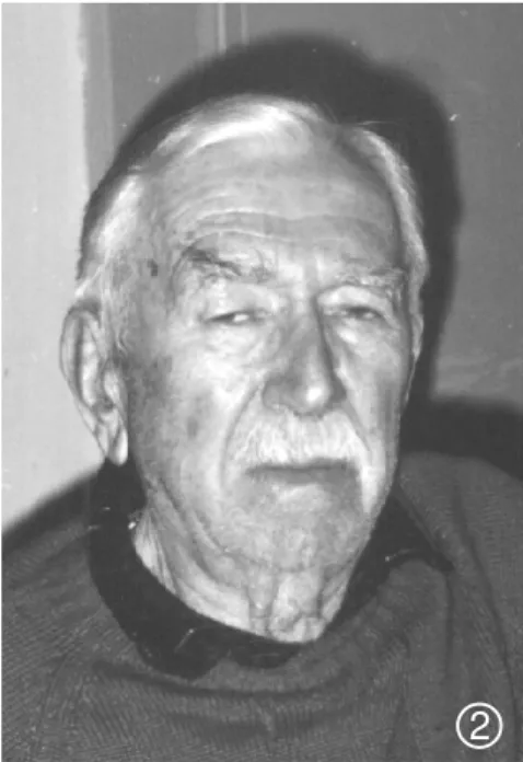
auscultation, only an hyperphonetic pulmonary second murmur being detected. No signs of heart failure. Lungs, kidney, spleen, liver and descendent colon were clinically normal. In the face, a very slight reduction of the right eyelid rift can be noted (Figure 2). Other subsidiary exams at the present time included: EKG: synusal rhythm, with rare ventricular and supraventricular ectopic beats and
Figure 2 - Mr. PER in 1998. Note reduction of the right eyelid rift. (photo by J.C. Pinto Dias).
1
Figure 1 - Mr. PER in 1947. Note Romaña’s sign (right eye). (photo by Prof. R. Talice).
*
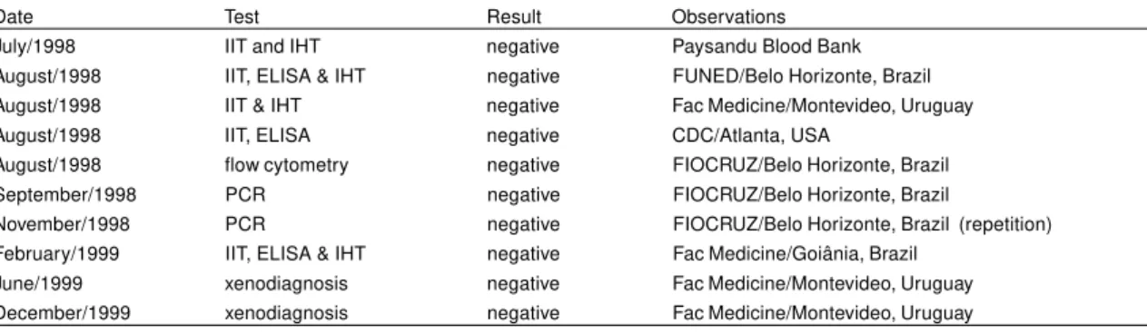
Indirect haemagglutination and indirect immunofluorescence.isolated complete bundle branch block (CRBBB), with a ventricular ÂQRS axis = 100º (positive). A cyclo-ergometric test was sufficient and normal for the age, reaching 600 kgmts/min. with an adequate elevation of arterial blood pressure (170/90mmHg and CF of 120 bpm) and no disturbances in the recuperation. During the test, basal ventricular extra-systoles disappear and no T waves alterations were revealed. The basal CRBBB remained unaltered. The interruption of the test was caused by leg muscle fatigue. Chest X-ray in PA position was normal (aor tic discrete hyper trophy), but esophageal X-ray showed a very discrete retention of contrast one minute after swallowing (borderline from normal to anectasic first degree esophagopathy). Several specific tests to detect T. cruzi infection were then performed on this patient between August 1998 and December 1999, all of them resulting negative
(Table 1). Observation. To meet ethical aspects, the medical examination was made with the full voluntary cooperation of the patient, who was completely informed about his case and the clinical and laboratory examinations to which he would be submitted, as well as the right he would have to leave the examination and/ or to refuse any medical procedure at any time, according to his will. Local and Ministerial Health Authorities as well PAHO Office (Montevideo) were also informed about this case study, none of them manifesting any doubts about the work to be done. Finally, Mr. PER continues to receive permanent medical attention from one of the authors (A.F.A.) until the date of this publication. In 2001 the patient was submitted to an inguinocrural hernia surgery in Paysandu with peridural anesthesia, presenting very good follow up and no intrasurgical complications.
Table 1 - Laboratory examinations for Trypanosoma cruzi infection in the patient PER between 1998 and 2000.
Date Test Result Observations
July/1998 IIT and IHT negative Paysandu Blood Bank
August/1998 IIT, ELISA & IHT negative FUNED/Belo Horizonte, Brazil August/1998 IIT & IHT negative Fac Medicine/Montevideo, Uruguay
August/1998 IIT, ELISA negative CDC/Atlanta, USA
August/1998 flow cytometry negative FIOCRUZ/Belo Horizonte, Brazil
September/1998 PCR negative FIOCRUZ/Belo Horizonte, Brazil
November/1998 PCR negative FIOCRUZ/Belo Horizonte, Brazil (repetition) February/1999 IIT, ELISA & IHT negative Fac Medicine/Goiânia, Brazil
June/1999 xenodiagnosis negative Fac Medicine/Montevideo, Uruguay December/1999 xenodiagnosis negative Fac Medicine/Montevideo, Uruguay IIT = indirect immunofluorescence test; IHT = indirect hemoagglutination test;
Xenodiagnosis: 40 Triatoma infestans nymphs , examined at 30 and 60 days
The parasite DNA research was made by PCR technique, utilizing the phenol-chloroform method for extraction, according to Diaz et al8.
DISCUSSION This case probably confirms prior very rare reports
of spontaneous cure of chronic Chagas’ disease described in Costa Rica, Brazil and Chile1 12 17 25.
According to the present consensus among HCD experts, a permanent absence of specific antibodies and parasitic evidence strongly suggests the nonexistence of chagasic infection, this criterion being utilized to indicate the cure of treated patients3 7 13 15 24 25.
Very singular cases of absence of antibodies in immunodeficient chagasic individuals have been reported, but generally such patients show high peripheral parasitaemia, easily detected by xenodiagnosis and/or PCR8 9 13. The evidence of an acute
phase of the disease 41 years ago in this patient was stated by direct examination (official records) and xenodiagnosis (referred by the patient), as well as the fact that his picture was published by Prof. Talice in a Medical textbook, to illustrate a typical case of acute HCD6 10 11 20. Unfortunately, no records of serology were
found in this patient’s antecedents. According to all available records and evidence, PER presented a typical
and benign acute phase of HCD and naturally entered in the chronic phase within a few weeks of clinical course, without specific treatment. The typical Romaña’s sign (Figure 1) and both information from the patient and Dr. Talice’s records leave no doubt that PER presently examined is the same person registered in 1947 and that he was contaminated by the vector route. This route is most probable since the patient had just arriving from Russia (a non-endemic country) and never received blood transfusion in his life. On the other hand, Paysandu region was highly infested by T. infestans at that time, with a general infection rate due to T. cruzi of 47%. According to Talice et al22. It should also be
underscored that Paysandu was the origin of the first case of HCD registered in Uruguay and that 35% of the cases reported by Talice et al originated from this region22. Electrocardiographical benign alterations in the
acute phase of this patient show a diffuse and slight myocardial involvement, with favorable prognosis, chiefly in the absence of clinical signs of acute heart failure5 11 20 24.
patient during the acute onset, since deaths and severe clinical pictures are always detected in younger patients and experimental animals1 4 10. To date, the clinical course
of this patient was very good and specific treatment was never initiated. It must be emphasized that at that time no effective drugs against T. cruzi were available and that Dr. Talice himself had tried in 1945 an arsenical drug in acute HCD cases, with no influence in clinical course and only slight influence in the parasitaemia of only a few cases studied 23. The central aspect of this
case concerns the complete and permanent disappearance of his IgG antibodies 41 years after the acute phase of HCD, which indicates the absence of the parasite in healthy individuals or, in other words, cure of the infection13 14 15 25. Such a hypothesis is also
reinforced here by the negative result of flow cytometry, a very specific test to detect the lytic protector antibodies which indicate the existence of live parasites3 13 14 15. The
cure was also supported by parasitological tests, namely two negative xenodiagnosis and two PCR assays8 12 15.
Clinically, the present examination of this patient shows an elderly man with slight heart and esophageal impairment which could be due to T. cruzi infection, according to several observations in Latin American endemic countries7 16 18 24. Nevertheless, the cardiac
involvement (CRBBB & extra-systolic beats) as well as the first degree esophageal dysperistalsis can also appear in elderly non-chagasic individuals, as a result of myocardial sclerosis and physiologic esophagus denervation (presby-oesophagus), thus making it almost impossible to determine the central etiological component of the present clinical picture7 18. As a general
rule and considering a series of longitudinal studies, the most probable is that both factors have been involved in this evolution, since the patient certainly had an acute phase of HCD, in which myocytolysis and autonomic denervation generally occurs1 2 5 10 20. The basic difference
between this case and the ones detected earlier in Costa Rica is just the present clinical picture, because those cases were in the chronic indeterminate form, with no cardiac and/or digestive detected disturbances25. Not
only the age of the patients can explain this clinical difference, since the Costa Rican individuals were much younger than PER5 25. Also geographical differences
might be involved, since the parasite strains are not the same, as well as the regional vectors (Triatoma dimidiata in Costa Rica: and T. infestans in Uruguay); since in general terms, it is accepted that the morbidity of HCD is usually more severe in the Southern Cone than in Central America7 20 24 25.
ACKNOWLEDGEMENTS
The authors are indebted to Dr. Olindo Martins Filho (FIOCRUZ), Dr. Alejandro Luquetti (Goiás University) and Dr. Roberto Salvatella (PAHO, Uruguay) for their valuable help in complementary serology and epidemiological data of this case.
REFERENCES
1. Andrade ZA. Patologia da Doença de Chagas. Trypanosoma cruzi e doença de Chagas, 2nd edition, Guanabara Koogan Editora, Rio de Janeiro, p. 201-230, 1999.
2. Arribada A, Apt W, Ugarte IM. A four year follow-up survey of chagasic cardiopathy in Chile.Bulletin of Pan American Health Organization 20: 240-266, 1986.
3. Cançado JR. Terapêutica Específica. In: Dias JCP, Coura JR (eds) Clínica e Terapêutica da Doença de Chagas. Uma Abordagem Prática para o Clínico Geral. Editora da Fundação Oswaldo Cruz, Rio de Janeiro, p. 323-342, 1997.
4. Chagas CRJ. Clinical and anatomo-pathologic aspects of American Trypanosomiasis. Medical Surgery Journal 12: 630-660, 1920.
5. Dias JCP. História Natural. In: Cançado JR, Chuster M (eds) Cardiopatia Chagásica. Editora da Fundação Carlos Chagas, Belo Horizonte, p. 99-113, 1984.
6. Dias JCP. Acute Chagas’ disease. Memórias do Instituto Oswaldo Cruz 79 (supl): 85-92, 1984.
7. Dias JCP, Coura JR. Epidemiologia. In: Dias JCP, Coura JR (eds) Clínica e Terapêutica da Doença de Chagas. Uma Abordagem Prática para o Clínico Geral. Editora da Fundação Oswaldo Cruz, Rio de Janeiro, p. 33-66, 1997.
8. Diaz C, Nussenzweig V, Gonzalez A. An improved polymerase chain reaction assay to detect Trypanosoma cruzi in blood. The American Journal of Tropical Medicine and Hygiene 46: 616-623, 1990.
9. Ferreira MS, Nishioka SA, Rocha A, Silva AM. Doença de Chagas e Imunossupressão. In: Dias JCP, Coura JR (eds) Clínica e Terapêutica da Doença de Chagas. Uma Abordagem Prática para o Clínico Geral. Editora da Fundação Oswaldo Cruz, Rio de Janeiro, p. 365-382, 1997.
10. Laranja FS, Dias E, Nóbrega GC, Miranda A. Chagas’ disease. A clinical, epidemiologic and pathologic study. Circulation 14: 1035-1060, 1956.
11. Lugones H, Ledesma O, Storino R, Marteleur A, Meneclier CR, Barbieri C. Chagas Agudo. In: Storino R, Milei J (eds) Enfermedad de Chagas. Doyma Argentina Editorial, Buenos Aires, p. 209-234, 1994.
12. Luquetti AO. Diagnóstico laboratorial da infecção pelo Trypanosoma cruzi. In: Brener Z, Andrade ZA, Barral-Neto M (eds) Trypanosoma cruzi e doença de Chagas, 2nd edition, Rio de Janeiro: Guanabara Koogan Editora, p. 344-378, 1999. 13. Luquetti AO, Rassi A. Tratamiento específico de la enfermedad
de Chagas en la fase crónica: criterios de cura convencionales: xenodiagnóstico, hemocultivos y serología. Revista de Patologia Tropical 27 (supl):37-50, 1998.
trypomastigote antibodies and monitor the efficacy of specific treatment in human Chagas’ disease. Clinical Diagnosis and Laboratorial Immunology 2: 569-573, 1995.
16. Prata AR. Abordagem geral do paciente chagásico. In: Dias JCP, Coura JR (eds) Clínica e Terapêutica da Doença de Chagas. Uma Abordagem Prática para o Clínico Geral. Editora da Fundação Oswaldo Cruz, Rio de Janeiro, p. 115-126, 1997. 17. Puigbó JJ, Nava Rhode JR. Barrios HG, Yepes CG. Cuatro años de
estudio longitudinal de una comunidad rural con endemia chagásica. Boletín de la Oficina Sanitaria Panamericana 48: 112-120, 1969. 18. Rezende JMO. O aparelho digestivo na doença de Chagas:
aspectos clínicos. In: Dias JCP, Coura JR (eds) Clínica e Terapêutica da Doença de Chagas. Uma Abordagem Prática para o Clínico Geral. Editora da Fundação Oswaldo Cruz, Rio de Janeiro, p. 153-176, 1997.
19. Salgado AA, Mayrink W, Dias JCP. Estudo comparativo entre a reação de fixação de complemento com os antígenos benzeno cloroformado e metílico e o xenodiagnóstico. Revista do Instituto de Medicina Tropical de São Paulo 12: 36-40, 1970.
20. Storino R, Milei J, Manzullo E, Darraidou M. Evolución Natural y Estudios Longitudinales. In: Storino R, Milei J (eds) Enfermedad de Chagas. Doyma Argentina Editorial, Buenos Aires, p. 593-604, 1994. 21. Talice RV. Epidemiologia y profilaxis de las tripanosomiasis (Enfermedad del Sueño. Enfermedad de Chagas). In: Salveraglio FJ (ed) Enfermedades Infecciosas. Inter-Médica Editorial, Buenos Aires, p. 390-399, 1949.
22. Talice RV, Costa S, Rial B, Osimani JJ. Enfermedad de Chagas (Tripanosomiasis americana). Instituto de Higiene de Montevideo Editora, Montevideo,1940.
23. Talice RV, Fernandez JL. Primeros ensayos de tratamiento de las formas agudas de enfermedad de Chagas con un nuevo arsenical. Archivos Uruguayos de Medicina, Cirurgía y Especialidades XXVII (1): 33-46, 1945.
24. World Health Organization. Chagas Disease Control. Technical Series Report no 811, 1991