Risk of persistent high-grade squamous intraepithelial
lesion after electrosurgical excisional treatment with
positive margins: a meta-analysis
Risco de persistência da lesão intraepitelial escamosa de alto grau após tratamento
excisional eletrocirúrgico com margens comprometidas: uma metanálise
Caroline Alves de Oliveira
I, Fábio Bastos Russomano
II, Saint Clair dos Santos Gomes Júnior
III, Flávia de Miranda Corrêa
IVInstituto Fernandes Figueira (IFF), Fundação Oswaldo Cruz (Fiocruz), Flamengo, Rio de Janeiro, Brazil
ABSTRACT
CONTEXT AND OBJECTIVE: Even if precursor lesions of cervical cancer are properly treated, there is a risk of persistence or recurrence. The aim here was to quantify the risks of persistence of high-grade intraepithelial squamous lesions, one and two years after cervical electrosurgical excisional treatment with positive margins.
DESIGN AND SETTING: Systematic review of the literature and meta-analysis at Instituto Fernandes Figueira.
METHODS: This meta-analysis was on studies published between January 1989 and July 2009 that were identiied in Medline, Scopus, Embase, Cochrane, SciELO, Lilacs, Adolec, Medcarib, Paho, Wholis, Popline, ISI Web of Science and Sigle. Articles were selected if they were cohort studies on electrosurgical excisional treatment of high-grade squamous intraepithelial lesions with a minimum follow-up of one year, a histo-pathological outcome of persistence of these lesions and a small risk of bias.
RESULTS: The search identiied 7,066 articles and another 21 in the reference lists of these papers. After ap-plying the selection and exclusion criteria, only four articles were found to have extractable data. The risk of persistence of high-grade intraepithelial lesions after one year was 11.36 times greater (95% conidence interval, CI: 5.529-23.379, P < 0.0001) in patients with positive margins and after two years, was four times greater (95% CI: 0.996-16.164), although without statistical signiicance.
CONCLUSION: This meta-analysis conirms the importance of positive margins as an indicator of incom-plete treatment after the irst year of follow-up and highlights the need for appropriately chosen electro-surgical techniques based on disease location and extent, with close surveillance of these patients.
RESUMO
CONTEXTO E OBJETIVO: As lesões precursoras do câncer de colo uterino, mesmo se tratadas adequada-mente, têm risco de persistirem ou recidivarem. O objetivo foi quantiicar o risco de persistência da lesão intraepitelial escamosa de alto grau (HSIL) em um e dois anos após tratamento excisional eletrocirúrgico do colo uterino com margens comprometidas.
TIPO DE ESTUDO E LOCAL: Revisão sistemática da literatura e metanálise no Instituto Fernandes Figueira.
METÓDO: Metanálise de estudos publicados entre janeiro de 1989 e julho de 2009 identiicados em Medline, Scopus, Embase, Cochrane, SciELO, Lilacs, Adolec, Medcarib, Paho, Wholis, Popline, Isis Web of Science e Sigle. Os artigos eram selecionados se fossem estudos tipo coorte sobre tratamento excisional eletrocirúrgico de HSIL com acompanhamento mínimo de um ano e tivessem como desfecho histopato-lógico a persistência de HSIL com pequeno risco de viés.
RESULTADOS: Foram identiicados 7.066 artigos e mais 21 nas listas de referências desses artigos. Após aplicação de critérios de seleção e de exclusão, somente quatro artigos ofereciam dados passíveis de extração. O risco de persistência da HSIL em um ano foi 11.36 vezes maior nas pacientes com margens comprometidas (intervalo de coniança, IC 95%: 5.529-23.379; P < 0,0001) e, em dois anos, chegou a quatro vezes, embora sem signiicância estatística (IC 95% 0.996-16.164).
CONCLUSÃO: Esta metanálise conirma a importância de margem comprometida como indicador de trata-mento incompleto no primeiro ano e ressalta a necessidade de uma adequada escolha da técnica eletrocirúr-gica em função da localização e extensão da doença e um acompanhamento adequado dessas pacientes.
IMD, MSc. Obstetrician and Gynecologist,
Hospital Federal de Bonsucesso, Bonsucesso, Rio de Janeiro, Brazil.
IIMD, PhD. Deputy Director, Department of
Education, and Head of the Colposcopy Sector, Instituto Fernandes Figueira (IFF), Fundação Oswaldo Cruz (Fiocruz), Flamengo, Rio de Janeiro, Brazil.
IIIPhD. Assistant Researcher, Clinical Research
Unit, Department of Neonatology. Instituto Fernandes Figueira (IFF), Fundação Osvaldo Cruz (Fiocruz), Flamengo, Rio de Janeiro, Brazil.
IVMD, MSc. Senior Analyst, Cancer Control
Program, Divisão de Apoio à Rede de Atenção Oncológica (DARAO), Instituto Nacional do Câncer (INCA), Rio de Janeiro, Brazil.
KEY WORDS:
Cervical intraepithelial neoplasia. Recurrence.
Prognosis. Electrosurgery.
Meta-analysis [publication type].
PALAVRAS-CHAVE:
Neoplasia intra-epitelial cervical. Recidiva.
INTRODUCTION
High-grade squamous intraepithelial lesions (HSIL) are consid-ered to be precursors of uterine cervical cancer, and even if ade-quately treated, they carry a risk of persistence or recurrence.1 Some factors highlighted in literature may increase this risk, especially in cases with positive surgical margins.2-5 However, the magnitude of the risk of persistent disease with positive surgical margins is a matter for debate.
Determining whether positive margins in a lesion are really a relevant predictor of persistence of the precursor disease of uter-ine cervical cancer may aid in managing these patients and thus in guiding new studies. From this, more efective therapeutic techniques and diferentiated strategies for follow-up treatment for women at greater risk of residual lesions might be derived.
To shed light on this issue, Ghaem-Maghami et al.6 con-ducted a meta-analysis in which they described the indings among women who were treated using electrosurgical, laser or cold knife excisional procedures. he studies included made it possible to consider that presence of low-grade intraepithelial lesions was an indicator for treatment and represented an out-come of persistence or recurrence. We did not ind any other meta-analysis that included the risk of persistence relating only to electrosurgical procedures and with consistent diagnostic cri-teria of residual precursor disease.
OBJECTIVE
Our study aimed to evaluate the risk of residual disease ater using the electrosurgical excision method, because this method is used preferentially nowadays for ectocervical lesions in our envi-ronment, as well as increasingly for endocervical lesions.7
We chose to diferentiate between residual and recurrent dis-ease and studied the irst, since we believe that it is more plausible for lesions to persist ater accomplishment of what can be consid-ered to be incomplete excision. Persistence of the lesion was con-sidered to be an outcome when diagnosed within two years ater the treatment, as described by van Hamont et al.8 All residual lesions were diagnosed by means of a biopsy. To this end, we con-ducted a systematic review of the literature and a meta-analysis in order to quantify the risk of persistence of HSIL with positive margins, one and two years ater electrosurgical excisional treat-ment on the uterine cervix.
METHODS
Identiication of studies
We conducted a systematic review of the literature and a meta-analysis, and followed the description known as preferred reporting items for systematic reviews and meta-analyses (PRISMA).9 At the end of the study, in order to ensure quality in the description of the meta-analysis, we used the checklist
for meta-analyses on observational studies in epidemiology (MOOSE): a proposal for reporting studies.10
Our search sources were electronic means (Medline, Sco-pus, Cochrane, Lilacs, SciELO, Embase, Popline, Adolec, Med-carib, ISI Web of Science, Wholis, Paho and Sigle), reference lists of reputable articles in master’s degree and PhD thesis databases (Coordenação de Aperfeiçoamento de Pessoal de Nível Superior [Capes], Fundação Oswaldo Cruz [Fiocruz] and Universidade de São Paulo [USP]) as well as contacts with authors within the ield for possible searches for relevant material not yet published.
he initial strategy using Medline sought to correlate three groups of articles with keywords relating to: (1) recurrence or persistence; (2) precursor disease for uterine cervical cancer; and (3) excisional treatment. hus, the MeSH (Medical Subject Heading) terms and other terms that the authors could use were listed as shown in Chart 1.
he following limits were applied: NOT “Breast Neoplasm” [MeSH] NOT “Neoplasm Invasiveness” [MeSH] NOT “BCG Vaccine” [MeSH] NOT “Urinary Bladder Neoplasm” [MeSH] NOT “Laryngeal Neoplasm” [MeSH] NOT “Vulvar Neoplasm” [MeSH] NOT “Lymphatic Metastasis” [MeSH] NOT “Lung Dis-eases” [MeSH] NOT “Sarcoidosis” [MeSH] NOT “Ovarian Neo-plasm” [MeSH] NOT “Lung NeoNeo-plasm” [MeSH] NOT “Skin Neoplasm” [MeSH] NOT “Melanoma” [MeSH] NOT” Stomach Neoplasm” [MeSH] NOT “Anus Neoplasm” [MeSH] NOT “hy-roid Neoplasm” [MeSH] NOT “Lymphoma” [MeSH].
he search was limited to articles published between 1989 and July 2009 that were qualiied as original articles (published papers classiied in Medline as clinical trial, randomized controlled trial, comparative study, controlled clinical trial, journal article or mul-ticenter study). here were no language constraints.
Selection of studies
Articles were selected if they were cohorts on electrosurgical excisional treatment of HSIL with a minimum follow-up of one
year11,12 and a histopathological outcome of HSIL persistence.13
he eligibility of the studies was evaluated blindly with regard to authors, journals and funding sources by two researchers. here was no disagreement among researchers. Studies were considered eligi-ble if they did not present a risk of selection bias (i.e. they used a well-deined and representative sample of the patient population), if the sample loss was less than 20% and if there were similar follow-ups among the independent comparisons of margin status.14
Data extraction
that might improve the analysis on the results (via colposcopy, electrosurgical technique and histopathological descriptions).
Statistical analysis
In order to evaluate clinical and methodological heterogeneity, we described the methodology, participants’ characteristics and inter-vention type and deined the outcomes from eligible articles. Statis-tical heterogeneity was evaluated by means of the chi-square test.
he meta-analysis was performed using the Stata 10.1 sotware, by means of the ixed-efect model,15 and the results were expressed as relative risks with 95% conidence intervals and absolute risks.
Since this was a systematic review (i.e. using a method in which scientiic publications are the subject of investigation, with-out relating to patients), there was no need for approval of the pres-ent study from the ethics committee, for it to be implempres-ented.
RESULTS
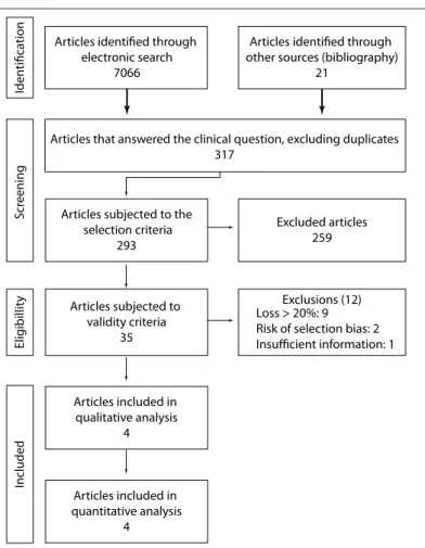
We identiied 7,066 articles using similar strategies in each electronic source. heir titles were analyzed, and 524 articles relating to the topic were retained. Among these, 227 were duplicates, thus leav-ing 297 for a more detailed analysis of titles and abstracts. A further 21 articles were extracted through manual searches in the reference lists of these 297 articles. One article was a duplicate, thus produc-ing a total of 317 articles. Twenty-four were excluded because they were reviews, replies to authors of published reviews or in languages that we were unable to translate into Portuguese (seven articles in the following languages: Bulgarian, Chinese, Finnish, Hebrew, Rus-sian and Serbian). herefore, 293 articles were subjected to the selection criteria. Out of these, only 35 went forward for eligibility evaluation. Twenty-three of these were considered to be eligible for extraction, but only four contained extractable data that could con-tribute towards conducting a meta-analysis (Figure 1). he reasons for the exclusions with regard to the impossibility of data extraction are listed below (with numbers of articles):
Insuicient data (4); no separation between electrosurgical tech-nique or cold knife conization (1); no diferentiation of compro-mised margins of positive endocervical curettage as a prognos-tic factor (1); impossible to separate lesion grade data regarding margins with recurrence, i.e. low or high-grade intraepithelial lesions (1); no data on the time of diagnosis to allow diferen-tiation between residual and recurrent disease (5); no data to conirm histology of recurrence (2); no separate data relating to HSIL (4); and margins considered to be compromised by human papillomavirus (HPV) infection alone (1).
We attempted to contact the 33 authors from whom we believed that we needed to obtain more information about their articles, by sending e-mails or letters to the addresses identiied in their papers. However, up to the time of inal writing of the present study, we had not obtained any response.
Articles that answered the clinical question, excluding duplicates 317
Excluded articles 259
Exclusions (12) Loss > 20%: 9 Risk of selection bias: 2 Insufficient information: 1
Articles included in quantitative analysis
4
Articles subjected to the selection criteria
293
Articles subjected to validity criteria
35
Articles identified through electronic search
7066
Articles identified through other sources (bibliography)
21
Articles included in qualitative analysis
4
Elig
ibillit
y
S
cr
eening
Iden
tifica
tion
Included
Figure 1. Flowchart for data search, with inclusion and exclusion of articles.
1st stage of strategy: identiication of a group of articles that dealt with
recurrence or persistence of disease:
#1 “Recurrence” [MeSH] OR “Neoplasm, Residual” [MeSH] #2 “Recurrence” OR “Residual” OR “Persistent”
#3 “Prognosis” [MeSH] #4 (#1 OR #2 OR #3)
2nd stage of strategy: identiication of a group of articles that dealt with
precursor disease through to uterine cervical cancer:
#5 “Uterine Cervical Neoplasm” [MeSH] OR “Cervical Intraepithelial Neoplasia” [MeSH] OR “Uterine Cervical Dysplasia” [MeSH] OR “Carcinoma in Situ” [MeSH] #6 “CIN” OR “SIL” OR “Squamous Intraepithelial Lesion”
#7 (#5 OR #6)
3rd stage of strategy: identiication of a group of articles that dealt with
excisional treatment:
#8 “Electrosurgery” [MeSH] OR “Therapeutics” [MeSH] OR “therapy” [Subheading] OR “Conization”[MeSH]
#9 “Treatment” OR “Management” OR “Excision” OR “LEEP” OR “Loop Electrosurgical Excision Procedure” OR “LLETZ” OR “Large Loop Excision of the Transformation Zone” OR “LLETZ-Conization” OR “LOOP-Conization” OR “LEEP- Conization” OR “SWETZ” OR “Straight Wire Excision of the Transformation Zone” OR “NETZ” OR “Needle Excision of the Transformation Zone” OR “SWETZ- Conization” OR “NETZ- Conization” OR “Conization” #10 (#8 OR #9)
4th stage of strategy: identiication of a subgroup of articles that satisied
the three abovementioned conditions (intersection of the groups):
#11 (#4 AND #7 AND #10).
he following studies contributed towards risk estimation ater one year:
• Chang et al.16: 172 patients who underwent conization
fol-lowed by hysterectomy, independent of the results relating to surgical margins, at the National University Hospital of Tai-wan. Our review only used the information from 129 cases of high-grade intraepithelial lesions. Sixteen patients had resid-ual disease, among 24 with positive margins, while seven had residual disease among 105 with negative margins.
• Goya-Canino et al.17: 382 patients who were followed up
through appointments at the cervical pathology clinic of the Mother-Child University Hospital of Canary, Spain. Among these, 305 underwent loopconization to treat high-grade intraepithelial squamous lesion (cancer cases had been excluded), and all of them underwent cyto-colposcopic fol-low-up. Four patients had residual disease among 44 with positive margins, while one had residual disease among 261 with negative margins.
he following studies contributed towards risk estimation ater two years:
• Gardeil et al.18: 583 patients who had been referred from other
hospitals, family doctors and family planning units, to Coombe Women’s Hospital in Dublin, Ireland. We only took into con-sideration the data on 225 patients who had a histopathological diagnosis of HSIL (NIC III) and had undergone LLETZ (large-loop excision of the transformation zone) by means of a dia-thermal loop of depths ranging from 10 mm to 20 mm. here was no information on how many patients underwent coniza-tion and how many underwent only excision of the transfor-mation zone. he follow-up was cyto-colposcopic. here was a loss of 42 patients. Out of the remaining 183 patients, four had residual disease among 84 with positive margins, while no patient had residual disease among 99 with negative margins.
• Verguts et al.19: 72 women with high-grade intraepithelial
squamous lesions who were treated by means of LLETZ in the Department of Gynecology of Gasthuisberg Univer-sity Hospital in Leuven, Belgium, with cyto-colposcopic fol-low-up. Two patients had residual disease among 14 with positive margins, while four had residual disease among 58 with negative margins.
he results extracted from the four selected articles were set up in an electronic spreadsheet, which was processed using the Stata 10.1 sotware for meta-analysis.
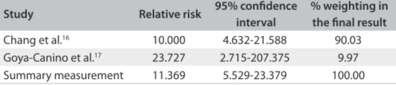
he risk of residual disease one year ater electrosurgical treatment was 11.369 times greater among the patients with posi-tive margins, as can be seen in Table 1 and Figure 2. he abso-lute risk of presenting residual disease one year ater a diagnosis of positive margins reached 29.4%, versus 2.1% in the cases with free margins.
Study Relative risk 95% conidence
interval
% weighting in the inal result
Chang et al.16 10.000 4.632-21.588 90.03
Goya-Canino et al.17 23.727 2.715-207.375 9.97
Summary measurement 11.369 5.529-23.379 100.00
Table 1. Risk of residual disease one year after electrosurgery excisional treatment with positive margins
P = 0.459 (chi-square test for heterogeneity); I-squared (variation of relative risk relating to heterogeneity) = 0.0%; relative risk test = 1; z = 6.61; P < 0.0001.
Study Relative risk 95% conidence
interval
% weighting in the inal result
Gardeil et al.18 10.588 0.578-193.861 22.80
Verguts et al.19 2.071 0.421-10.197 77.20
Summary measurement 4.013 0.996-16.164 100.00
Table 2. Risk of residual disease two years after electrosurgery excisional treatment with positive margins
P = 0.297 (chi-square test for heterogeneity); I-squared (variation of relative risk relating to heterogeneity) = 8.2%; relative risk test = 1; z = 1.96; P = 0.051.
Study
Chang et al.16
Goya-Canino et al.17
Summary measure (P = 0.961) 10.13 (5.59; 18.37) 100.00
26.2
RR (95% CI) Weight %
1 0.0381
10.31 (4.05, 26.23) 42.81 10.00 (4.63; 21.59) 57.1
RR = relative risk; CI = conidence interval.
Figure 2. Forest plot including the two studies that analyzed the risk of residual disease one year after electrosurgical excisional treatment with positive margins.
Figure 3. Forest plot including the two studies that analyzed the risk of residual disease two years after electrosurgical excisional treatment with positive margins.
Study
Gardeil et al.19
Verguts et al.19
Summary measure (P = 0.297) 4.01 (1.00; 16.16) 100.00
194
RR (95% CI) Weight %
1 0.00516
2.07 (0.42, 10.20) 77.20 10.59 (0.58; 193.86) 22.80
he risk of residual disease two years ater electrosurgical treatment was four times greater among the patients with posi-tive margins, but without reaching statistical signiicance (Table 2 and Figure 3). he absolute risk of presenting residual disease two years ater a diagnosis of positive margins was 6%, versus 2.5% in the cases with free margins.
DISCUSSION
We observed that there was a notable risk of residual disease one year ater electrosurgical treatment in patients with posi-tive margins. he estimated risk was about 11 times higher than among patients with free margins. he conidence intervals show that this risk can be estimated as at least 5.5 times higher and at most 23.3 times higher. he absolute risk of presenting residual disease over the irst year ater a diagnosis of positive margins reached 29.4%, versus 2.1% in the cases with free margins. he risk calculation ater two years showed that the estimated risk was about four times greater than among patients with free mar-gins, although this did not reach statistical signiicance.
his association is more important with regard to resid-ual disease over the irst year of follow-up, which reinforces the premise that this factor may be more connected to incomplete treatment than to recurrent disease.
he studies that were evaluated showed notable variation in the percentage of positive margins. In Gardeil et al.,18 almost half of the sample had positive margins (45%) and 4.7% presented resid-ual disease. On the other hand, in Verguts et al.,19 19% had posi-tive margins and 2.77% had residual disease. In the 1997 study,18 it was reported that only seven cases presented thermal artifacts that hindered margin evaluation. he percentage of positive mar-gins of 45% is extremely high and allows us to infer that it is pos-sible that some cases may have been misevaluated with regard to choosing the excisional method or with regard to the margins.
None of the studies mentioned any well-deined diagnostic crite-ria for positive margins. However, it is known that at this phase, there are problems that might also have inluenced the variation of percent-ages of positive margins observed among the studies. Although none of the studies mentioned the anatomical site of the positive margin (endocervical or ectocervical), it is important to report which mar-gin is compromised, in order to provide better follow-up for patients with endocervical margin involvement. In this latter type of case, it may be more diicult to diagnose a residual lesion.
Chang et al.16 gave a detailed description of how the specimen was processed. hey reported that the specimen was cut at 12 o’clock, parallel to the axis of the cervical canal, ixed onto a cork plate and let in formaldehyde until the next day. Following this, the specimen was cut every 3-4 mm and then received perpendicular cuts to the surface of the mucous membrane. However, Gardeil et al.18 only reported that the specimens were opened at 12 o ’ clock and put in formaldehyde. he other articles did not mention the specimen processing.
All the problems relating to the histopathological analysis (diagnostic criteria for positive margins and thermal damage) and to the surgical technique (surgical excision of more than one segment and specimen processing) might explain the discrepancy in the results relating to positive margins and residual disease.
Ater performing this meta-analysis, we checked the list of the 26 articles included in the treatment type “electrosurgery” in the study by Ghaem-Maghami et al.6 (compared with the four articles included in our study). We noted that sixteen articles included in that study had been excluded in the selection phase of our work. Moreover, one article on cold conization20 was wrongly included in the follow-up group of studies ater electrosurgery. Among the remaining ten articles, two were excluded in the validity evalua-tion. Six were excluded in our study at the data extraction phase and thus, out of the initial list of 26 articles included in the meta-analysis of Ghaem-Maghami et al.,6 only two18,19 were considered suitable for inclusion in our meta-analysis.
It needs to be highlighted that the risk shown in the meta-analysis by Ghaem-Maghami et al.6 was almost 50% lower (RR 6.09) than what we observed in our work. Ghaem-Maghami et al.6 collected data from articles reporting all therapeutic meth-ods (cold conization, laser and electrosurgery). he lower asso-ciation that they found between the studied factor and the out-come may be translated into diiculty in diagnosing positive margins, especially in cases undergoing laser excision, and may lead to underestimated risk. However, when calculating the risk in articles on electrosurgery alone, Ghaem-Maghami et al.6 took both high and low-grade lesions together as the outcome, thereby reaching a risk rate that was approximately one third of what we observed in our work (RR 3.34). his may have been due to inclu-sion of articles with lower levels of validity and thus greater like-lihood of bias. Another possibility is that by including low-grade lesions within the outcome, their greater frequency may lead to a lower diference in incidence between the comparison groups, thus not representing the residual disease properly.
Another issue to be addressed is that the meta-analysis by Ghaem-Maghami et al.6 used Medline as the only data source. Not only are the other research databases mentioned earlier all of importance, but also one article included in our meta-anal-ysis17 was only retrieved from Scopus in the Spanish language. his article was not considered in the meta-analysis by Ghaem-Maghami et al.,6 and it made an important contribution towards our results, since it achieved a relative risk of ten, thereby increas-ing the value of our summary measurement.
In one of the studies included, Gardeil et al.18 provided data on patients who were reviewed at 6 and 24 months, but not one year. hus, absence of a simple piece of information meant that we were prevented from using this study to contribute towards the cutof point.
On several occasions in the studies included, the data described in the text difered from what was presented in the tables. At times, the information regarding procedures was vague. Another limita-tion of the present study was that we did not have access to the data-base of each article. Hence, the calculated data was not adjusted, and the possibility of confounding thus cannot be dismissed.
We aimed to study the risk of residual disease separately for exci-sion of the transformational zone and electrosurgical conization. However, all the studies included related to conization or did not present any diferentiation regarding the technique applied. Further studies should be conducted, with better description of the surgical technique applied, in order to establish whether there is any difer-ence in the risk. Moreover, we suggest that objective standardized criteria should be deined for the diagnosis of positive margins.
Another limitation of our work was that it was impossible for us to translate into Portuguese seven articles in the following languages: Bulgarian, Chinese, Finnish, Hebrew, Russian and Serbian.
Despite these limitations, ater conducting a systematic review and then a meta-analysis with stringent methodologi-cal testing that is repeatable, we achieved a signiicant result in which the estimated risk of detecting residual disease one year ater electrosurgery was approximately eleven times greater when there are positive margins.
hus, we may assert that there is signiicantly higher likeli-hood that the disease will persist during the irst year, in cases in which the surgical specimen shows positive margins, and accord-ingly, these patients should have a diferentiated follow-up. We highlight the importance of choosing an appropriate electrosur-gical technique, with due regard to the location and extent of the lesion, in order to reduce the risk of incomplete treatment of pre-invasive cervical lesions.
Further studies should be conducted to make it clear what the best possible follow-up for such patients would be.
CONCLUSIONS
he risk of residual disease one year ater electrosurgical exci-sional treatment with positive margins is about 11 times greater than among patients with free margins. he risk of residual dis-ease two years ater such treatment is four times greater, but with-out statistical signiicance.
he absolute risk of presentation of residual lesions over the irst year is 29.4% among patients whose specimen showed posi-tive margins whereas over the second year, it is 6%.
Attention is required regarding proper indications, appro-priate surgical procedures, correct processing for the excised
specimen and appropriate choice of technique, which needs to be individualized for each case, in order to reduce the risk of residual disease. Despite the small number of studies included in this meta-analysis and the limitations mentioned above, this study clearly shows the importance of the risk of treatment fail-ure when there are reports of positive margins. Further studies should be conducted to determine the best strategy for following up these patients, especially during the irst year ater treatment.
REFERENCES
1. American College of Obstetricians and Gynecologists. ACOG Practice Bulletin No. 99: management of abnormal cervical cytology and histology. Obstet Gynecol. 2008;112(6):1419-44.
2. Lu CH, Liu FS, Kuo CJ, Chang CC, Ho ES. Prediction of persistence or recurrence after conization for cervical intraepithelial neoplasia III. Obstet Gynecol. 2006;107(4):830-5.
3. Tyler LN, Andrews N, Parrish RS, Hazlett LJ, Korourian S. Signiicance of margin and extent of dysplasia in loop electrosurgery excision procedure biopsies performed for high-grade squamous intraepithelial lesion in predicting persistent disease. Arch Pathol Lab Med. 2007;131(4):622-4. 4. Park JY, Lee SM, Yoo CW, et al. Risk factors predicting residual disease
in subsequent hysterectomy following conization for cervical intraepithelial neoplasia (CIN) III and microinvasive cervical cancer. Gynecol Oncol. 2007;107(1):39-44.
5. Manchanda R, Baldwin P, Crawford R, et al.Efect of margin status on cervical intraepithelial neoplasia recurrence following LLETZ in women over 50 years. BJOG. 2008;115(10):1238-42.
6. Ghaem-Maghami S, Sagi S, Majeed G, Soutter WP. Incomplete excision of cervical intraepithelial neoplasia and risk of treatment failure: a meta-analysis. Lancet Oncol. 2007;8(11):985-93.
7. Brasil. Ministério da Saúde. Instituto Nacional de Câncer. Nomenclatura Brasileira para Laudos Cervicais e Condutas Preconizadas: recomendações para proissionais de saúde [Brazilian Nomenclature for Cervical Cytology Reports and Guidelines]. J Bras Patol Med Lab. 2006;42(5):351-73. 8. van Hamont D, van Ham MA, Struik-van der Zanden PH, et al.
Long-term follow-up after large-loop excision of the transformation zone: evaluation of 22 years treatment of high-grade cervical intraepithelial neoplasia. Int J Gynecol Cancer. 2006;16(2):615-9.
9. Moher D, Liberati A, Tetzlaf J, Altman DG, PRISMA Group. Preferred reporting items for systematic reviews and meta-analyses: the PRISMA statement.PLoS Med. 2009;6(7):e1000097.
10. Stroup DF, Berlin JA, Morton SC, et al. Meta-analysis of observational studies in epidemiology: a proposal for reporting. Meta-analysis Of Observational Studies in Epidemiology (MOOSE) group. JAMA. 2000;283(15):2008-12.
11. Luesley D, Leeson S. Colposcopy and Programme Management. Guidelines for the NHS Cervical Screening Programme. NHSCSP Publication no 20. Sheield: NHS Cancer Screening Programmes;
12. Wright TC Jr, Massad LS, Dunton CJ, et al. 2006 consensus guidelines for the management of women with cervical intraepithelial neoplasia or adenocarcinoma in situ. Am J Obstet Gynecol. 2007;197(4):340-5. 13. Grimes DA, Schulz KF. Bias and causal associations in observational
research. Lancet. 2002;359(9302):248-52.
14. Sacket DL, Straus SE, Richardson WS, Rosenberg W, Haynes RB. Prognóstico. In: Sacket DL, Straus SE, Richardson WS, Rosenberg W, Haynes RB, eds. Medicina baseada em evidências. Prática e ensino. Porto Alegre: Artmed; 2003. p. 109-17.
15. Petitti DB. Statistical methods in meta-analysis. In: Petitti DB, editor. Meta-Analysis, decision analysis and cost-efectiveness analysis. Methods for quantitative synthesis in medicine. New York: Oxford University Press; 2000. p. 94-118.
16. Chang DY, Cheng WF, Torng PL, Chen RJ, Huang SC. Prediction of residual neoplasia based on histopathology and margin status of conization specimens. Gynecol Oncol. 1996;63(1):53-6.
17. Goya-Canino MM, Falcón-Santana JM, Arencibia-Sánchez O, et al. Follow-up of high risk intraepithelial lesions after loop excision. Prog Obstet Ginecol. 2006;49(2):72-6. Available from: http://www.elsevier. es/en/revistas/progresos-obstetricia-ginecologia-151/follow-up-of- high-risk-intraepithelial-lesions-after-13084345-articulos-originales-2006. Accessed in 2011 (Nov 4).
18. Gardeil F, Barry-Walsh C, Prendiville W, Clinch J, Turner MJ. Persistent intraepithelial neoplasia after excision for cervical intraepithelial neoplasia grade III. Obstet Gynecol. 1997;89(3):419-22.
19. Verguts J, Bronselaer B, Donders G, et al. Prediction of recurrence after treatment for high-grade cervical intraepithelial neoplasia: the role of human papillomavirus testing and age at conisation. BJOG. 2006;113(11):1303-7.
20. Bodner K, Bodner-Adler B, Wierrani F, et al. Is therapeutic conization suicient to eliminate a high-risk HPV infection of the uterine cervix? A clinicopathological analysis. Anticancer Res. 2002;22(6B):3733-6. 21. Fletcher RW, Fletcher SE. Revisões sistemáticas. In: Epidemiologia
clínica: elementos essenciais. Porto Alegre: Artmed; 2006. p. 240-57.
Sources of funding: None
Conlict of interest: None
Date of irst submission: May 3, 2011
Last received: November 21, 2011
Accepted: November 28, 2011
Address for correspondence: Caroline Alves de Oliveira
Rua César Lattes, 260 — apto 301 — bloco 1 Barra da Tijuca — Rio de Janeiro (RJ) — Brasil CEP 22793-329