Rev Odontol UNESP. 2017 July-Aug; 46(4): 220-226 © 2017 - ISSN 1807-2577
ORIGINAL ARTICLE
Doi: http://dx.doi.org/10.1590/1807-2577.22916
Comparison between aluminum chloride and tetryzoline
hydrochloride for control of vertical gingival displacement and
crevicular luid
Comparação entre cloreto de alumínio e o cloridrato de tetrizolina no controle do afastamento gengival
vertical e luído crevicular
Clóvis Lamartine de MORAES MELO NETO
a*, Helder Fernando BORGES JUNIOR
a,
Yasmin FIRMINO DE SOUZA
a, Gabriela Cristina SANTIN
a, Sérgio SÁBIO
aaUEM – Universidade Estadual de Maringá, Maringá, PR, Brasil
Resumo
Introdução: A utilização de fio de afastamento gengival com uma substância de afastamento gengival é um procedimento comum para se realizar uma moldagem com qualidade do término cervical em dentes com finalidade protética. Objetivo: Avaliar se o método mecânico-químico com cloridrato de tetrizolina a 0,05% ou cloreto de alumínio a 25% são capazes de reduzir o fluido crevicular e afastar o tecido gengival verticalmente em uma quantidade maior, estatisticamente significante, em comparação com o método mecânico de afastamento (sem substâncias químicas). Material e método: Dez pacientes foram selecionados, e os fios Ultrapak n° 000 e 1 foram posicionamos de maneira randomizada nos dentes 13, 21 e 23. Grupo I: fio sem substância química; Grupo II: fio impregnado com cloridrato de tetrizolina; Grupo III: fio impregnado com cloreto de alumínio. Utilizando modelos de gesso, foram capturadas trinta imagens com uma câmera acoplada a uma lupa, para análise do grau de afastamento. O fluxo crevicular foi quantificado através de uma balança de alta precisão, a partir de tiras de papel absorvente personalizadas para cada dente que recebeu afastamento gengival. Resultado: Não houve diferença estatística entre os três grupos quanto a quantidade de afastamento gengival vertical (Anova, p=0,26). Quanto a redução de fluido crevicular, não houve diferença entre os grupos teste e controle (Wilcoxon e Friedman, p < 0,05). Conclusão: A utilização do cloridrato de tetrizolina ou cloreto de alumínio em fio de afastamento gengival não melhorou a quantidade de afastamento vertical da gengiva e não reduziu a quantidade de fluido crevicular.
Descritores: Retração gengival; líquido do sulco gengival; sulco gengival.
Abstract
Introduction: The use of a gingival displacement cord with a gingival displacement substance is a common procedure for taking a quality impression of the cervical terminal in teeth for prosthetic purposes. Objective: To evaluate whether the mechanical-chemical method with 0.05% tetryzoline hydrochloride or 25% aluminum chloride is capable of reducing crevicular fluid, and displacing a statistically significant larger quantity of gingival tissue vertically, compared with the mechanical method (without chemical substances). Material and method: Ten patients were selected, and then No. 000 and 1 Ultrapak cords were randomly positioned on teeth 13, 21 and 23. Group I – cord with no chemical substance; Group II – cords impregnated with tetryzoline hydrochloride and Group III – cords impregnated with aluminum chloride. Using dental stone models, thirty images were captured with a camera coupled to a loupe to analyze the degree of gingival displacement. Crevicular fluid was quantified using a high-precision scale and individualized strips of absorbent paper for each tooth on which gingival displacement was performed. Result: There was no statistical difference between the three groups relative to the amount of vertical gingival displacement (Anova, p=0.26). As regards reduction in crevicular fluid, there was no difference between the test and control groups (Wilcoxon and Friedman, p < 0.05). Conclusion: The use of tetryzoline hydrochloride or aluminum chloride to impregnate the gingival displacement cord did not improve the quantity of vertical gingival displacement and did not reduce the amount of crevicular fluid.
to construct prosthetic crowns, facets, contact lenses and onlay / inlay restorations in the laboratory1. his step can frequently be diicult to perform, because the position of the gingival tissue prevents the impression material from penetrating into the gingival sulcus and correctly copying the cervical terminal2.
Most of the impression materials such as addition silicones and polyether (elastomers) have hydrophobic3 characteristics and this is a problem when there is a large volume of crevicular luid within the gingival sulcus during impression-taking. herefore, in order to achieve a quality impression, it is necessary to use gingival displacement methods and crevicular luid control1.
Both horizontal and vertical displacement of the gingival tissue must be achieved, as this is essential for obtaining an exact copy of the cervical terminal; furthermore, it is necessary to promote homeostasis of the tissues that may eventually become ulcerated1,4.
Several methods of gingival displacement have been described in literature, among them mechanical, chemical, mechanical-chemical and surgical methods (gingival curettage and electrosurgery)2,5. he method most commonly used in clinical practice is the mechanical-chemical type2, in which a substance is associated with a gingival displacement cord, for example. Chemicals for reducing the low of crevicular luid can be classiied as astringent or hemostatic. he astringent types promote localized tissue contraction through chemical reaction with proteins, generating reduction in mucous secretions and bleeding. he hemostatic types are agents that reduce blood low through the formation of clots and may stop hemorrhage5.
Chemical agents that may, for example, be used for the mechanical-chemical method are: 0.1% or 8% racemic epinephrine; 5% or 25% aluminum chloride (AlCl3); 13.3% ferric sulphate; 8% or 40% zinc chloride4. However, many of these chemical agents promote side efects or disadvantages to the patient and / or impression material, such as: racemic epinephrine, one of the agents that cause systemic1 alterations, as it increases the systolic blood pressure and pulse rate6,7; ferric or ferrous salts may be corrosive and harmful to sot tissues, enamel and can stain the teeth8; zinc chloride, a caustic and chagasic substance5, may cause damage to sot tissues and bone8; and AlCl
3, an anhydrous
5 astringent salt
widely used in contemporary dentistry that has fewer adverse efects than other substances that can be used for this purpose8. In spite of this, AlCl3 used at concentrations of over 10%6 may cause tissue destruction and permanent gingival recession of 0.1 mm9. According to Tarighi, Khoroushi8 (2014), AlCl
3 may interact in the polymerization process of polyvinyl siloxane-based (addition silicone) materials, however, this fact has been refuted by some authors who claim that there is no such correlation10.
herefore, research on gingival displacement techniques should develop methods or materials that allow correct copying of the cervical terminal. Many new substances have been tested in literature in an endeavor to ind an agent capable of stagnating
hese substances are the active ingredients in various ophthalmic solutions and nasal decongestants, such as, for example, 0.05% tetryzoline hydrochloride. Additionally, according to Sábio et al.11, the Afrin decongestant (oxymetazoline hydrochloride) and Vislin
collyrium (tetryzoline hydrochloride) did not chemically afect the impression materials tested.
he objective of this study was to evaluate whether gingival displacement cords impregnated with chemical substances such as 0.05% tetryzoline hydrochloride or 25% AlCl3 would be able to reduce crevicular low and vertically displace a larger, statistically signiicant quantity of the gingival tissue, compared with a mechanical displacement method (without chemical substances).
MATERIAL AND METHOD
Patients from the dental clinic of the State University of Maringá (Brazil) participated in this study. All patients received clariications regarding the objectives and procedures involved in the study, and signed a term of free and informed consent. he study received approval from the Research Ethics Committee on Studies with Human Beings of the university under process number 20183214.2.0000.0104.
he study was conducted with a convenience sample of patients attending the Integrated Adult Clinic of the undergraduate course in Dentistry, allocated during a period of eight months. he inclusion criteria were: men or women aged between 18 and 40 years; with good general systemic health status; good periodontal status (absence of gingivitis and periodontitis), and thick gingival biotype. Excluded from the study were: smokers; those with dental caries; abrasion; erosion; bleeding on probing; periodontal pockets; gingival recession; prosthetic posts or unsatisfactory restorations of the maxillary incisors or canines. hose who itted the inclusion criteria – 36 patients - were invited to participate in the research, but only 10 (6 women and 4 men) signed the term of free and informed consent, and attended the scheduled procedure. he procedures were divided into two steps: Evaluation of vertical gingival displacement and the amount of luid in the gingival sulcus.
In the irst step, relative isolation of the area was performed with cotton rolls and a previously cut piece of adhesive tape (Con-tact)
was attached to the vestibular surface of teeth 13, 21 and 23, level with the gingival margin to record the initial position of the gingival tissue. he gingival displacement cords were then positioned. A size 000 (Ultrapak, Ultradent, USA) cord measuring 10 mm long was irst placed in the gingival sulcus of each tooth. Ater this, one displacement cord No.1 (Ultrapak, Ultradent, USA) measuring 10 mm long was soaked in 0.05% tetryzoline hydrochloride (Mirabel, Allergan Produtos Farmacêuticos LTDA, São Paulo,
displacement cord was placed on a tooth over the displacement cord No.000 (Ultrapak, Ultradent, USA).
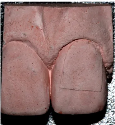
Ater four minutes, the cords were removed and the area was dried with compressed air. Impressions were taken with addition silicone (3D - Angelus, Londrina, Brazil) and the impression was removed from the oral cavity ater the silicone had cured. Ater two hours, the impression was illed with special type IV dental stone (Asfer, São Paulo, Brazil). he casts were cut into small blocks and each tooth submitted to gingival displacement was photographed using a camera coupled to a loupe (Olympus SZ-STS, Shinjuku-ku, Tokyo, Japan) (Figure 2). hirty images were acquired and analyzed with the aid of the Image-Pro Plus program (version 4.5), which was used to measure the distance between the adhesive tape indicating the initial position of the gingival tissue up to the gingival margin ater displacement. hese measurements were performed by a diferent researcher (not the examiner). All images were acquired 24 to 48 hours ater the dental stone had been cast.
In the second step, the impression from the previous procedure was used to prepare a strip of paper (Figure 3) adapted individually to the vestibular surface of teeth 13, 21 and 23 with the aim of quantifying crevicular luid. Two identical paper strips were made for each tooth and each strip was stored in a duly labeled receptacle. With the lids open, the receptacles were placed in a hothouse at 37 °C to dry for 24 hours. Ater this, the receptacles were sealed and weighed using a precision scale (Mettler Toledo, model XS205, Greifensee, Switzerland).
Relative isolation was performed with cotton roles and a cheek retractor. he selected teeth were dried with compressed air and the irst individualized strips of absorbent paper were inserted into the gingival sulcus to measure the amount of crevicular luid prior to the displacement procedure. he paper strips were held in position for 60 seconds and placed in the respective receptacles. he displacement cords (Ultrapak sizes 000 and 1) were then positioned, as in the previous step, let for four minutes and removed. he teeth were dried with compressed air and the second individualized strips of absorbent paper were inserted into the gingival sulcus, held in position for 60 seconds and stored in the respective receptacles. he receptacles with the paper strips were weighed on a precision scale (Mettler Toledo, model XS205). he diference between the initial and inal weight of each paper strip was determined, corresponding to the weight of the luid expelled by the gingival sulcus and absorbed by the paper (Figure 4).
Figure 1. (a) Adhesive tape (Com-tact) attached to vestibular surface of tooth 21; (b) Displacement cord (Ultrapak No. 000) positioned in
gingival sulcus; (c) Displacement cord (Ultrapak No. 1) positioned over irst cord.
Figure 2. Model in special type IV dental stone cut into block.
group and the Friedman, for inter-group comparing the amount of crevicular luid. he level of signiicance accepted was < 5%.
RESULT
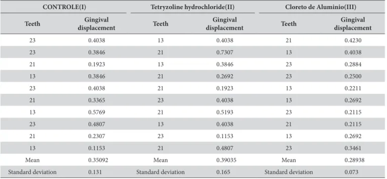
he ANOVA test (Table 1) revealed no statistically signiicant diference among groups regarding the amount of vertical gingival displacement (p = 0.26).
In Table 2, the initial and inal crevicular luid quantity values within each group were compared by the Wilcoxon test (alfa = 0.05),
Figure 4. Strip of absorbent paper inserted into gingival sulcus.
Table 1. Quantity of vertical gingival displacement (mm)
CONTROLE(I) Tetryzoline hydrochloride(II) Cloreto de Aluminio(III)
Teeth Gingival
displacement Teeth
Gingival
displacement Teeth
Gingival displacement
23 0.4038 13 0.4038 21 0.4230
23 0.3846 21 0.7307 13 0.4038
21 0.1923 13 0.3846 23 0.2884
13 0.3846 21 0.2692 23 0.2500
23 0.4038 21 0.1923 13 0.2211
21 0.3365 23 0.4038 13 0.2692
13 0.5769 21 0.5193 23 0.2115
23 0.4807 13 0.4038 21 0.2115
21 0.2307 23 0.1153 13 0.2692
13 0.1153 21 0.4807 23 0.3461
Mean 0.35092 Mean 0.39035 Mean 0.28938
Standard deviation 0.131 Standard deviation 0.165 Standard deviation 0.073
ANOVA test. he level of signiicance, p=0.26.
Table 2. Quantity of luid in gingival sulcus before (initial) and ater (inal) gingival displacement process
Substance Control(I) Tetryzoline hydrochloride (II) Aluminum chloride(III) Initial (mg) Final (mg) Diference Initial (mg) Final (mg) Diference Initial (mg) Final (mg) Diference
Patient 1 1 0.7 -0.3 0.6 0.3 -0.3 0.7 0.9 0.2
Patient 2 0.4 0.2 -0.2 0.5 0.4 -0.1 0.2 0.3 0.1
Patient 3 0.6 0.9 0.3 0.4 0.7 0.3 0.6 0.8 0.2
Patient 4 0.8 0.4 -0.4 0.4 0.2 -0.2 0.6 0.4 -0.2
Patient 5 0.7 0.6 -0.1 0.6 0.4 -0.2 0.4 0.5 0.1
Patient 6 0.4 0.8 0.4 1.2 0.6 -0.6 0.7 0.3 -0.3
Patient 7 0.4 0.3 -0.1 0.7 0.5 -0.2 0.5 0.4 -0.1
Patient 8 0.4 0.4 0 0.7 0.4 -0.3 0.7 0.5 -0.2
Patient 9 0.9 0.5 -0.4 0.6 0.3 -0.3 0.7 0.6 -0.1
Patient 10 0.7 0.6 -0.1 0.5 0.3 -0.2 0.4 0.5 -0.1
Median (mg) 0.65 0.55 -0.1 0.6 0.4 -0.2 0.55 0.52 -0.1
with a statistically signiicant diference only for tetryzoline hydrochloride.
he Friedman test revealed no statistically signiicant diference regarding the quantity of crevicular luid between the groups I (without chemical substance) and Group II (cord impregnated with tetryzoline hydrochloride) (p = 0.92) or between Group I and Group III (cord impregnated with aluminum chloride) (p = 0.83). However, a statistically signiicant diference was found between Groups II and III (p = 0.037).
DISCUSSION
he elastomers used as impression materials alone do not have suicient consistency to displace the gingival tissues. his is necessary to enable the impression material to penetrate into the gingival sulcus and overlap the cervical terminal2. In esthetic regions, vestibular surface and part of the proximal surfaces, the union between the crown and the cervical terminal should be camoulaged. For this purpose, the cervical terminal is positioned between 0.5 (ideal position)2 or 1.0 mm inside the gingival sulcus. To precisely copy this tooth preparation, we must obtain a template that exposes the cervical terminal. he gingiva should be capable of being displaced between 0.6 or 1.1 mm vertically.
According to Donovan, Chee2, Baharav et al.12, Laufer et al.13 and Chandra et al.14, a horizontal width of 0.2 mm (approximately) can be obtained for the material to perform this task2,12-14. However, this width is maintained for a maximum of 60 seconds ater removal of the cord, and later the gingival sulcus returns to its original position14. his is a factor that can hinder the impression-taking process, since the polymerization time of rubber-based materials, for example, can vary from 2 to 7 minutes13. he present study did not measure the amount of displacement obtained in the horizontal direction, however, this distance was observed to be insigniicant, since neither the impression nor the plaster model showed horizontal displacement. he polymerization time of the impression material was probably longer than the recovery time and accommodation of the gingival tissue.
he amount of vertical gingival displacement obtained in this study showed mean values of 0.35 mm in Group (I) with the cord without chemical substance; 0.39 mm in Group (II) with cord impregnated with tetryzoline hydrochloride, and 0.28 mm in Group (III) with cord impregnated with AlCl3; and in this situation there was no statistically signiicant diference between the groups. None of the methods was able to achieve gingival displacement beyond 0.5 mm, so that none of the situations tested in the present study would be able to displace a suicient quantity of gingiva to expose the cervical terminal to a subgingival depth of 0.5 mm of a tooth prepared to receive a ceramic crown, for example.
Chaudhari et al.15 compared three substances for gingival displacement. Two of them, by the mechanical-chemical method (AlCl3 / tetryzoline) and one substance by the chemical method (Expasyl - 15% AlCl3). he results showed that the gingival displacement between the AlCl3 group and the tetryzoline hydrochloride group was comparable. In our study, there was no statistically signiicant diference in relation to gingival displacement
between the two groups (II and III), nor were these groups able to provide a statistically signiicant gingival displacement value higher than that obtained in Group I (control).
During impression taking, other factors to consider are the hydrophobic3 properties of the majority of impression materials, because the interior of the gingival sulcus is constantly irrigated by the crevicular luid. In the case of inlamed gingival tissues - a frequent condition in teeth that will receive prosthetic crowns, this irrigation is more intense and may interfere in the penetration of the material during impression-taking. he gingival displacement process generally promotes changes in the microcirculation of periodontal tissues, causing inlammation and an increase in the amount of crevicular luid production16. Phatale et al.17 observed that junctional epithelium of teeth ater gingival displacement with Ultrapak cord 00 impregnated with 5% AlCl3 underwent major changes, such as intracellular degeneration and desquamation due to the chemical displacement methods used17. herefore, the technique with gingival displacement cord, either associated with a chemical agent, or not, may damage the periodontal tissue and increase the amount of crevicular luid16-18. herefore, a sensitive technique associated with correct manipulation of the sot tissues17 during the introduction of the gingival displacement cord can be a very important and efective factor to avoid exacerbated tissue irritations1. Moreover, it is important for the gingiva to be healthy and for the cervical terminal of the preparation not to be placed at exaggerated2 depths.
he study of Wöstmann et al.18, compared three methods of gingival displacement: namely, the mechanical-chemical (racemic epinephrine), chemical (Expasyl-15% AlCl3) and mechanical (with cord only) for measuring the reduction in crevicular luid. he cited authors observed that use of the mechanical method alone (with cotton yarn only) was not eicient for controlling the crevicular luids; they found a greater increase in gingival luid low in this method. hese indings were not in agreement with the results of the present study, since the mechanical method (Group I) showed no increase luid production; furthermore, 25% AlCl3 or 0.05% tetryzoline hydrochloride did not signiicantly reduce the crevicular luid low, when compared with Group I (Control). hese data alone appeared to show that the mechanical barrier of the displacement cord was as capable of decreasing the crevicular luid low, as was the action of chemicals.
obtained, thereby justifying their use.
In addition to correct impression-taking, excellent laboratory work is necessary for correct adaptation of a prosthesis to a dental preparation. herefore the computer-assisted design and computer-assisted manufacturing (CAD/CAM) technology, such as, for example, the Cerec (Sirona), Lava (3M Espe) and Pro-cera (Nobel Biocare) systems have been increasingly used in dentistry. Even when copying the cervical terminal correctly, dentists depend on a quality dental prosthesis, and CAD/CAM may provide advantages in relation to the classical method of fabricating a prosthesis, such as eiciency (cost and time) use of materials such as Zirconia and the precision of prosthetic work, which are requisites for the longevity of a dental prosthesis21. Greater precision of prostheses in relation to the cervical terminal of the preparation means avoiding exaggerated marginal gaps that may generate dissolution of the cement, thereby increasing the potential of leakage, caries and periodontal disease. Moreover, this may avoid exaggerated space between the surface
during rehabilitation with dental prostheses.
Although a convenience sample was used, (which was a limitation of the study), the patients received the three types of treatment, in a randomized manner among teeth 13, 21 and 23, thus reducing the sample bias. However, the authors suggest that new clinical trials should be conducted.
CONCLUSION
Impregnating the gingival displacement cord with tetryzoline hydrochloride or AlCl3 did not reduce the amount of gingival crevicular luid produced in comparison with the gingival displacement cord without a chemical substance.
he diferent procedures used in the present study did not difer signiicantly with regard to the amount of gingival displacement. herefore, there is no justiication for the use of chemical substances during gingival displacement procedures.
REFERENCES
1. Sábio S, Correa GO, Sábio SS, Andrade HM. Utilização do cloridrato de tetrizolina para afastamento gengival em moldagens com silicona de adição. Rev Dental Press Estét. 2008 Jul-Set;5(3):45-54.
2. Donovan TE, Chee WW. Current concepts in gingival displacement. Dent Clin North Am. 2004 Apr;48(2):vi, 433-44. PMid:15172609. http://dx.doi.org/10.1016/j.cden.2003.12.012.
3. Wassell RW, Barker D, Walls AW. Crowns and other extra-coronal restorations: impression materials and technique. Br Dent J. 2002 Jun;192(12):679-84, 687-90. PMid:12125794. http://dx.doi.org/10.1038/sj.bdj.4801456.
4. Bowles WH, Tardy SJ, Vahadi A. Evaluation of new gingival retraction agents. J Dent Res. 1991 Nov;70(11):1447-9. PMid:1960257. http:// dx.doi.org/10.1177/00220345910700111101.
5. Nemetz EH, Seibly W. The use of chemical agents in gingival retraction. Gen Dent. 1990 Mar-Apr;38(2):104-8. PMid:2132221.
6. Polat NT, Ozdemir AK, Turgut M. Effects of gingival retraction materials on gingival blood flow. Int J Prosthodont. 2007 Jan-Feb;20(1):57-62. PMid:17319365.
7. Donovan TE, Gandara BK, Nemetz H. Review and survey of medicaments used with gingival retraction cords. J Prosthet Dent. 1985 Apr;53(4):525-31. PMid:3889287. http://dx.doi.org/10.1016/0022-3913(85)90640-7.
8. Tarighi P, Khoroushi M. A review on common chemical hemostatic agents in restorative dentistry. Dent Res J (Isfahan). 2014 Jul-Aug;11(4):423-8. PMid:25225553.
9. Benson BW, Bomberg TJ, Hatch RA, Hoffman W Jr. Tissue displacement methods in fixed prosthodontics. J Prosthet Dent. 1986 Feb;55(2):175-81. PMid:3514852. http://dx.doi.org/10.1016/0022-3913(86)90336-7.
10. Machado CE, Guedes CG. Effects of sulfur-based hemostatic agents and gingival retraction cords handled with latex gloves on the polymerization of polyvinyl siloxane impression materials. J Appl Oral Sci. 2011 Nov-Dec;19(6):628-33. PMid:22230998. http://dx.doi. org/10.1590/S1678-77572011000600014.
11. Sábio S, Franciscone PA, Mondelli J. Effect of conventional and experimental gingival retraction solutions on the tensile strength and inhibition of polymerization of four types of impression materials. J Appl Oral Sci. 2008 Jul-Aug;16(4):280-5. PMid:19089261. http://dx.doi. org/10.1590/S1678-77572008000400010.
12. Baharav H, Laufer BZ, Langer Y, Cardash HS. The effect of displacement time on gingival crevice width. Int J Prosthodont. 1997 May-Jun;10(3):248-53. PMid:9484057.
13. Laufer BZ, Baharav H, Langer Y, Cardash HS. The closure of the gingival crevice following gingival retraction for impression making. J Oral Rehabil. 1997 Sep;24(9):629-35. PMid:9357742. http://dx.doi.org/10.1046/j.1365-2842.1997.00558.x.
15. Chaudhari J, Prajapati P, Patel J, Sethuraman R, Naveen YG. Comparative evaluation of the amount of gingival displacement produced by three different gingival retraction systems: an in vivo study. Contemp Clin Dent. 2015 Apr-Jun;6(2):189-95. PMid:26097353. http://dx.doi. org/10.4103/0976-237X.156043.
16. Zhulev EN, Zolotukhina EV, Saakian MI. The impact of retraction cord design on marginal gingiva status in patients with fixed dentures. Stomatologia. 2013;92(6):51-2.
17. Phatale S, Marawar PP, Byakod G, Lagdive SB, Kalburge JV. Effect of retraction materials on gingival health: A histopathological study. J Indian Soc Periodontol. 2010 Jan;14(1):35-9. PMid:20922077. http://dx.doi.org/10.4103/0972-124X.65436.
18. Wöstmann B, Rehmann P, Balkenhol M. Influence of different retraction techniques on crevicular fluid flow. Int J Prosthodont. 2008 May-Jun;21(3):215-6. PMid:18548958.
19. Woody RD, Miller A, Staffanou RS. Review of the pH of hemostatic agents used in tissue displacement. J Prosthet Dent. 1993 Aug;70(2):191-2. PMid:8371184. http://dx.doi.org/10.1016/0022-3913(93)90018-J.
20. Kopac I, Batista U, Cvetko E, Marion L. Viability of fibroblasts in cell culture after treatment with different chemical retraction agents. J Oral Rehabil. 2002 Jan;29(1):98-104. PMid:11844038. http://dx.doi.org/10.1046/j.1365-2842.2002.00790.x.
21. Karl M. In vitro studies on CAD/CAM restorations fabricated with Procera technology: an overview. Quintessence Int. 2015 Jul-Aug;46(7):561-74. http://dx.doi.org/10.3290/j.qi.a33937. PMid:25918760.
22. Mously HA, Finkelman M, Zandparsa R, Hirayama H. Marginal and internal adaptation of ceramic crown restorations fabricated with CAD/CAM technology and the heat-press technique. J Prosthet Dent. 2014 Aug;112(2):249-56. PMid:24795263. http://dx.doi.org/10.1016/j. prosdent.2014.03.017.
CONFLICTS OF INTERESTS
he authors declare no conlicts of interest.
*CORRESPONDING AUTHOR
Clóvis Lamartine de Moraes Melo Neto, Clínica Odontológica, UEM – Universidade Estadual de Maringá, Av. Mandacaru, 1550, Centro, 87080-000 Maringá - PR, Brasil, e-mail: [email protected]