Contents lists available at
ScienceDirect
Progress in Neuropsychopharmacology
& Biological Psychiatry
journal homepage:
www.elsevier.com/locate/pnp
IDO chronic immune activation and tryptophan metabolic pathway: A
potential pathophysiological link between depression and obesity
Adriano José Maia Chaves Filho
a,b, Camila Nayane Carvalho Lima
a,b,
Silvânia Maria Mendes Vasconcelos
a,b, David Freitas de Lucena
a,b, Michael Maes
c,d,e,
Danielle Macedo
a,b,⁎aNeuropsychopharmacology Laboratory, Drug Research and Development Center, Faculty of Medicine, Universidade Federal do Ceará, Fortaleza, CE, Brazil bDepartment of Physiology and Pharmacology, Faculty of Medicine, Universidade Federal do Ceará, Fortaleza, CE, Brazil
cImpact Strategic Research Center, Deakin University, Geelong, Australia dFaculty of Medicine, Chulalongkorn University, Bangkok, Thailand
eHealth Sciences Graduate Program, Health Sciences Center, State University of Londrina, Londrina, Brazil
A R T I C L E I N F O
Keywords:
Indoleamine 2,3-dioxygenase Tryptophan catabolites Obesity
Depression
Comorbid depression/obesity
A B S T R A C T
Obesity and depression are among the most pressing health problems in the contemporary world. Obesity and depression share a bidirectional relationship, whereby each condition increases the risk of the other. By in-ference, shared pathways may underpin the comorbidity between obesity and depression. Activation of cell-mediated immunity (CMI) is a key factor in the pathophysiology of depression. CMI cytokines, including IFN-γ, TNFαand IL-1β, induce the catabolism of tryptophan (TRY) by stimulating indoleamine 2,3-dioxygenase (IDO) resulting in the synthesis of kynurenine (KYN) and other tryptophan catabolites (TRYCATs). In the CNS, TRYCATs have been related to oxidative damage, inflammation, mitochondrial dysfunction, cytotoxicity, ex-citotoxicity, neurotoxicity and lowered neuroplasticity. The pathophysiology of obesity is also associated with a state of aberrant inflammation that activates aryl hydrocarbon receptor (AHR), a pathway involved in the de-tection of intracellular or environmental changes as well as with increases in the production of TRYCATs, being KYN an agonists of AHR. Both AHR and TRYCATS are involved in obesity and related metabolic disorders. These changes in the TRYCAT pathway may contribute to the onset of neuropsychiatric symptoms in obesity. This paper reviews the role of immune activation, IDO stimulation and increased TRYCAT production in the pa-thophysiology of depression and obesity. Here we suggest that increased synthesis of detrimental TRYCATs is implicated in comorbid obesity and depression and is a new drug target to treat both diseases.
1. Introduction
Obesity and depression are two of the most pressing and costly
health problems faced today. Several studies indicate that the
pre-valence of obesity has increased alarmingly in recent decades. For
ex-ample, from 1980 to 2008, the overall prevalence of obesity has more
than doubled, with 10% of men and 14% of women around the world
being considered obese (i.e., body mass index > 30) (
Preiss et al., 2013;
Schneider et al., 2010
). These data have a serious impact since obesity
is a major risk factor associated with chronic diseases such as
hy-pertension, coronary artery disease, type 2 diabetes mellitus and
cancer, as well as with the increased risk of premature death (
Fontaine
et al., 1997; Hrabosky and Thomas, 2008; Allison et al., 2009
).
Depression, in turn, is the leading cause of disability worldwide
http://dx.doi.org/10.1016/j.pnpbp.2017.04.035
Received 31 October 2016; Received in revised form 3 April 2017; Accepted 10 April 2017
⁎Corresponding author at: Drug Research and Development Center, Department of Physiology and Pharmacology, Federal University of Ceará, Rua Cel. Nunes de Melo 1000,
60430-270 Fortaleza, CE, Brazil.
E-mail address:[email protected](D. Macedo).
Abbreviations:1-MT, 1-methyltryptophan; 3-HK, 3-hydroxykynurenine; 5-HT, serotonin; 5-HTP, 5-hydroxy-L-tryptophan; 5-HIAA, 5-hydroxyindoleatic acid; ACTH,
adrenocortico-trophic hormone; AADC, aromaticL-amino acid descarboxylase; AA-NAT, aralkylamineN-acetyltransferase; AHR, aryl hydrocarbon receptor; AM, autobiographical memory; BMI, body
mass index; BCG, bacille Calmette–Guérin; BDNF, brain derived neurotrophic factor; CNS, central nervous system; COX-2, cyclooxygenase-2; CRP, C-reactive protein; fMRI, functional magnetic resonance imaging; HIOMT, hydroxyindoleO-methyltransferase; HFD, high fat diet; HPA, hypothalamic-pituitary-adrenal axis; IDO, indoleamine 2,3-dioxygenase; IFN, in-terferon; IL, interleukin; LPS, lipopolysaccharide; KAT, kynurenine aminotransferase; KMO, kynurenine 3-monooxigenase; KYN, kynurenine; KYNA, kynurenic acid; MDD, major de-pressive disorder; MRL, medial temporal lobe; MT2, melatonergic receptor 2; NAD, nicotinamide adenine dinucleotide; NAS,N-acetylserotonin; NFκB, nuclear factorκB; NMNAT,
nicotinamide mononucleotide adenyltransferase; NOS, nitric oxide synthase; QUIN, quinolic acid; QPRT, quinolinate phosphoribosyltransferase; SNP, single nucleotide polymorphism; TDO, tryptophan 2,3-dioxygenase; TNF, tumor necrosis factor; TRYCATs, tryptophan catabolites
Available online 12 June 2017
0278-5846/ © 2017 Elsevier Inc. All rights reserved.
being the major contributor to the overall global burden of disease.
Recently, the World Health Organization (WHO) recognizes depression
as the main cause of disability and loss of productive life years
world-wide (
WHO, 2015; Kessler et al., 2015
). In the USA 2013, it was
esti-mated that > 15.7 million people had episodes of major depression,
accounting for > 6.6% of adult population of this country (
Substance
Abuse and Mental Health Services Administration, 2014
). In addition,
similarly to obesity, depression has been associated with increased risk
of developing severe chronic diseases such as atherosclerotic heart
disease, type 2 diabetes mellitus and cancer and increased mortality
rates (
Clarke and Currie, 2009; Grippo, 2009; Preiss et al., 2013
).
An increasing body of evidence has pointed to an important
bi-directional link between obesity and depression (
Miller et al., 2003;
Rosmond, 2004; Hryhorczuk et al., 2013; Castanon et al., 2015
). In this
context, population-based analysis revealed that obese people have an
increased incidence of depressive symptoms (> 30%) compared to
healthy subjects (
Pan et al., 2012; Lin et al., 2013
). Furthermore,
longitudinal studies have demonstrated a prospective link between
obesity and depression with obese individuals having a higher risk for
developing depression (about 55%) over time. Conversely, individuals
with depression present a higher risk to become obese (about 58%)
(
Lupp et al., 2007; Preiss et al., 2013
). There is also a significant
as-sociation between obesity and the onset of mood changes and cognitive
deficits in older adults (
Cournot et al., 2006; Roberts et al., 2010; Dahl
et al., 2013
). Of note, depression significantly impacts the quality of life
and social skills of obese people. Depression also impairs adherence to
treatment and beneficial changes in lifestyle, representing an additional
risk factor for the worsening of obesity and its pathological
complica-tions, particularly cardiovascular disease (
Roberts et al., 2003; Simon
et al., 2006; Zhao et al., 2011; Hamer et al., 2012
).
1.1. Neurobiology of depression: an overview
For many years, since the introduction of the
first antidepressants,
the pathophysiology of depression was restricted to a deficit in biogenic
amines (
López-Muñoz et al., 2007; López-Muñoz and Alamo, 2009
). In
this regard, the promising antidepressant effects of the class of
anti-depressant drugs called serotonin (5-HT) uptake inhibitors gave rise to
the development of the so-called serotonin hypothesis of depression.
Based on this hypothesis depression was primarily associated with a
decrease in 5-HT synthesis and action on its receptors (
Fangmann et al.,
2008; López-Muñoz and Alamo, 2009
). Nevertheless, although drugs
with mechanism of action based on the serotonergic theory of
depres-sion have shown efficacy in the treatment of a subgroup of individuals,
its pharmacological potential is currently limited. This is due to the fact
that a large population of patients seems to be refractory or to have a
late onset of action when prescribed these drugs. Thus, the
mono-aminergic theory of depression, as proposed, presents some limitations.
Modern theories are proposed to better explain the pathophysiology of
this mental disorder (
Maes et al., 2011b; Salazar et al., 2012; Réus
et al., 2015
). Activated immune-inflammatory pathways are associated
with depression and may be induced by common trigger factors of
depression, including psychosocial stressors, exogenous stressors and
medical comorbidities (
Maes et al., 2011a, 2011b
). Indeed, physical
and psychological stressors can activate the immune system in both the
periphery and Central Nervous System (CNS) thereby releasing
in-flammatory cytokines leading to neurotransmitter and behavioral
changes (
Maier and Watkins, 1998, Koo and Duman, 2008
).
In fact, high levels of pro-inflammatory cytokines, such as interferon
(IFN)-
γ
, tumor necrosis factor (TNF)-
α
, and interleukin (IL)-1
β
have
been consistently reported in plasma and brain samples of depressive
patients (
Maes, 1995a, 1995b; Kling et al., 2007; Song et al., 2009;
Dowlati et al., 2010; Felger and Lotrich, 2013
). This is reinforced by the
findings that in humans and animal models a pro-inflammatory state
induced by exogenous cytokines, including IL-6 (
Sukoff
Rizzo et al.,
2012; Kong et al., 2015
), TNF
α
(
Reichenberg et al., 2001; Simen et al.,
2006
), IFN-
α
(
Raison et al., 2005, 2013
) and bacterial endotoxins or
lipopolysaccharides (LPS) (
Grigoleit et al., 2011; Custódio et al.,
2013;Tomaz et al., 2014
) may cause depression and depression-like
symptoms, such as lethargy, anhedonia, anorexia, decreased sexual
activity and sleeping disorders. Therefore, it is now considered that
neuro-immune mechanisms play a key role in the pathogenesis and
pathophysiology of depression (
Maes, 1995a, 1995b; Schiepers et al.,
2005; Maes et al., 2011b; Rosenblat et al., 2014
).
1.2. Pathophysiology of obesity: an overview
Obesity is not only a metabolic disease, but also a chronic
in-flammatory condition, in which both innate and acquired immune
re-sponses are affected (
Dandona et al., 2004; Bastard et al., 2006;
Cancello and Clément, 2006
). Elevated serum levels of inflammatory
markers e.g. IL-1
β
, TNF
α
and IL-6 have been observed in obese patients
(
Kopp et al., 2005; Park et al., 2005; Capuron et al., 2011a
) and in
animal models of obesity (
Bigorgne et al., 2008; Cani et al., 2009; Pistell
et al., 2010; Lawrence et al., 2012; Dinel et al., 2014b
). Aberrant
flammation activates aryl hydrocarbon receptor (AHR), a pathway
in-volved in the detection of intracellular or environmental changes,
sensing light, oxygen and redox potential (
Gu et al., 2000
). Thus, based
on the fact that genetic contribution to obesity is estimated by 25
–
70%,
while environmental factors (consumption of the high-calorie, high-fat,
low-fiber Western diet) contribute by 30
–
75% (
Baillie-Hamilton, 2002
),
AHR seems to be the biological entity that tightly links genes and the
environment in the pathophysiology of obesity (
Moyer et al., 2016
).
Interestingly, a significant association between systemic
pro-in-flammatory status and the emergence of depressive symptoms (
Capuron
et al., 2008; Castanon et al., 2014
) and cognitive deficits (
Sweat et al.,
2008; Sellbom and Gunstad, 2012
) has been observed in obese
in-dividuals. In addition, an important elevation of pro-inflammatory
cy-tokines is found in brain areas associated with mood disorders, such as
the hippocampus and hypothalamus, in experimental obesity (
Pistell
et al., 2010; André et al., 2014; Miller and Spencer, 2014
). Of note,
these
findings were positively associated with the onset of anxiogenic
and depressive-like behaviors (
Pistell et al., 2010; André et al., 2014;
Dinel et al., 2014
).
1.3. An overview of the pro-in
fl
ammatory state in depression and obesity
and tryptophan catabolites (TRYCATs) pathway
The overproduction of pro-inflammatory cytokines may activate a
major enzyme involved in tryptophan (TRY) metabolism, namely
in-doleamine 2,3-dioxygenase (IDO), taking away TRY from 5-HT
synth-esis thereby driving the production of tryptophan catabolites
(TRYCATs), including kynurenine (KYN), 3-hydroxykynurenine (3-HK),
kynurenic acid (KYNA), xanthurenic acid, quinolinic acid (QUIN),
pi-colinic acid and anthranilic acid (
Connor et al., 2008; Maes et al., 2008;
Maes, 2011; Dinel et al., 2014; Réus et al., 2015
). These TRYCATs have
different biological and neurobehavioral actions. For example, KYNA in
physiological levels seems to present antioxidant and neuroprotective
properties mainly based on its ability to bock
N-methyl-
D-aspartate
(NMDA) receptors. On the other hand, 3-HK and QUIN have noxious
effects including neurotoxic, excitotoxic, cytotoxic and pro-oxidative
effects (
Guillemin et al., 2001; Maes et al., 2007, 2011
).
between IDO activation or increased serum TRYCATs and the onset and
severity of mood symptoms in depressive patients (
Maes et al., 2002;
Wichers et al., 2005; Mackay et al., 2009; Vignau et al., 2009; Gabbay
et al., 2010; Meier et al., 2016; Savitz et al., 2015c
).
Recently, an imbalance of TRY metabolism and increased
circu-lating levels of detrimental TRYCATs have been reported in obese
pa-tients and related metabolic disorders, such as heart atherosclerotic
disease and type 2 diabetes mellitus (
Brandacher et al., 2006, 2007;
Oxenkrug, 2010, 2013; Mangge et al., 2014a, 2014b; Favennec et al.,
2015
). Of note, KYN and less prominently KYNA, 3-HK,
3-hydro-xyanthranilic acid and QUIN are endogenous AHR agonists
Mezrich
et al. (2010)
, which directly activated AHR gene expression (
Sallée
et al., 2014; Oxenkrug et al., 2016
). Recently, a study showed that the
inhibition of AHR prevents Western diet-induced obesity (
Moyer et al.,
2016
). Furthermore, an interesting correlation has been proposed
be-tween the levels of detrimental TRYCATs and the worsening of the
prognosis and pro-inflammatory status of these patients (
Sulo et al.,
2013; Mangge et al., 2014b; Eussen et al., 2015
). Despite this evidence,
it is not clear whether detrimental TRYCATs are also produced in the
CNS of obese individuals or may impact the natural history of obesity
including the onset of neuropsychiatric symptoms.
1.4. Aims
The objective of this paper is to review the current body of evidence
that IDO activation and the resulting production of detrimental
TRYCATs play a role in the pathophysiology of comorbid depression
and obesity. We hypothesize that the production of detrimental
TRYCATs could be a potential biological link between obesity and
de-pression and thus a new drug target for treating comorbid obesity and
depression.
2. Search strategy
A comprehensive literature search was conducted with the PubMed/
MEDLINE database to identify studies that were relevant to this current
review. The search terms
“
inflammation
”
[MeSH] OR
“
cytokines
”
[Mesh] OR
“
tryptophan
”
[MeSH] OR
“
kynurenine
”
[MeSH] OR
“
ky-nurenine pathway
”
OR
“
indoleamine 2,3-dioxygenase
”
[MeSH] OR
“
tryptophan 2,3-dioxygenase
”
OR
“
kynurenic acid
”
[MeSH] OR
“
qui-nolinic acid
”
[MeSH] OR
“
serotonin
”
[MeSH] OR
“
cognition
”
[MeSH]
OR
“
cognitive functions
”
[MeSH] OR
“
melatonin
”
[MeSH] were
cross-referenced with
“
Depression
”
[MeSH] OR
“
Depressive Disorder
”
[MeSH] OR
“
Depressive Disorder, Major
”
[MeSH] AND
“
Obesity
”
[MeSH] OR
“
Metabolic Syndrome
”
[MeSH] OR
“
Abdominal Fat
”
[MeSH]. We included papers published in English language until April
1st 2017. The inspection of reference lists of the included studies and
tracking citations of included papers in Google Scholar augmented this
search strategy. Observational, experimental studies in human and
animal models and literature reviews addressing the role of components
of the TRYCAT pathway in the pathophysiology of depression and
obesity were included. The overall methodological quality of retrieved
references was considered for
final inclusion.
3. Results
3.1. IDO activation and increased TRY metabolism in depression
Tryptophan is an essential amino acid relevant to many
physiolo-gical processes, in particular to the CNS. Tryptophan has two distinct
metabolic pathways: the methoxyindole or 5-HT pathway and the
oxidative or TRYCAT pathway (
Fernstrom, 1983; Fernstrom and
Fernstrom, 1995; Oxenkrug, 2007
). The main products of
methox-yindole pathway are serotonin and melatonin. Tryptophan is converted
to 5-HT by the enzyme tryptophan hydroxylase being this, the
rate-limiting step in the synthesis of the neurotransmitter 5-HT.
Accordingly, 5-HT levels are limiting for melatonin synthesis. On the
other hand, the oxidative pathway leads ultimately to the production of
nicotinamide and generation of energy through glutarate. The
first step
of KYN pathway is the conversion of TRY to KYN (rate-limiting step),
catalyzed by two enzymes, tryptophan 2,3-dioxygenase (TDO) or IDO.
Subsequently other metabolites are generated, the TRYCATS, which
have important biological effects, both in the CNS and in peripheral
organs (
Lapin, 2003; Mackay et al., 2009; Maes et al., 2011b
).
Tryptophan levels are significantly reduced in depressed patients
(
Joseph et al., 1984; Maes et al., 1987a, 1987b, 1990a, 1991b; Capuron
et al., 2011b; Liu et al., 2015b
). In these patients, TRY plasma levels
may be inversely associated with severity of depression, anxiety,
so-matization, suicidal ideation, neuromuscular symptoms and paranoia
(
Lehmann, 1972; Curzon et al., 1979; Hoes et al., 1981; Joseph et al.,
1984; Capuron et al., 2011b; Maes et al., 2011b; Blankfield, 2013;
Flores-Ramos et al., 2014; Gostner et al., 2015; Hüfner et al., 2015
).
Besides this, TRY plasma levels may constitute a predictor of
ther-apeutic response to serotonergic antidepressants, particularly selective
inhibitors of the 5-HT uptake (
Møller, 1985
). Conversely, a favorable
response to serotonergic antidepressants usually is followed by
nor-malization of TRY levels (
Møller, 1985; Healy and Leonard, 1987;
Badawy and Morgan, 1991
). Therefore the reduction in TRY
avail-ability in depressive patients seems to be explained by metabolic
pro-cesses (
Maes, 1995a,b; Maes et al., 2011b
).
TDO is one of the main enzymes involved in the peripheral
cat-abolism of TRY. This enzyme is predominantly present in the liver and
some stimuli such as glucocorticoids and excessive levels of TRY can
increase its activity. Generally, TDO maintains the circulating levels of
TRY in homeostatic equilibrium, but may also increase the catabolism
of TRY in case of increased energy demand and elevated nicotinamide
synthesis (
Wolf, 1974; Hoes and Sijben, 1981; Oxenkrug, 2007
).
Pre-clinical studies have shown that treatment with dexamethasone
in-creases TDO activity and lowers the levels of TRY in the plasma, liver
and brain, indicating that glucocorticoids induce TDO activity (
Green
et al., 1975; Young, 1981; Morgan and Badawy, 1989
). Also
dex-amethasone administration to humans lowers the brain availability of
TRY (
Maes et al., 1990a,b,c, d
).
Increased baseline hyperactivity of the
hypothalamic-pituitary-adrenal axis (HPA) and the loss of the negative feedback loop of
glu-cocorticoids, leading to increased basal serum levels of
adrenocortico-trophic hormone (ACTH) and cortisol is one of the biological hallmarks
of severe depression (
Du and Pang, 2015; Kabia et al., 2015
). Patients
on prolonged use of glucocorticoids or with Cushing's syndrome, who
chronically presents high levels of TDO activity, often experience
neuropsychiatric disorders, such as depressive symptoms (
Kelly et al.,
1980, 1983
). Furthermore, TRY plasma levels are negatively related to
ACTH and cortisol levels in depressive patients (
Maes et al. 1987a,
1990b
). Therefore, it has been suggested that TDO activation in the
context of HPA axis hyperactivity may be a potential contributing factor
to the increased TRY catabolism and decreased bioavailability of this
amino acid in depression (
Maes et al., 1990a; Fukuda, 2014; Gibney
et al., 2014
). Another important point is that an excessive activation of
HPA axis is a core alteration observed in patients suffering from
psy-chotic depression (
Belanoff
et al., 2001; Keller et al., 2006
). Indeed,
TDO activation leads to KYNA synthesis, a NMDA receptor antagonist
(
Wu et al., 2013
). NMDA blockade is related to the emergence of
psy-chotic symptoms (
Krystal et al., 1994
).
Th (CD4+) and T suppressor/cytotoxic (CD8+) cells, and the
pro-duction of cytokines, such as IFN-
γ
and IL-2. These cytokines, in turn,
can activate monocytes/macrophages, stimulating the production of the
“
monocytic
”
cytokines, like IL-1
β
, IL-6, IL-12 and TNF
α
, that can
fur-ther activate T lymphocytes, forming a positive feedback circuit (
Rocha
et al., 2008; Maes, 2011
). Furthermore, there is a population of T
lymphocytes, designated Th3-type, which action is suppressing Th1
responses. Th3 cells primarily produce transforming growth factor beta
(TGF-
β
) seeming to be fundamental to maintain Th1 and Th2 balance in
several organs, including CNS (
MyInt et al., 2005
).
Additionally, in the last years, it was identified a new family of
cytokines named IL-17 cytokines. This originated a new subset of Th
cells, the Th17-type cells. Similar to Th1 and Th2 cells, Th17 cells
re-quire specific cytokines and transcription factors, of note, TGF-
β
and
IL-21 for differentiation and IL-23 for growth and stabilization. While the
function of this cell subtype is not completely elucidated, emerging data
suggest that Th17 cells may play an important role in host defense
against extracellular pathogens, which are not efficiently cleared by
Th1-type and Th2-type immunity (
Bettelli et al., 2007; Korn et al.,
2009
). Also, considerable data proposes that Th17 cytokines play
highly pro-inflammatory actions and that Th17 cells mediate immune
responses against self-antigens in autoimmunity disorders
Lee K et al.
2012; Sigdel et al., 2016
).
In patients with depression, an abnormal exacerbation of Th1
im-mune responses has been repeatedly reported. In this context, previous
evidence showed an increased production of IFN-
γ
(
Maes et al., 1993,
1994; Seidel et al., 1995
) and high rates of IFN-
γ
/IL-4 (indicative of
Th-1/Th-2 balance) and IFN-
γ
/TGF-
β
(indicative of Th-1/Th-3 balance)
(
MyInt et al., 2005; Kim et al., 2007; Song et al., 2009
) in depressed
patients. Furthermore, increased levels of T-cell activation markers,
such as the count of T cell CD25
+, HLA-DR, IL-2 soluble receptor
(sIL-2R); and CMI markers, including neopterin and IL-12 levels, have been
demonstrated (
Maes et al., 1991a, 1992; Miller et al., 2009
). Therefore,
a state of CMI activation marked by a mutual stimulation of T
lym-phocytes and monocytic cells seems to be a key factor in the
in-flammatory pathophysiology of depression (
Maes, 1995a,b, Maes,
2011; Maes et al., 2011b
).
The IDO enzyme, similarly to TDO, participates in the
first step of
KYN metabolic pathway. Differentially from TDO, IDO is distributed in
human tissues, such as brain, lung, kidney, intestine, beyond monocytic
cells, being its expression very low in physiological conditions. The
inflammatory cytokine IFN-
γ
is the main inductor of IDO expression
(
Oxenkrug, 2007, 2010
). Other inflammatory cytokines, such as TNF
α
,
IL-2, IL-1
β
and prostaglandin PGE2, may also induce IDO expression,
while anti-inflammatory cytokines, e.g. IL-4, IL-10 and TGF
β
, may
in-hibit IDO (
Liebau et al., 2002; Oxenkrug, 2010
). Thus, in a state of cell
immune activation with overproduction of pro-inflammatory cytokines,
the determining enzyme of TRY metabolism is IDO. Moreover, during
the inflammatory responses, while IDO is activated, TDO appears to be
suppressed (
Takikawa et al., 1986, 1988; Brandacher et al., 2007
).
Oxidative and nitrosative stress, related to depression and other chronic
inflammatory conditions (
Maes et al., 2011a; Du and Pang, 2015
) may
also contribute to the IDO induction (
Daley-Yates et al., 1988; Thomas
and Stocker, 1999; Maes et al., 2007
).
Consistent with these
findings, TRY levels in depressed patients are
inversely related to serum concentrations of pro-inflammatory markers,
such as IL-6 and haptoglobin (a positive acute phase protein), and
po-sitively related to the levels of anti-inflammatory markers, such as
transferrin (a negative acute phase inflammatory protein) (
Maes et al.,
1993; Seidel et al., 1995
). Furthermore, reduced TRY levels have been
frequently associated with increased levels of IFN-
γ
and neopterin,
important markers of Th-1 immune response (
Maes et al., 1994; Widner
et al., 2002; Celik et al., 2010
). The pro-inflammatory status in
de-pression thus may stimulate IDO and consequently TRY catabolism
(
Maes et al., 2007, 2011b; Oxenkrug, 2010
).
3.2. IDO activation and increased TRY metabolism in obesity
Recent studies have suggested that TRY metabolic imbalance may
also be involved in the development of metabolic disorders, particularly
obesity (
Brandacher et al., 2007; Oxenkrug, 2013; Mangge et al.,
2014a, 2014b
). Classically, excessive nutrient intake and lack of
ex-ercise are key factors in the natural history of obesity. In obese
in-dividuals and related metabolic disorders the circulating levels of TRY
are often reported as being reduced (
Caballero et al., 1988; Wurtman
et al., 2003; Oxenkrug, 2010; Sulo et al., 2013; Mangge et al., 2014b;
Raheja et al., 2015
) and as in depression, an imbalance of TRY
meta-bolism has been proposed (
Oxenkrug, 2010; Mangge et al., 2014b
).
Obesity has been consistently considered as a chronic
in-flammatory disease characterized by increased levels of Th1 cytokines,
such as IFN-
γ
and TNF
α
as well as other T lymphocytic and monocytic
cell markers (
Cancello and Clément, 2006; Rocha et al., 2008; Liu et al.,
2014; Donma et al., 2015; Zahorska-Markiewicz et al., 2000
). There is
evidence supporting that Th1 cytokines, including IFN-
γ
, play a key role
in the immune-inflammatory pathophysiology of obesity (
Svec et al.,
2007; Strissel et al., 2010; Lee and Lee, 2014
). This is reinforced by
preclinical
findings showing that genetically or diet-induced obese mice
produce more pro-inflammatory cytokines, including IFN-
γ
and TNF
α
,
than control mice (
Kawanishi et al., 2010; Yamada et al., 2016; Zhou
et al., 2015
). Furthermore, administration of TNF
α
or IL-6 to pregnant
rats results in considerable expansion of adipose tissue in their offspring
and increased vulnerability to obesity (
Dahlgren et al., 2001
). Higher
serum levels of IFN-
γ
and IL-6 were observed in obese individuals when
compared to healthy individuals. This increase in serum levels of
pro-inflammatory cytokines was related to circulating levels of leptin, a
hormone derived from adipose tissue related to differentiation of T
naive cells towards a Th1 phenotype (
Park et al., 2005; Pacifico et al.,
2006; Rocha et al., 2008
).
In this context, genetic factors, diet and physical inactivity among
others are mechanism proposed to explain the systemic inflammatory
status associated with obesity. Recently, diet is calling great attention
since it can modulate the status of the resident gut
flora and the
translocation of toxigenic bacterial products through intestinal
epithe-lium (
Turnbaugh et al., 2008; Cani et al., 2009; Silventoinen et al.,
2010; Lopresti et al., 2013; Lecomte et al., 2015
). In fact, it was shown
that high fat diets can affect the profile of microbial community
fa-voring the growing of more toxigenic bacteria and facilitating the
transport of bacterial products, especially the endotoxin LPS, to
sys-temic circulation (
Moreira et al., 2012
). Furthermore, saturated fatty
acids (SFA), an important component of diet-induced obesity, directly
induce pro-inflammatory changes in adipocytes and adipose tissue
macrophages. These fatty acids can activate nuclear factor-
κ
B (NF-
κ
B),
a transcription factor necessary for the expression of several
in-flammatory cytokines like IFN-
γ
and TNF
α
as well as for the stimulation
of the secretion of macrophage chemotactic factors, such as monocyte
chemoattractant protein 1 (MCP-1/CCL2). Interestingly, the action of
SFA seems to be mediated, similarly to LPS, by toll-like receptor 4
(TLR4). This is confirmed by
findings showing that the inhibition of
TLR4 prevents the development of pro-inflammatory changes in
adi-pocytes (
Yeop Han et al., 2010; Caesar et al., 2015
).
2013
). Also, morbidly obese patients seem to present high serum levels
of neopterin and KYN and reduced serum levels of TRY. Interestingly,
these alterations remain even after bariatric surgery (
Brandacher et al.,
2006
). Furthermore, a recent study analyzing adipose tissue and liver
samples found increased IDO expression, pro-inflammatory cytokines
(IL-18, TNF
α
) and T cell activation markers (CD14, CD68, and CD3
ε
) in
samples of obese patients (
Wolowczuk et al., 2012
). Therefore,
simi-larly to depression, systemic immune activation in obesity may
stimu-late IDO thereby inducing an imbalance of TRY catabolism.
3.3. TRYCAT pathway in depression and obesity: 5-HT depletion and the
production of detrimental TRYCATs
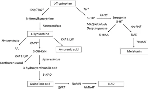
Kynurenine is a substrate for the production of KYNA and
nicoti-namide-adenine dinucleotide (NAD). KYNA synthesis is regulated by
the enzymes kynurenine aminotransferase (KAT) I, II and III. The
en-zymes kynurenin-3-monooxygenase enen-zymes (KMO), kynureninase and
kynurenine hydroxylase generate NAD as
final product. Together, these
routes can generate > 30 intermediate metabolites, collectively called
TRYCATs, with multiple biological actions, some of which deleterious
(
Maes et al., 2007; Oxenkrug, 2007
).
In the CNS, the TRYCATs can mediate important effects. For
ex-ample, QUIN and picolinic acid, both derived from NAD pathway, are
endogenous agonists of glutamate NMDA receptors. In addition, QUIN
can inhibit glutamate uptake by astrocytes potentiating the toxic effects
of the overactivation of NMDA receptors (
Tavares et al., 2002, 2005
).
QUIN can cause the destruction of postsynaptic neural elements and the
death of hippocampal and granular progenitor cells (
Khaspekov et al.,
1989; Santamaría et al., 2001; Steiner et al., 2011; Lugo-Huitrón et al.,
2013; Meier et al., 2016
). Other metabolites of NAD pathway, such as
3-HK and 3-hydroxyanthranilic acid, generate free radicals and induce
pro-apoptotic effects in neuronal cells being associated with the
de-velopment of neurodegenerative diseases, e.g. Huntington's and
Alz-heimer's disease (
Goldstein et al., 2000; Yan et al., 2005; Smith et al.,
2009; Gulaj et al., 2010; Tan et al., 2012; Reyes-Ocampo et al., 2015
).
On the other hand, KYNA is an endogenous NMDA receptor antagonist
presenting antioxidant properties. These antioxidant properties of
KYNA can counteract the excitotoxicity induced by the toxic
metabo-lites of NAD pathway (
Henderson et al., 1990; Ma
ł
aczewska et al.,
2014; Savitz et al., 2015a
). However, KYNA was also related to the
antagonism of
α
7 nicotinic receptors. This nicotinic receptor has a key
role in central cholinergic anti-inflammatory response (
Hilmas et al.,
2001
) (an overview of TRYCATs pathway can be seen in
Fig.1
).
3.3.1. Increased detrimental TRYCATs in depression
Previous research has indicated that some TRYCATs can affect
an-imal behavior towards anxiogenic- and depressive-like phenotypes. The
first evidence of this effect came from studies demonstrating that
per-ipheral or intracerebroventricular administration of KYN and QUIN
could induce anxiogenic-like effect in rodents in the open
field and
elevated plus maze tests (
Vécsei and Beal, 1990; Lapin et al., 1996
).
More recently,
O'Connor et al. (2009b)
reported that the peripheral
administration of L-KYN triggers depressive-like behavior in rodents in
a dose-dependent manner. These investigators also observed that the
behavioral alterations caused by peripheral administration of LPS
de-pend on the central activation of IDO being reversed by
1-methyl-tryptophan, a competitive IDO inhibitor, or by minocycline, a
tetra-cycline with notable anti-inflammatory effects (
O'Connor et al., 2009b
).
Immune activation triggered by LPS is capable of inducing enzyme
activities of IDO and kynurenine hydroxylase (one of the main enzymes
involved in NAD pathway), but not kynurenine aminotransferases (the
main enzymes involved in the KYNA pathway). These
findings suggest
that immune challenge with LPS selectively stimulates TRY catabolism
towards NAD pathway resulting in the production of neurotoxic
TRYCATs (
Connor et al., 2008
). A possible explanation for this
phe-nomenon is that, in situations of brain immune challenge, microglia
orchestrate neuro-inflammatory changes in the brain. Microglia mainly
express the enzymes of NAD pathway, as KMO and kynurenine
hy-droxylase. The opposite occurs in astrocytes, in which KAT
pre-dominates. Consequently, in animal models of depression induced by
LPS, microglia is responsible for the production of neurotoxic
meta-bolites derived from NAD pathway, such as QUIN and 3-HK (
Guillemin
et al., 2001; O'Connor et al., 2009b; Steiner et al., 2011; Salazar et al.,
2012
).
Clinical evidence supports the hypothesis that the increased
production of neurotoxic TRYCATs may occur in depression (
Mackay
et al., 2009; Gabbay et al., 2010; Meier et al., 2016; Savitz et al.,
2015c
). In this context,
Gabbay et al. (2010)
found high rates of KYN/
TRY (indicator of IDO activity) and of 3-HK/KYN in adolescent patients
with depression, which reflects the occurrence of TRY metabolism
through NAD pathway in depression. Additionally, these increased
ra-tios were positively associated with the severity of depressive symptoms
(
Gabbay et al., 2010
). High serum levels of hydroxyanthranilic acid and
QUIN were also demonstrated in adult patients with depression and
being associated with the worsening of mood symptoms (
Mackay et al.,
2009; Savitz et al., 2015c
). Furthermore, some evidence indicates that
not only TRY levels are reduced, but also KYNA levels are compromised
in patients with depression (
Myint et al., 2007; Savitz et al., 2015c
).
Some of the most interesting
findings about the participation of
Fig. 1.Overview of the tryptophan catabolite (TRYCAT)
TRYCATs in depression came from studies with patients using
im-munotherapy. Immunotherapy mimics the systemic immune activation
observed in chronic inflammatory disorders, such as depression. In
these patients, the onset and severity of depressive symptoms correlates
strongly with IDO activity and TRYCATs production (
Bonaccorso et al.,
2001; Bonaccorso et al., 2002; Wichers et al., 2005; Vignau et al., 2009;
Raison et al., 2010; Maes et al., 2011b
).
Bonaccorso et al. (2002)
and
Wichers et al. (2005)
demonstrated that the onset of depressive
symp-toms during IFN-
α
-based immunotherapy was positively associated
with the KYN/TRY and KYN/KYNA ratios. Since KYN is the substrate
for QUIN synthesis these results allow us to better visualize the
asso-ciation between the increase in detrimental TRYCATs and the
devel-opment of depressive symptoms over time (
Bonaccorso et al., 2002;
Wichers et al., 2005
). Also,
Raison et al. (2010)
reported a significant
association between serum levels of QUIN and the emergence of
de-pressive symptoms in patients undergoing IFN-
α
-based immunotherapy
(
Raison et al., 2010
). Therefore, systemic immune activation in
de-pression may selectively induce TRY metabolism towards the
produc-tion of potentially detrimental TRYCATs, which are associated with the
onset of depressive symptoms.
3.3.2. Increased detrimental TRYCATs in obesity
Besides central effects, TRYCATs also exert effects on peripheral
organs. Previous studies have shown an increase in the production of
detrimental TRYCATs in the course of obesity and related metabolic
disorders (
Oxenkrug, 2010, 2013; Mangge et al., 2014a, 2014b
).
En-zymes involved in TRYCAT synthesis are constitutively expressed in key
metabolic organs, such as liver, pancreas and adipose tissue and are
activated by pro-inflammatory cytokines (
Däubener and MacKenzie,
1999; Fujigaki, 2006; Wolowczuk et al., 2012; Liu JJ et al., 2015
). It
was also reported that KYN, QUIN and picolinic acid can stimulate
nitric oxide synthase (NOS) activity resulting in an increased
produc-tion of nitric oxide and nitrous free radicals in macrophages and
en-dothelial cells (
Melillo et al., 1994; Chiarugi et al., 2000
). In pancreatic
cells, TRYCATs such as 3-hydroxyanthranilic, picolinic and xanthurenic
acid can promote a cascade of arachidonic acid reactions, increasing the
production of pro-inflammatory factors, such as prostaglandins and
leukotrienes (
Melillo et al., 1993; Alberati-Giani et al., 1997; Bosco
et al., 2000, 2003; Cesario et al., 2011
). On the other hand, KYNA has
anti-inflammatory and immunomodulatory actions in peripheral
im-mune cells (
Ma
ł
aczewska et al., 2014
).
Some diet components may contribute to the synthesis of
detri-mental TRYCATs in key metabolic organs, including the pancreas. For
example,
Liu et al. (2015a
,
b
) reported that high concentrations of
glucose and SFA stimulate the expression of IDO and KMO leading to an
increased KYN/KYNA ratio in cultured
β
-pancreatic cells. Oxidative
stress and glucocorticoids also induce the expression of these enzymes
and TRYCATs production in pancreatic cells (
Liu JJ et al., 2015
).
Im-portantly, TRYCATs have important metabolic effects. For example,
acute exposure to TRYCATs, including hydroxykynurenine and
3-hydroxyanthranilic acid, inhibits the secretion of insulin by
β
-pan-creatic cells (
Rogers and Evangelista, 1985; Liu et al., 2015a
).
Ad-ditionally, xanthurenic acid may compromise the biological activity of
insulin, forming an antigenic complex with this hormone. Together,
these mechanisms may be potentially involved in the onset of insulin
resistance in obesity and metabolic syndrome (
Kotake et al., 1975;
Meyramov et al., 1998; Oxenkrug, 2013
).
There is some evidence that increased TRYCATs production may
also be causally associated with the onset of obesity and related
me-tabolic disorders (
Mangge et al., 2014b; Favennec et al., 2015
). In this
context,
Favennec et al. (2015)
demonstrated increased levels of KYN,
KYNA and QUIN in serum of obese patients as compared to healthy
controls. Positive correlations between the serum levels of KYN and
QUIN and body mass index (BMI) as well as insulin resistance were
found. Analyzing the omental adipose tissue of obese patients, a
con-siderable expression of several enzymes involved in the TRYCAT
pathway was found, including IDO, kynureninase, KMO and KTA III
(
Favennec et al., 2015
). Other recent studies demonstrate high serum
levels of TRYCATs, including KYNA and xanthurenic acid, in patients
with type 2 diabetes mellitus and coronary atherosclerosis. The
in-creased levels of detrimental TRYCATs in metabolic disorders were also
associated with a worse outcome of these metabolic diseases (
Eussen
et al., 2015; Oxenkrug, 2015
).
The link between the activation of TRYCATs pathway and the
de-velopment of weight gain/obesity has been reinforced by recent studies
using genetic-based approaches. For example,
Nagano et al., 2013
re-ported that
Ido1
−/−mice present less weight gain compared with
wild-type mice when submitted to Western diet (for 26 weeks). This was
confirmed by the study of
Moyer et al. (2016)
, that additionally
de-monstrated the relevance of aryl hydrocarbon receptor (AHR) pathway
to the IDO/TRYCATs effects in the context of diet-induced obesity. Of
note, it was demonstrated that AHR antagonists, such as
α
-naphtho-flavone or CH-223191, prevent the development of obesity and
adip-osity and ameliorates liver steatosis in mice fed with Western diet
(
Moyer et al., 2016
). Interestingly, Moyer and coworkers demonstrated
that the genetic or pharmacological blockade of IDO1 reduces the
ex-pression of AHR in mouse hepatocytes. Other relevant pathways, such
as TGF
β
1 and TLR2/4 signaling also exert their effects on AHR
ex-pression through IDO induction and KYN production (
Moyer et al.,
2016
). Therefore, AHR activation through TRYCATs synthesis seems to
be a unifying mechanism for the effects of inflammatory-related
path-ways in diet-induced weight gain.
Obesity is not just associated with chronic inflammation in
per-ipheral tissues, but also with central inflammation. Upregulated levels
of IL-6, TNF
α
and NF-
κ
B are detected in the brain of genetically obese
rodents or rodents subjected to diet-induced obesity (
Zhang et al., 2008;
Boitard et al., 2014; Maric et al., 2014; Dorfman and Thaler, 2015
).
Reactive gliosis with infiltration of peripheral immune cells and
pro-liferation of resident glial cells is found in the brain of obese animals
(
Buckman et al., 2014; Dorfman and Thaler, 2015
). Clinical
postmortem
studies corroborate these
findings, showing considerable glial
activa-tion and neuronal injury in brain tissue of obese individuals (
Thaler
et al., 2012
).
There is compelling evidence that central neuroinflammation is an
important factor in the onset of neuropsychiatric symptoms related to
obesity (
Soczynska et al., 2011; Miller and Spencer, 2014; Castanon
et al., 2015
). Among the several neuroinflammatory processes involved
in obesity, IDO activation and subsequent TRYCAT production in the
CNS may be relevant to the onset of mood and anxiety (
Lin et al., 2007;
Swardfager et al., 2009; Dinel et al., 2011, 2014
). Preclinical studies
demonstrate that in genetically obese mice the induction of
depressive-like behavior following systemic LPS administration is intrinsically
re-lated to IDO activation following increases in pro-inflammatory
cyto-kines (IL-1
β
and TNF
α
) in the hippocampus (
Dinel et al., 2014
). A
significant reduction in hippocampal expression of brain derived
neu-rotrophic factor (BDNF), which has been consistently related to
de-pression pathophysiology (
Moylan et al., 2013; Dinel et al., 2014
).
Si-milarly, other recent evidence demonstrated that animals subjected to
diet-induced obesity present significant behavioral changes,
depressive-like and anxiogenic-depressive-like behavior, concomitantly to the increase in the
expression of several pro-inflammatory cytokines and IDO activity in
the hypothalamus and hippocampus (
André et al., 2014
).
et al., 2014; Strassnig et al., 2015; Vu
č
i
ć
Lovren
č
i
ć
et al., 2015
).
Nevertheless, as far as we know there are no studies showing IDO
ac-tivation and TRYCATs synthesis in the brain of obese patients as well as
their relationship with the emergence of psychiatric symptoms (
Fig. 2
).
This could be a potential mechanism in this comorbid being thus an
important issue to be addressed in further studied.
3.4. TRYCAT pathway and cognitive de
fi
cits in depression and obesity
Cognitive functions in humans comprise different domains such as
perception, attention, memory and executive function (
Cohen et al.,
1996; Guan et al., 2016
). Several studies have shown that depression is
associated with impairments in cognitive functions, especially
frontal-temporally mediated cognitive domains, including memory, executive
functioning and planning. Furthermore, some deficits present early in
the course of the disorder and may worsen with staging (
Papakostas,
2006; Bora et al., 2013
). In this context, a recent meta-analysis,
fo-cusing in the cognitive deficits present in early depression, analyzed the
data from 13 different studies involving > 640 patients (
Lee RSC et al.,
2012
). These researchers found that patients with MDD had in the
first
mood episode significant impairment in psychomotor speed (effect size
0.48), attention (effect size 0.36), and visual learning and memory
(effect size 0.53). Also, within the domain of executive functions,
at-tentional switching (effect size 0.22), verbal
fluency performance
(ef-fect size 0.59) and cognitive
flexibility (effect size 0.53) were worse in
these patients (
Lee RSC et al., 2012
). Declarative memory and
psy-chomotor speed deficits frequently become more severe with long-term
illness duration, and sometimes associate with relapses or recurrences
following antidepressant treatment (
MacQueen et al., 2002; Trivedi and
Greer, 2014; Kim et al., 2016
). In fact, a study conducted with older
individuals showed that patients with poor executive function
mea-sured at the beginning of antidepressant treatment were more likely to
have a relapse over a 16-week continuation phase and more likely to
experience a recurrence over a 2-year maintenance phase as compared
to those with normal executive functions (
Alexopoulos et al., 2000
).
Cognitive deficits have an important impact on functionality and
Fig. 2.Mechanisms underlying chronic systemic in-flammation involved in obesity and depression leading to central and peripheral IDO activation and production of potentially detrimental TRYCATs. Etiological risk factors, e.g., fat/sugar diet, physical inactivity, social/psychological stress or chronic infections can originate a state of systemic inflammation involved in the development ofobesity-de-pression comorbid. The peripheral release of
proin-flammatory cytokines can activate IDO pathway in key
metabolic organs, such as adipose tissue, as well as access the brain via several pathways (e.g., neural, humoral, and cellular routes). In the CNS cytokines can induce
neuroin-flammatory processes, primarily by activating microglia and after by promoting the induction of IDO activity and synthesis of neuroactive/neurotoxic TRYCATs. Deregulations of the gut–brain axis, by changes in gut mi-crobiota composition and permeability, can also participate in induction of systemic inflammatory state and activation