Time-course effects of aerobic exercise training on
cardiovascular and renal parameters in 2K1C
renovascular hypertensive rats
R.C.A. Maia
2*, L.E. Sousa
1,3*, R.A.S. Santos
4, M.E. Silva
2,3, W.G. Lima
1,2,3,
M.J. Campagnole-Santos
4and A.C. Alzamora
1,2,3 1Departamento de Ciências Biológicas, Instituto de Ciências Exatas e Biológicas, Universidade Federal de Ouro Preto, Ouro Preto, MG, Brasil 2Programa de Pós-Graduac
¸ão em Saúde e Nutric¸ão, Universidade Federal de Ouro Preto, Ouro Preto, MG, Brasil
3Programa de Pós-Graduac
¸ão em Ciências Biológicas, NUPEB, Universidade Federal de Ouro Preto, Ouro Preto, MG, Brazil
4Departamento de Fisiologia e Biofísica, Instituto de Ciências Biológicas, Universidade Federal de Minas Gerais,
Belo Horizonte, MG, Brasil
Abstract
Exercise training (Ex) has been recommended for its beneficial effects in hypertensive states. The present study evaluated the time-course effects of Ex without workload on mean arterial pressure (MAP), reflex bradycardia, cardiac and renal histology, and oxidative stress in two-kidney, one-clip (2K1C) hypertensive rats. Male Fischer rats (10 weeks old; 150–180 g) underwent surgery (2K1C or SHAM) and were subsequently divided into a sedentary (SED) group and Ex group (swimming 1 h/day, 5 days/week for 2, 4, 6, 8, or 10 weeks). Until week 4, Ex decreased MAP, increased reflex bradycardia, prevented concentric hypertrophy, reduced collagen deposition in the myocardium and kidneys, decreased the level of thiobarbituric acid-reactive substances (TBARS) in the left ventricle, and increased the catalase (CAT) activity in the left ventricle and both kidneys. From week 6 to week 10, however, MAP and reflex bradycardia in 2K1C Ex rats became similar to those in 2K1C SED rats. Ex effectively reduced heart rate and prevented collagen deposition in the heart and both kidneys up to week 10, and restored the level of TBARS in the left ventricle and clipped kidney and the CAT activity in both kidneys until week 8. Ex without workload for 10 weeks in 2K1C rats provided distinct beneficial effects. The early effects of Ex on cardiovascular function included reversing MAP and reflex bradycardia. The later effects of Ex included preventing structural alterations in the heart and kidney by decreasing oxidative stress and reducing injuries in these organs during hypertension.
Key words: 2K1C renovascular hypertension; Swimming; Baroreflex bradycardia; Heart and kidney adaptations; Oxidative stress
Introduction
Structural and functional alterations in the heart and kidney are involved in the development of arterial hypertension by hyperactivity of the sympathetic nervous system and renin-angiotensin system (RAS) as well as their contributions to high blood pressure and reduced sensitivity of the baroreflex control of the heart rate (HR) (1,2). Many studies (1–4) have used the two-kidney, one-clip (2K1C) Goldblatt hypertensive model in an attempt to understand the mechanisms of development and main-tenance of renovascular hypertension. The time course of the 2K1C hypertensive model has been divided into several phases after clipping of the renal artery: at about
4 weeks, blood pressure rises in association with increases in the plasma renin activity and circulating angiotensin II (Ang II) concentration. In weeks 5 to 8, hypertension is associated with increases in tissue RAS components despite a fall in plasma renin activity and circulating Ang II. At week 9 and after, hypertension is maintained by increases in the tissue RAS activity, plasma volume, and sympathetic tone (1). Moreover, evidence has shown increased generation of reactive oxygen species (ROS) in specific organs such as the brain, heart, and kidneys during renovascular hypertension (3,4).
Correspondence: A.C. Alzamora:<[email protected]>.
*R.C.A. Maia and L.E. Sousa are co-first authors.
Ang II, aldosterone, and catecholamines are involved in the development of ventricular hypertrophy under pathological (5,6) and physiological (7,8) conditions, reflected as worsening or improvement of cardiac function, respectively. Ventricular hypertrophy can be concentric in certain pathological conditions, such as arterial hypertension, or concentric and eccentric in physiological cardiac hypertrophy induced by static or dynamic physical exercise training (Ex), which induces two different types of intermittent chronic cardiac workload (6,8,9).
Ex induces adaptive cardiovascular benefits in hypertensive conditions by reducing the sympathetic outflow, vascular resistance, and plasma Ang II levels and improving the sensitivity of the baroreflex (2,10–13). Additionally, during endurance Ex, the increase in oxygen consumption results in increased generation of ROS, which is involved in the adaptive up-regulation of antioxidant gene expression (14). Moreover, evidence has shown that low-intensity Ex (50–60% of maximal exercise capacity) more effectively decreases blood pressure in hypertensive patients and rats than does high-intensity Ex (7,15–17). However, to maintain these benefits over time, close monitoring by healthcare professionals is required to adjust the Ex intensity to avoid possible adverse effects of more vigorous exercise, especially in hypertensive states, considering that risk factors such as age and cardiac disease could be associated with this pathology (18–20).
In the present study, our hypothesis was that Ex performed without adjusting the workload over time, even if it does not effectively reduce the blood pressure, could have beneficial effects on organs that participate in the control of blood pressure and thus reduce the cardiovascular risk. In view of these considerations, we evaluated the time-course effects of Ex without workload on the mean arterial pressure (MAP), reflex bradycardia, cardiac and renal histology, and oxidative stress at different stages of development of 2K1C hypertension.
Material and Methods
Ethics approval
All experiments were performed on 123 male Fischer rats (10 weeks of age; 150–180 g) from ENUT, Universi-dade Federal de Ouro Preto, MG, Brasil. The animals were housed in separate cages in groups of four (2K1C or SHAM) with free access to rat chow and tap water in a temperature- and light-controlled room (24±1°C; 12:12 h light-dark cycle). All animal procedures were in accor-dance with the Guidelines for Ethical Care of Experimental Animals and performed as approved by the Institutional Ethics Committee of the Universidade Federal de Ouro Preto (Protocol #022/2007).
Induction of renovascular hypertension
Renovascular hypertension was induced as described by Goldblatt et al. (21). Briefly, the rats were anesthetized with a mixture of ketamine (50 mg/kg) and xylazine (10 mg/kg,ip), and a silver clip (inner diameter, 0.20 mm) was placed around the left renal artery through a midline incision (2K1C). The other rats were submitted to similar procedures but without the renal artery clip placement (SHAM group or normotensive rats).
Physical Ex protocol
Four days after surgery (SHAM or 2K1C), the rats were subjected to swimming Ex without a workload for 2, 4, 6, 8 or 10 weeks for 1 h/day, 5 days/week. For adaptive purposes, the rats swam for 20 min on day 1, 40 min on day 2, and 1 h from day 3 until the end of training period. The Ex was performed in groups of four or five rats in a 38- 60- 50-cm tank. Water temperature was maintained at approximately 30±2°C, controlled by a thermostat. Sedentary (SED) rats were placed in the swimming apparatus with shallow water for 1 h/day, 5 days/week to mimic the water stress associated with the experimental protocol. The Ex protocol was performed according to a previously described method (22).
Arterial pressure measurements
Forty-eight hours after the end of the Ex and SED protocols, the rats were anesthetized with urethane (1.2 g/kg body weight, ip; Sigma-Aldrich, USA). Next, a polyethylene catheter was inserted into the abdominal aorta through the femoral artery to measure the arterial pressure, and another catheter was inserted into the inferior vena cava through the femoral vein for injection of drugs to evaluate the baroreflex sensitivity (23). Anesthe-sia was intravenously supplemented thereafter. The adequate depth of anesthesia was determined by observ-ing the corneal and paw pinch reflexes. Pulsatile arterial pressure was monitored by a Gould pressure transducer (PM-1000; CWE, USA) coupled to a blood pressure signal amplifier (UIM100A PowerLab System; ADInstruments, New Zealand). MAP and HR were determined from the arterial pressure wave. All variables were continuously recorded with a PowerLab digital acquisition system (Power Lab 4/20; ADInstruments) with an 800-Hz sampling rate.
Evaluation of baroreflex bradycardia
different doses of phenylephrine for each animal. The slope of the regression line was used as an index of baroreflex sensitivity (baroreflex gain), as in previous studies (23).
Analysis of cardiac and renal structures
For the histopathological analysis, hearts and kidneys were collected andfixed in 10% neutral-buffered formalin solution. After 72 h offixation, the hearts and kidneys were dehydrated, cleared, and embedded in paraffin. The paraffin block was cut into 4- to 5-mm-thick sections, and adjacent sections were stained with either hematoxylin/ eosin for evaluation of general myocardial and renal damage or Masson’s trichrome for quantification of collagen-tissue deposition. Morphometric evaluations were made in tissue sections under an optical microscope (DM5000; Leica, Germany) and analyzed with QWin Image Processing and Analysis Software (Leica) in 20 optical microscope images at 40 magnification for each animal. In the hearts, the cardiomyocyte diameter was measured by a previously described method (2) in 20 optical microscope images at 40 magnification. The left ventricle wall thickness (Wt) and ventricle lumen (L) were measured on sections at 5 magnification, and the degree of cardiac hypertrophy was calculated as the Wt/L ratio. Higher Wt/L ratios indicated concentric hypertrophy, and lower Wt/L ratios indicated eccentric hypertrophy. Because the SHAM SED rats did not show changes in these ratios, the Wt/L ratio of these animals was used as a control. The cardiac and renal inflammatory process and tissue collagen deposition were also quantified as previously described (2).
Analysis of oxidative damage
In the other groups of animals, the level of thiobarbituric acid-reactive substances (TBARS) and catalase (CAT) activity were analyzed at the end of 4 or 8 weeks of the Ex protocol. The left ventricle was perfused with 0.9% saline, and the heart and kidneys were collected and stored on crushed ice in labeled tubes. The organs were then homogenized in 1 mL of potassium phosphate buffer, pH 7.5, and centrifuged at 1500gfor 10 min. The supernatant was collected and thefinal volume of all samples adjusted to 1.5 mL with phosphate buffer. The samples were stored in a freezer for later biochemical analysis (24).
We used the formation of TBARS during an acid-heating reaction as an index of lipid peroxidation (25). Briefly, the samples from homogenates were mixed with 1 mL of 10% trichloroacetic acid and 1 mL of 0.67% thiobarbituric acid and then heated in a boiling water bath for 30 min. The TBARS level was determined by the absorbance at 532 nm and reported as malondialdehyde equivalents (U/mg protein).
The organ homogenates were used to determine the CAT activity, which was measured by the rate of decrease of H2O2at 240 nm. The total protein content in the organ
homogenate samples was determined using the Bradford method (26).
Statistical analysis
The results are reported as means±SE. TBARS and catalysis data were analyzed using the Shapiro-Wilk normality test. Other data were analyzed for Kolmo-gorov-Smirnov normality and followed the standard normal distribution; they were subsequently assessed by two-way ANOVA followed by the Bonferroni post-test. Pearson correlation coefficients were used for correlation analysis. Statistical analyses were performed with the software GraphPad Prism (version 5.0; GraphPad Software, USA). The criterion for statistical significance was set at Po0.05.
Results
Baseline MAP and HR
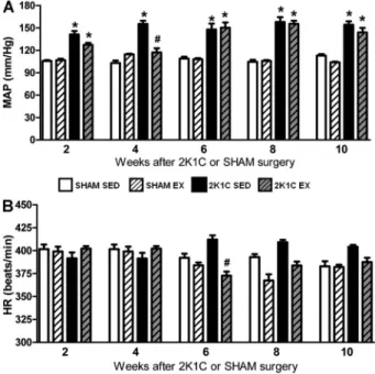
The baseline MAP of all 2K1C SED rats was higher than the baseline MAP of all SHAM SED rats from week 2 to week 10 (Po0.05). A significant interaction between
hypertension and Ex was observed in weeks 2 and 4 (Po0.05). The MAP in 2K1C Ex rats was significantly
lower than that in 2K1C SED rats (Po0.05) and reached
a level similar to that in the SHAM SED group only in
week 4. In weeks 6, 8, and 10, the 2K1C Ex rats had a baseline MAP similar to that of the 2K1C SED rats (P40.05) and higher than that of the SHAM SED rats (Figure 1A). In all weeks, there was no difference between the SHAM SED and SHAM Ex rats (P40.05) (Figure 1A).
The baseline HR of SHAM Ex rats was similar to that of the SHAM SED rats in all weeks (P40.05). In week 6, however, the baseline HR of the 2K1C Ex rats was significantly lower than that of the 2K1C SED rats (Po0.05) (Figure 1B). Furthermore, the HR was lower at
6, 8, and 10 weeks than at 2 weeks for both groups submitted to Ex (SHAM Ex and 2K1C Ex).
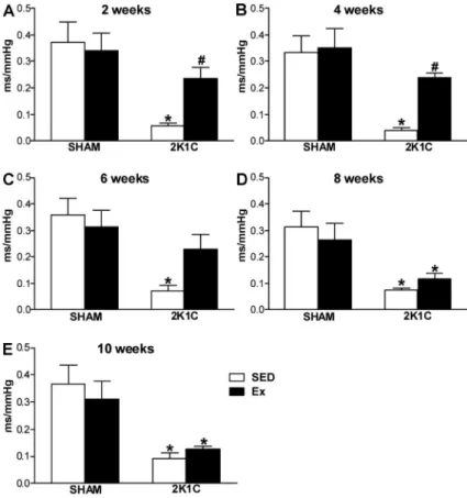
Evaluation of baroreflex bradycardia
As expected, the sensitivity of reflex bradycardia in 2K1C SED rats was lower than that in SHAM SED rats in all weeks (Po0.05). However, the reflex bradycardia in
the 2K1C Ex rats was higher than that of 2K1C SED rats (Po0.05) and similar to that of SHAM SED rats (P40.05)
at weeks 2 and 4 (Figure 2A and B). At week 6, the reflex bradycardia in 2K1C Ex rats was similar to that in 2K1C SED and SHAM SED rats (P40.05) (Figure 2C). Furthermore, at weeks 8 and 10, Ex did not improve the reflex bradycardia in 2K1C rats compared with 2K1C SED rats (P40.05) (Figure 2D and E). No
change was observed in the sensitivity of reflex bradycardia between SHAM SED and SHAM Ex rats (P40.05) (Figure 2A-D). Our data also showed that the baseline blood pressure correlated inversely (Po0.05)
with the sensitivity of the reflex at weeks 2 (r=–0.9768), 4 (r=–0.9524), and 8 (r=–0.9806).
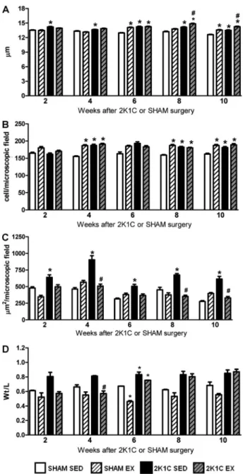
Analysis of cardiac structure
From week 2 to week 10, the wet relative heart weights of 2K1C SED rats (0.39±0.01 g/100 g body weight, n=27) were significantly higher than those of the SHAM SED rats (0.31±0.01 g/100 g body weight, n=30; Po0.05) and
similar to those of the 2K1C Ex rats (0.39±0.02 g/100 g body weight, n=45; P40.05). Also from week 2 to week 10, the cardiomyocyte diameter in the 2K1C SED rats was significantly greater than that of the SHAM SED rats (Po0.05). At week 6, the SHAM Ex and 2K1C Ex rats
showed a significantly greater cardiomyocyte diameter than that of the SHAM SED rats (Po0.05). However,
at weeks 8 and 10, the 2K1C Ex rats showed a significantly greater cardiomyocyte diameter than that of the SHAM SED and 2K1C SED rats (Po0.05)
(Figure 3A). Additionally, at weeks 4, 8, and 10, the 2K1C SED, 2K1C Ex, and SHAM Ex rats showed higher numbers of myocardial inflammatory cells than did the SHAM SED rats (Figures 3B and 4).
Figure 2. Index of the sensitivity of baroreflex bradycardia (ms/mmHg) induced by injection of phenylephrine (4.0 mg,iv) in normotensive (SHAM, n=5–8) and hypertensive rats (2K1C, n=5–8), either sedentary (SED) or subjected to physical exercise training (Ex), at 2 (A), 4 (B), 6 (C), 8 (D), and 10 weeks (E) of swimming. *Po0.05 com-pared to SHAM SED rats.#P
The tissue deposition of collagen in the myocardium of 2K1C SED rats was significantly higher than that of SHAM SED rats from week 2 to week 10 (Po0.05).
However, Ex prevented myocardial collagen deposition in 2K1C rats because these values remained significantly
lower than those in the 2K1C SED rats (Po0.05) and
similar to those in the SHAM SED rats (P40.05) at weeks 4, 8, and 10 (Figures 3C and 4). At week 4, the Wt/ L ratio of the left ventricle was significantly lower in 2K1C Ex rats than in 2K1C SED rats (Po0.05) and similar
between 2K1C Ex and SHAM Ex rats (P40.05). However, at weeks 6, 8, and 10, the Wt/L ratio of the left ventricle was similar between the 2K1C Ex and 2K1C SED rats (P40.05). At week 6, the Wt/L ratio of the left ventricle was significantly lower in SHAM Ex rats than in SHAM SED rats (Po0.05), suggesting eccentric
hyper-trophy (Figure 3D).
Analysis of renal structure
From week 2 to week 10, the relative wet weight of the left kidney (clipped) of 2K1C SED rats (0.27±0.01 g/100 g body weight, n=27) and 2K1C Ex rats (0.26±0.01 g/100 g body weight, n=45) was sig-nificantly lower than that of the left kidney of SHAM SED rats (0.33±0.01 g/100 g body weight, n=30; Po0.05).
Also from week 2 to week 10, the relative wet weight of the right kidney (non-clipped) of 2K1C SED rats (0.41±0.02 g/100 g body weight, n=27) and 2K1C Ex rats (0.41±0.03 g/100 g body weight, n=45) was significantly higher than that of SHAM SED rats (0.35±0.02 g/100 g body weight, n=30; Po0.05).
There was no difference between the right and left kidney of Sham Ex and SED rats. In weeks 6 and 10, the number of inflammatory cells in the left kidney was larger in 2K1C rats (SED and Ex) than that in SHAM SED rats (Figure 5). However, the Ex protocol sig-nificantly decreased the number of inflammatory cells in the right kidney of 2K1C rats compared with 2K1C SED rats in weeks 4 and 10 (Po0.05) (Figure 5).
Collagen deposition in the left (clipped) kidney of 2K1C SED rats was significantly higher than that in SHAM SED rats from week 2 to week 10 (Po0.05). The
Ex protocol significantly reduced collagen deposition in the left kidney of 2K1C rats compared with 2K1C SED rats in weeks 2, 4, and 10 (Po0.05). However, in week
10, collagen deposition in the left kidney of 2K1C Ex rats was similar to that of SHAM SED rats (P40.05) (Figures 5B and 6). In the right kidney, the collagen deposition in 2K1C SED rats was also significantly higher than in SHAM SED rats from week 2 to week 10 (Po0.05). However, collagen
deposition in 2K1C Ex rats was significantly lower than that in 2K1C SED rats from week 2 to week 10 (Po0.05)
(Figures 5D and 7).
The MAP was positively correlated with the area of collagen deposition in the myocardium (r=0.9821), left kidney (r=0.9648), and right kidney (r=0.9597) for all animals at week 4 (Po0.05). We also observed an
inverse correlation between the sensitivity of reflex bradycardia and the area of collagen deposition in the left kidney (r=–0.9988) and right kidney (r=–0.9807) for all animals at week 4 (Po0.05).
Figure 3.Cardiomyocyte diameter (mm,A), number of myocardial inflammatory cells per microscopicfield (B), myocardial collagen deposition (mm2/microscopic field, C), and left ventricle wall thickness/lumen ratio (Wt/L,D) of normotensive (SHAM, n=3–7) and hypertensive (2K1C, n=3–5) rats, either sedentary (SED) or subjected to physical exercise training (Ex), for 2, 4, 6, 8, and 10 weeks of swimming. *Po0.05 compared to SHAM SED rats. #
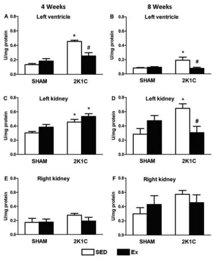
Analysis of oxidative damage and formation of TBARS
After 4 weeks of the Ex protocol, the level of TBARS, an indicator of lipid peroxidation, was signifi-cantly higher in the left ventricle and left kidney of 2K1C SED rats than SHAM SED rats (Po0.05). However,
the level of TBARS in the left ventricle was signi-ficantly lower in 2K1C Ex than 2K1C rats (Po0.05) and
similar between 2K1C Ex and SHAM SED rats (Figure 8A and C).
After 8 weeks of the Ex protocol, the level of TBARS in the left ventricle and left kidney was significantly higher in 2K1C SED than SHAM SED rats (Po0.05). However, the
level of TBARS in the left ventricle and left kidney was significantly lower in 2K1C Ex than 2K1C SED rats (Po0.05) (Figure 8B and D). No difference in the level of
TBARS was observed in the right and left kidneys in
SHAM and 2K1C (SED and Ex) rats (P40.05) (Figure 8E and F).
CAT activity
After 4 weeks of the Ex protocol, the activity of CAT (an antioxidant enzyme) in the left ventricle, left kidney, and right kidney was significantly lower in 2K1C SED rats than in SHAM SED rats (Po0.05).
However, the CAT activity in the left ventricle, left kidney, and right kidney was significantly higher in 2K1C Ex rats than in 2K1C SED rats (Po0.05)
(Figure 9A, C and E).
significantly higher in the 2K1C Ex than 2K1C SED rats (Po0.05) (Figure 9D and F).
Discussion
In the present study, swimming without a workload effectively reduced MAP; restored reflex bradycardia and prevented concentric ventricular hypertrophy; restored the level of TBARS, an indicator of lipid peroxidation, in the left ventricle; and increased the activity of CAT, an antioxidant, in the left ventricle and both kidneys to levels similar to those in the SHAM SED rats for up to 4 weeks. Although Ex did not completely reverse MAP and reflex bradycardia at 8 weeks, it effectively decreased the TBARS level in the left ventricle and left kidney and increased the CAT activity in left and right kidneys. Ex also prevented collagen deposition in the myocardium and clipped kidney in 2K1C rats and maintained these levels similar to those observed in the SHAM SED rats up to week 10 of swimming.
The literature indicates the effectiveness of Ex in reducing MAP and improving baroreflex sensitivity in hypertensive humans (15) and animals (2,16,22,27). Previous studies (26,29) have demonstrated the impor-tant role of baroreceptors in the cardiovascular and autonomic adaptations induced by Ex. The sinoaortic
denervation in spontaneously hypertensive rats sub-jected to treadmill running attenuated the adaptations induced by Ex, such as reduction of MAP and HR, reestablishment of baroreflex sensitivity, increased diameter of the left ventricular chamber, and reduced collagen deposition in the myocardium. However, other studies (7,16,18,20) have suggested that adjustment of the Ex intensity is necessary to maintain these benefits over time.
The results of the present study show that Ex effectively reduced the MAP and restored the reflex bradycardia up to week 4 of swimming, in agreement with previous studies from our laboratory that used the same model of hypertension and the same swimming protocol for 4 weeks (22) and 5 weeks (2). However, from week 6 to week 10, the basal MAP in the 2K1C Ex rats was similar to that in the 2K1C SED rats. Never-theless, the reflex bradycardia of the 2K1C Ex rats was similar to that of the 2K1C SED and SHAM SED rats at week 6 and lower than that of the SHAM SED rats at weeks 8 and 10, suggesting that the adaptive benefits of Ex for improving reflex bradycardia until week 6 are compensated by hypertensive factors, especially by Ang II, which can modulate glutamatergic neurons in the rostral ventrolateral medulla and worsen the reflex bradycardia (30). A previous study from our Figure 5.AandC, number of inflammatory cells per microscopicfield in left (clipped) and right (non-clipped) kidneys, respectively. BandD, collagen deposition (mm2/microscopic
laboratory (2) showed that Ex with a 0% or 3% workload for 5 weeks had beneficial effects on high blood pressure, high HR, and cardiac dysfunction in 2K1C rats. Nevertheless, Ex with a 0% workload more effectively improves the cardiac alterations observed in renovascular hypertensive rats; Ex with a 3% workload did not change the relatively lower sensitivity of the reflex bradycardia or prevent the cardiac lesions induced by hypertension. A possible explanation for these data is that Ex without workload until week 4 should induce adjustments of the activity of the sympathetic and parasympathetic nervous systems (7,17,29,31) and reduce oxidative stress (13), probably due to decreased levels of Ang II (11).
Concentric ventricular hypertrophy has been de-scribed under conditions of hypertension and myocardial infarction. (5,8), while eccentric ventricular hypertrophy
increased metabolic demand in response to Ex. Together, these data suggest that from week 4 onward, the volume, frequency, or intensity of Ex should be adjusted to maintain these benefits. Furthermore, Ex in the present study effectively reduced the TBARS level in the left ventricle at weeks 4 and 8, while the CAT activity was higher in 2K1C Ex than 2K1C SED rats only at week 4. The excessive production of TBARS and decreased CAT activity in the left ventricle and left (clipped) kidney in 2K1C SED rats was probably due to the increased levels of Ang II that develop in this model of hypertension. This could also explain the higher collagen deposition in the myocardium in 2K1C SED rats than in SHAM SED rats from week 2 to week 10. Moreover, 2K1C Ex rats showed a level of myocardial collagen deposition similar to that of SHAM SED rats from week 4 to week 10, indicating that these beneficial effects of Ex on the left ventricle were maintained during
the entire 10 weeks, possibly by a decrease in oxidative stress. In fact, Ang II is a strong inductor of NADPH oxidase-induced reactive nitrogen and ROS generation in the plasma, heart, and kidney (32,33). Previously studies have demonstrated that high levels of Ang II-induced ROS are important for increased mRNA expression of collagen and fibronectin (34,35) and production of transforming growth factor-b1 (36). Conversely, the increase in oxygen consumption during Ex causes elevation of ROS, which is involved in the adaptive up-regulation of antioxidant gene expression (14).
in the clipped than non-clipped kidney. Filho et al. (11) showed that Ex increased tissue Ang- (1–7) levels and decreased plasma Ang II levels in hypertensive rats. In agreement, our 2K1C SED rats showed greater deposi-tion of collagen in both kidneys, clipped and non-clipped, from week 2 to week 10; however, Ex effectively reduced collagen deposition in the non-clipped kidneys of 2K1C rats from week 2 to week 10 and in the clipped kidney only at week 10 compared with 2K1C SED rats. The CAT activity in the left and right kidneys at weeks 4 and 8 was higher in 2K1C Ex rats than in 2K1C SED rats, while the TBARS level in the left kidney at week 8 was lower in 2K1C Ex rats than in 2K1C SED and similar to that in SHAM SED rats, suggesting a late beneficial effect of Ex over time, preventing collagen deposition likely due to the reduc-tion of oxidative stress.
A limitation of the present study was the use of urethane anesthesia. Although urethane acts similarly on the sympathetic and parasympathetic nervous systems (38,39), the reduction of parasympathetic
activity by this anesthetic could explain the unchanged HR in the SHAM Ex rats for each week. However, a lower HR was observed at 6, 8, and 10 weeks for both groups submitted to Ex (SHAM Ex and 2K1C Ex) than at week 2 of Ex. Additionally, we cannot rule out the possibility that the effects of anesthesia may differ between normotensive and hypertensive rats. Another limitation of this study was that reflex tachycardia was not evaluated.
In summary, our data indicate that Ex reduced the HR at 6, 8, and 10 weeks for both groups of rats submitted to Ex (SHAM Ex and 2K1C Ex) and that in 2K1C rats, Ex performed without a workload for 10 weeks provided distinct beneficial effects over time. Ex induced early effects on cardiovascular function by decreasing blood pressure and increasing reflex brady-cardia and induced late effects by decreasing the oxidative stress and reducing the worsening of injuries that occur in the heart and kidneys during renovascular hypertension, thus preventing structural alterations in these organs.
Acknowledgments
This study was supported by Universidade Federal de Ouro Preto (UFOP), Pró-Reitoria de Pós-Graduac¸ão (PROPP-UFOP), FAPEMIG (Fundac¸ão de Amparo à Pesquisa do Estado de Minas Gerais)-RedeToxifar,
CNPq, FAPEMIG-Universal, Pronex Project Grant (FAPEMIG/CNPq-2010) and Instituto Nacional de Ciência e Tecnologia em Nanobiofarmacêutica (INCT-Nanobio-far)-FAPEMIG-CNPq. R.C.A. Maia received a UFOP fellowship (Master’s degree) in the Programa de Pós-Graduac¸ão em Saúde e Nutric¸ão, UFOP.
References
1. Martinez-Maldonado M. Pathophysiology of renovascular hypertension.Hypertension1991; 17: 707–719, doi: 10.1161/ 01.HYP.17.5.707.
2. Soares ER, Lima WG, Machado RP, Carneiro CM, Silva ME, Rodrigues MC, et al. Cardiac and renal effects induced by different exercise workloads in renovascular hypertensive rats.Braz J Med Biol Res2011; 44: 573–582, doi: 10.1590/ S0100-879X2011007500049.
3. Nishi EE, Campos RR, Bergamaschi CT, de Almeida V, Ribeiro DA. Vitamin C prevents DNA damage induced by renovascular hypertension in multiple organs of Wistar rats. Hum Exp Toxicol 2010; 29: 593–599, doi: 10.1177/ 0960327109358267.
4. Oliveira-Sales EB, Dugaich AP, Carillo BA, Abreu NP, Boim MA, Martins PJ, et al. Oxidative stress contributes to renovascular hypertension.Am J Hypertens2008; 21: 98–104. 5. Sun Y, Weber KT. Angiotensin II and aldosterone receptor binding in rat heart and kidney: response to chronic angiotensin II or aldosterone administration. J Lab Clin Med1993; 122: 404–411.
6. Wakatsuki T, Schlessinger J, Elson EL. The biochemical response of the heart to hypertension and exercise.Trends Biochem Sci 2004; 29: 609–617, doi: 10.1016/j.tibs.2004. 09.002.
7. Medeiros A, Oliveira EM, Gianolla R, Casarini DE, Negrao CE, Brum PC. Swimming training increases cardiac vagal Figure 9.Catalase activity (U/mg protein) in the left ventricle (AandB), left kidney (CandD), and right kidney (EandF) of normotensive (SHAM, n=10) and hypertensive (2K1C, n=10) rats, either sedentary (SED) or subjected to 4 or 8 weeks of physical exercise training (Ex). *Po0.05 com-pared to SHAM SED group.#P
activity and induces cardiac hypertrophy in rats.Braz J Med Biol Res 2004; 37: 1909–1917, doi: 10.1590/S0100-879X2004001200018.
8. Swynghedauw B. Molecular mechanisms of myocardial remodeling.Physiol Rev1999; 79: 215–262.
9. Fernandes T, Soci UP, Oliveira EM. Eccentric and concentric cardiac hypertrophy induced by exercise training: microRNAs and molecular determinants.Braz J Med Biol Res2011; 44: 836–847, doi: 10.1590/S0100-879X2011007500112. 10. Marcus KD, Tipton CM. Exercise training and its effects with
renal hypertensive rats.J Appl Physiol1985; 59: 1410–1415. 11. Filho AG, Ferreira AJ, Santos SH, Neves SR, Silva Camargos ER, Becker LK, et al. Selective increase of angiotensin (1–7) and its receptor in hearts of sponta-neously hypertensive rats subjected to physical training.Exp Physiol2008; 93: 589–598.
12. Donley DA, Fournier SB, Reger BL, DeVallance E, Bonner DE, Olfert IM, et al. Aerobic exercise training reduces arterial stiffness in metabolic syndrome.J Appl Physiol2014; 116: 1396–1404, doi: 10.1152/japplphysiol.00151.2014. 13. Masson GS, Costa TS, Yshii L, Fernandes DC, Soares PP,
Laurindo FR, et al. Time-dependent effects of training on cardiovascular control in spontaneously hypertensive rats: role for brain oxidative stress and inflammation and baroreflex sensitivity. PLoS One 2014; 9: e94927, doi: 10.1371/journal.pone.0094927.
14. Radak Z, Zhao Z, Koltai E, Ohno H, Atalay M. Oxygen consumption and usage during physical exercise: the balance between oxidative stress and ROS-dependent adaptive signaling.Antioxid Redox Signal2013; 18: 1208–1246, doi: 10.1089/ars.2011.4498.
15. Laterza MC, de Matos LD, Trombetta IC, Braga AM, Roveda F, Alves MJ, et al. Exercise training restores baroreflex sensi-tivity in never-treated hypertensive patients. Hypertension 2007; 49: 1298–1306, doi: 10.1161/HYPERTENSIONAHA. 106.085548.
16. Veras-Silva AS, Mattos KC, Gava NS, Brum PC, Negrao CE, Krieger EM. Low-intensity exercise training decreases cardiac output and hypertension in spontaneously hyper-tensive rats.Am J Physiol1997; 273: H2627–H2631. 17. Brum PC, Da Silva GJ, Moreira ED, Ida F, Negrao CE,
Krieger EM. Exercise training increases baroreceptor gain sensitivity in normal and hypertensive rats. Hypertension 2000; 36: 1018–1022, doi: 10.1161/01.HYP.36.6.1018. 18. Fletcher GF, Balady GJ, Amsterdam EA, Chaitman B, Eckel
R, Fleg J, et al. Exercise standards for testing and training: a statement for healthcare professionals from the American Heart Association.Circulation2001; 104: 1694–1740, doi: 10.1161/hc3901.095960.
19. Hagberg JM, Park JJ, Brown MD. The role of exercise training in the treatment of hypertension: an update. Sports Med 2000; 30: 193–206, doi: 10.2165/00007256-200030030-00004.
20. Pescatello LS, Franklin BA, Fagard R, Farquhar WB, Kelley GA, Ray CA. American College of Sports Medicine position stand. Exercise and hypertension. Med Sci Sports Exerc2004; 36: 533–553, doi: 10.1249/01.MSS.0000115224. 88514.3A.
21. Goldblatt H, Lynch J, Hanzal RF, Summerville WW. Studies on experimental hypertension: I. The production of persistent
elevation of systolic blood pressure by means of renal ischemia.J Exp Med1934; 59: 347–379.
22. Rodrigues MC, Campagnole-Santos MJ, Machado RP, Silva ME, Rocha JL, Ferreira PM, et al. Evidence for a role of AT (2) receptors at the CVLM in the cardiovascular changes induced by low-intensity physical activity in renovascular hypertensive rats. Peptides 2007; 28: 1375–1382, doi: 10.1016/j.peptides.2007.06.001.
23. Alzamora AC, Santos RA, Campagnole-Santos MJ. Barore-flex modulation by angiotensins at the rat rostral and caudal ventrolateral medulla. Am J Physiol Regul Integr Com, hysiol 2006; 290: R1027–R1034, doi: 10.1152/ajpregu. 00852.2004.
24. Santos-Silva MA, Nagato AC, Trajano ET, Alves JN, Bandeira AC, Porto LC, et al. The oxidative response of mouse hearts is modulated by genetic background. Arq Bras Cardiol 2013; 100: 157–163, doi: 10.5935/abc. 20130029.
25. Draper HH, Squires EJ, Mahmoodi H, Wu J, Agarwal S, Hadley M. A comparative evaluation of thiobarbituric acid methods for the determination of malondialdehyde in biological materials.Free Radic Biol Med1993; 15: 353–363, doi: 10.1016/0891-5849(93)90035-S.
26. Bradford MM. A rapid and sensitive method for the quantitation of microgram quantities of protein utilizing the principle of protein-dye binding. Anal Biochem 1976; 72: 248–254, doi: 10.1016/0003-2697(76)90527-3.
27. Krieger EM, Brum PC, Negrao CE. State-of-the-Art lecture: influence of exercise training on neurogenic control of blood pressure in spontaneously hypertensive rats. Hypertension1999; 34: 720–723, doi: 10.1161/01.HYP.34. 4.720.
28. Ceroni A, Chaar LJ, Bombein RL, Michelini LC. Chronic absence of baroreceptor inputs prevents training-induced cardiovascular adjustments in normotensive and sponta-neously hypertensive rats.Exp Physiol2009; 94: 630–640, doi: 10.1113/expphysiol.2008.046128.
29. Moraes-Silva IC, De La Fuente RN, Mostarda C, Rosa K, Flues K, Damaceno-Rodrigues NR, et al. Baroreflex deficit blunts exercise training-induced cardiovascular and autonomic adaptations in hypertensive rats.Clin Exp Pharma-col Physiol2010; 37: e114–e120, doi: 10.1111/cep.2010.37. issue-3.
30. Carvalho TH, Bergamaschi CT, Lopes OU, Campos RR. Role of endogenous angiotensin II on glutamatergic actions in the rostral ventrolateral medulla in Goldblatt hypertensive rats. Hypertension 2003; 42: 707–712, doi: 10.1161/01. HYP.0000086524.35251.2D.
31. Liu JL, Kulakofsky J, Zucker IH. Exercise training enhances baroreflex control of heart rate by a vagal mechanism in rabbits with heart failure.J Appl Physiol2002; 92: 2403–2408. 32. Chade AR, Rodriguez-Porcel M, Herrmann J, Zhu X,
Grande JP, Napoli C, et al. Antioxidant intervention blunts renal injury in experimental renovascular disease. J Am Soc Nephrol2004; 15: 958–966, doi: 10.1097/01.ASN. 0000117774.83396.E9.
34. Brilla CG, Scheer C, Rupp H. Renin-angiotensin system and myocardial collagen matrix: modulation of cardiacfibroblast function by angiotensin II type 1 receptor antagonism. J Hypertens Suppl 1997; 15: S13–S19, doi: 10.1097/ 00004872-199715066-00004.
35. Crawford DC, Chobanian AV, Brecher P. Angiotensin II inducesfibronectin expression associated with cardiacfibrosis in the rat. Circ Res 1994; 74: 727–739, doi: 10.1161/01. RES.74.4.727.
36. Purnomo Y, Piccart Y, Coenen T, Prihadi JS, Lijnen PJ. Oxidative stress and transforming growth factor-beta1-induced cardiacfibrosis.Cardiovasc Hematol Disord Drug Targets2013; 13: 165–172.
37. Prieto MC, Gonzalez-Villalobos RA, Botros FT, Martin VL, Pagan J, Satou R, et al. Reciprocal changes in renal ACE/ ANG II and ACE2/ANG 1–7 are associated with enhanced collecting duct renin in Goldblatt hypertensive rats. Am J Physiol Renal Physiol 2011; 300: F749–F755, doi: 10.1152/ajprenal.00383.2009.
38. Maggi CA, Meli A. Suitability of urethane anesthesia for physiopharmacological investigations. Part 3: Other sys-tems and conclusions.Experientia1986; 42: 531–537, doi: 10.1007/BF01946692.