Abstract
Objective: To correlate forced expiratory volume in 1 second (VEF1) and peak expiratory flow (PEF) with clinical parameters in children with moderate and severe asthma.
Methods: This was a non-concurrent cohort study, carried out at a pediatric pneumology clinic, in Belo Horizonte, MG, Brazil, between March and October 2002. The study enrolled children aged 5 to 16 years, with persistent asthma, being treated with a minimum of 500 µg/day beclomethasone, and with symptoms under control for at least 3 months. Seventy-five patients (96.1%) were selected by simple randomization and monitored for 3 months, via a clinical severity scale and pulmonary function tests (PEF and VEF1). Results were analyzed using Pearsons coefficient.
Results: Correlations between absolute and percentage PEF figures and clinical severity score, were negative and very close to zero, signifying a weak correlation with no statistical significance. The same relationship was observed between VEF1 and clinical severity score. The correlation between VEF1 and PEF had a positive value with statistical significance (p = 0.000).
Conclusions: Since the best parameter for evaluating airway obstruction is VEF1, the finding that there is a positive correlation between this measure and absolute PEF reinforces the importance of its use and allows for the recommendation that PEF be measured as part of the management of asthmatic children, particularly in severe cases.
J Pediatr (Rio J). 2006;82(6):465-9: Asthma, monitoring, peak expiratory flow.
O
RIGINALA
RTICLE1. Especialista em Pneumologia Pediátrica. 2. Doutora em Medicina.
3. Doutora.
4. Doutor. Professor titular, Departamento de Pediatria, Faculdade de Medicina, Universidade Federal de Minas Gerais (UFMG), Belo Horizonte, MG, Brasil. Coordenador, Unidade de Pneumologia Pediátrica, Hospital das Clínicas, Faculdade de Medicina, UFMG, Belo Horizonte, MG, Brasil.
Manuscript received Mar 22 2006, accepted for publication Aug 02 2006.
Suggested citation: Fonseca AC, Fonseca MT, Rodrigues ME, Lasmar LM, Camargos PA. Peak expiratory flow monitoring of asthmatic children. J Pediatr (Rio J). 2006;82:465-9.
Introduction
The prevalence of asthma among children and adolescents varies from 0 to 30% in different populations.1 In Brazil, it is the third greatest cause of
hospital admissions among children and young adults, generating significant financial burden for the health service and elevated individual costs.2
Furthermore, many patients are not capable of recognizing their own clinical deterioration.3 This reduced
perception of their worsening state correlates with more
severe asthma crises and increased risk of death.3,4 In
Brazil, for example, it is estimated that in 1996 around 70% of deaths from asthma were of patients who did not recognize clinical decline and/or received inadequate treatment.4
The Global Initiative for Asthmas management guide (GINA) recommends objective measurements of pulmonary function, such as spirometry or peak expiratory flow (PEF), for assessing the severity of asthma and response to treatment. Due to its simplicity and ease of assessment, daily monitoring of PEF at home has been recommended by GINA for patients with moderate and severe asthma, in order to help with management of symptoms and to alert of periods of exacerbation.1
Peak expiratory flow represents the maximum flow generated during a forced expiration, at maximum intensity, starting from maximum lung inflation, i.e. total lung capacity. It is considered an indirect indicator of major airway obstruction and is affected by the degree of pulmonary inflation, thoracic elasticity, abdominal
465 Copyright © 2006 by Sociedade Brasileira de Pediatria
doi:10.2223/JPED.1566
Peak expiratory flow monitoring in asthmatic children
Ana Cristina C. F. Fonseca,1 Maria Tereza M. Fonseca,2
musculature and the patients muscular strength.5,6 It is
effort-dependent and, because of this, requires the patients collaboration. The measurement of PEF can be performed using spirometers or portable measurement units that are available at an accessible cost and are relatively simple to operate.
International reference values have been published for PEF, according to age, stature and sex. However, the best way to evaluate them is by comparing patients with their previous best levels.7
Some studies have questioned the true role of PEF measurements in the reduction of morbidity among asthmatic patients.8-10Since asthma primarily affects the
smaller airways, PEF will only be altered during later phases, after a significant increase in airway resistance. For this reason, it is argued that the best functional index for assessing small-airway function is the measurement of forced expiratory volume in 1 second (VEF1), which is measured using a spirometer.9 A spirometer is a relatively
high-cost piece of equipment that must be operated by a trained professional.
Several authors have attempted to correlate PEF measurements with the alterations observed in asthmatic childrens symptoms. A majority of these studies analyzed children with moderate and severe asthma, controlled with inhaled corticoids. They demonstrated that there is a weak correlation (Pearsons r varying from -0.34 to 0.5) between alterations to PEF and symptoms suggestive of deteriorating pulmonary function.3,8,10,11 However,
analogous studies are scarce in Brazil and Latin America.11
The objective of the work reported here was to attempt to correlate VEF1 and PEF measurements with clinical parameters in clinically stable children and adolescents with moderate and severe asthma on inhaled corticoids, in addition to verifying the contribution that serial PEF measurements have to offer for the outpatients follow-up of these patients.
Methods
This was a non-concurrent cohort study, carried out at the pediatric pneumology clinic at the Posto de Atendimento Médico Campos Sales. This is a secondary care clinic where around 400 asthmatic children are treated per year, referred by municipal health centers in Belo Horizonte. A sample of convenience was used, for which the participants were selected by simple randomization and followed-up for 3 consecutive months.
Inclusion and exclusion criteria
The study enrolled children and adolescents aged from 5 to 16 years old, with persistent asthma, on a minimum of 500 µg/day of beclomethasone and with symptoms under control for at least 3 months.
Exclusion criteria were exacerbations of symptoms, PEF and/or VEF1 below 59% of predicted, use of systemic corticoids during the 4 weeks prior to enrollment, infectious diseases of the upper airways or severe disease of any nature.
Children classified as having moderate or severe asthma, according to clinical parameters, were included in the study, as long as their pulmonary function was above 59% of predicted.
Operational definitions
Asthma diagnosis was confirmed by a history of recurrent wheezing crises, coughing and dyspnea, clinically reversible with rapid-acting inhaled beta-2 agonist, combined or not with systemic corticoids.
Asthma was classified as peristent mild. moderate or severe, according to GINA.1 These criteria are based on
the severity of exacerbations, intensity of symptoms, PEF and VEF1 measurements and inhaled corticoid dosage.
The maximum dosages of beclomethasone or its equivalent that are recommended by GINA are 400 µg, 800 µg, or over 800 µg for the treatment of persistent mild, moderate and severe asthma, respectively. Since the presentation available in Brazil contains 250 µg of beclomethasone per spray, we considered doses of 500 µg, 750 µg, and over 750 µg/day for persistent mild, moderate and severe asthma respectively.
The occurrence and severity of exacerbations, nocturnal symptoms, limitations to physical activity and use of fast-acting beta 2-agonist were evaluated and classified according to the clinical severity score validated and published by Rosier et al.12 The scale ranges from 2 to 19
points depending upon the severity of the parameters assessed, as follows: scores from 2 to 8 correspond to mild asthma; 9 to 14 to moderate asthma; and 15 to 19 to severe asthma. Questionnaires were completed by parents, guardians or the children themselves.
Monitoring of patients
After enrollment, patients were monitored for 3 months, with clinical severity scores and pulmonary function test results (PEF and VEF1) produced by independent examiners who were unaware of the study objectives. Clinical functional assessments were undertaken every 2 weeks for the first 2 months and patients were reassessed once more at the end of he third month. Patients were kept on the same inhaled corticoid medication they had been using before enrollment, given via a 650 mL plastic spacer with a valve (Flumax®, Flumax Equipamentos Médicos Ltda.,
Belo Horizonte).13-15
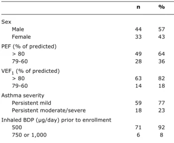
Table 1 - Descriptive characteristics of the patients studied
BDP = beclomethasone dippropionate; PEF = peak expiratory flow; VEF1 = forced expiratory volume in 1 second.
n %
Sex
Male 44 57
Female 33 43
PEF (% of predicted)
> 80 49 64
79-60 28 36
VEF1 (% of predicted)
> 80 63 82
79-60 14 18
Asthma severity
Persistent mild 59 77
Persistent moderate/severe 18 23
Inhaled BDP (µg/day) prior to enrollment
500 71 92
750 or 1,000 6 8
Figure 1 - Scatterplot and regression line for the linear correlation between absolute PEF values and clinical severity scores
PEF = peak expiratory flow.
Clinical severity score
0 0 50 100 150 250
P
E
F
350 450
200 300 400 500
2 4 6 8 10 12
y = -3.8613x + 279.71 = 0.0104 r = -0.03; p = 0.08 R2
measure PEF for the functional assessment, with the highest result chosen from three consecutive measurements for analysis, and adopting the reference values published by Polgar & Promadhat,16 these being
accepted internationally.
Spirometry was performed with bronchodilation using 400 µg of salbutamol for all patients at enrollment, at week eight and at the end of the third month, in accordance with American Thoracic Society recommendations, with reference values calculated using equations published by Polgar & Promadhat.17
At each visit, compliance with treatment and correct inhalation technique were assessed. Patients who had failed to use 25% or more of the inhaled corticoid dosage recommended for 2 weeks were excluded from the results.
Statistical aspects
Descriptive statistics were used to characterize the study population, and linear regression employed to evaluate any correlation between the variables studied (VEF1, PEF and clinical severity score).
Data were correlated using Pearsons coefficient, which varies from -1 to +1. Values close to -1 signify negative or inverse correlations, and values close to zero indicate an absence of correlation, while values close to +1 signify positive correlation. Results were considered significant when p < 0.05.
Ethical considerations
The research protocol and informed consent form were approved by the Research Ethics Committee at the Universidade Federal de Minas Gerais.
Results
The initial selection returned 87 children, of whom nine were excluded in accordance with the studys exclusion criteria. Seventy-eight patients began the observation period, but three were excluded later: one patient had been incorrectly enrolled (VEF1 < 59%), another reported clinical deterioration, and a third did not take sufficient medication, being excluded for partial compliance with treatment. Seventy-five patients therefore remained (96.1%) and were followed for 12 weeks.
Table 1 contains the characteristics of the 77 patients enrolled on the study.
There was a discrete predominance of the male sex, but without statistical significance (p = 0.58), in addition to the asthma of the majority of the children being under good control, which can be confirmed with reference to their PEF and VEF1 of over 80%.
Figure 1 is a scatterplot of correlations between absolute PEF measurements and the clinical severity score, based on data obtained at all six assessments made during the study period.
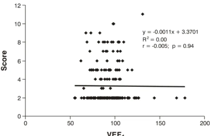
Figure 2 - Scatterplot and regression line for the linear correlation between VEF1 and clinical severity scores
VEF1 = forced expiratory volume in 1 second. VEF1
0 50 100 150 200
0 2 4 6 8 10 12
S
c
o
re
y = -0.0011x + 3.3701 = 0.00
r = -0.005; p = 0.94 R2
Figure 3 - Scatterplot and regression line for the linear correlation between VEF1 and absolute PEF values
PEF = peak expiratory flow; VEF1 = forced expiratory volume in 1 second.
0 100
P
E
F
200 300 400 500
0 50 100 150 200
VEF1
y = 0.9823x + 178.16 = 0.0527 r = 0.228; p = 0.0008 R2
Figures 2 is a scatterplot for the correlation between VEF1 and clinical severity score and Figure 3 shows the correlation between VEF1 and absolute PEF values, both generated from the analysis of all data collected during the study.
As was the case in the previous diagram, it will be observed that in Figure 2 the regression line is parallel to the x axis, demonstrating that there is no correlation between the variables. This can be confirmed by the r value, which is very close to zero, and by the p value, which demonstrates that there is no statistical significance (r = -0.005, p = 0.94).
In contrast, in Figure 3 the regression line for the correlation between VEF1 and absolute PEF values has a positive incline, which results in a positive correlation coefficient, with statistical significance (r = 0.23, p = 0.0008).
Discussion
There were no correlations observed between the functional parameters PEF and VEF1 and the clinical scale in the data published here. In contrast there was a positive correlation between absolute PEF values and VEF1 reference values. The absence of any correlation may be because the children studied here were under control, from a clinical point of view, on medication (the majority scored between two and eight points on the clinical severity scale adopted), or due to the difficulty in recognizing deteriorated pulmonary function resulting from patients adapting to the limitations of their disease.
Correlations between VEF1/PEF and clinical severity score did not have any statistical significance (p > 0.05). While, in the majority of cases, r was negative and close to zero, the distribution of points on the graphs demonstrated that there was no correlation between these variables. Patients classified with a score of 2 (mild asthma) exhibited PEF below 40% of predicted and VEF1 of 50%. It is possible, as is observed with other chronic diseases, that these children have adapted to the limitations imposed by their asthma, leading to a reduced perception of the symptoms associated with airway obstruction.
There was a positive correlation between VEF1 and PEF, demonstrating that if VEF1 increases, PEF also increases (and vice-versa). This being so, PEF should be a useful indicator for assessing small airway obstruction, since VEF1 is used as the gold standard in our country.
Clinical assessment is subjective and the results reveal their obvious limitations. This is why, before being relied on in isolation, it should be accompanied by objective measurements, such as PEF and spirometry.
This work was performed using a sample that is comparable to others in the literature, both in terms of number and age of participants and in terms of the follow-up period. The statistical analysis employed was also similar to other studies, in order to facilitate comparison and discussion. In contrast with the studies published to date, here all PEF measurements were taken by a trained professional who was unaware of the study objectives, which helped to control for possible measurement errors. In studies hitherto published, data were obtained, not just in the laboratory, but also from records of measurements taken by the patients themselves.
Brand et al. assessed 102 children aged 7 to 14 years over 2 weeks and found a weak correlation coefficient for PEF and a clinical severity score (r = -0.34, p < 0.01). The correlation observed for PEF and VEF1 was r = 0.15, but p did not denote significance.10
References
1. National Heart, Lung, and Blood Institute, National Institutes for Health. Global strategy for asthma management and prevention: NHLBI/WHO workshop report. Bethesda: National Institutes of Health; revised 2002.
2. NHLBI/WHO Workshop Report/Global strategy for asthma management and prevention. Bethesda (MD): NIH; 1995. Publication Nº 02-3659.
3. Sly SD. Relationship between change in PEF and symptoms: questions to ask in paediatric clinics. Eur Respir J Suppl. 1997;24:80S-3.
4. Noronha MF, Campos HS. Hospitalizações por asma no Brasil. Pulmão RJ. 2000;9:10-30.
5. Newr N, Yandell B, Howell L, Eddy M, Sheikh S. Can peak expiratory flow predict airflow obstruction in children with asthma? Pediatrics. 2000;105:354-58.
6. Paggiaro PL, Moscato G, Giannini D, Franco AD, Gherson G. Relationship between peak expiratory flow (PEF) and FEV1. Eur Respir J Suppl. 1997;24:39S-41.
7. Quanjer PH, Lebowitz MD, Gregg I, Miller MR, Pedersen OF. Peak expiratory flow: conclusions and recommendations of a Working Party of the European Respiratory Society. Eur Respir J Suppl. 1997;24:2S-8.
8. Brand PL, Duiverman EJ, Waalkens HJ, van Essen-Zandvliet EE, Kerrebijn KF. Peak flow variation in childhood asthma: correlation with symptoms, airways obstruction, and hyperresponsiveness during long term treatment with inhaled corticosteroids. Dutch CNLSD Study Group. Thorax. 1999;54:103-7.
9. Brand PL, Roorda RJ. Usefulness of monitoring lung function in asthma. Arch Dis Child. 2003;88:1021-25.
10. Brand PL, Duiverman EJ, Postma DS, Waalkens HJ, Kerrebijn KF, van Essen-Zandvliet EE, et al. Peak flow variation in childhood asthma: relationship to symptoms, atopy, airways obstruction and hyperresponsiveness. Eur Respir J. 1997;10:1242-7. 11. Cabral AL, Conceição GM, Saldiva PH, Martins MA. Effect of
asthma severity on symptom perception in childhood asthma. Braz J Med Biol Res. 2002;35:319-27.
12. Rosier MJ, Bishop J, Nolan T, Robertson CF, Carlin JB, Phelan PD. Measurement of functional severity of asthma in children. Am J Respir Crit Care Med. 1994;149:1434-41.
13. Rubim JA, Simal CR, Lasmar LM, Camargos PA. Deposição pulmonar de radioaerossol e desempenho clínico verificados com espaçador fabricado no Brasil. J Pediatr (Rio J). 2000;76: 434-42.
14. Camargos PA, Rubim JA, Lasmar LM. Beclomethasone diproprionate delivered through a new spacer developed in Brazil. Eur Respir J. 2000;16 Suppl. 31:541S.
15. Ates NB, Esposito-Festen JE, Tiddens HAWM, van der Mark TW. Asthma therapy for young children in developing countries: in search for a cost effective pMDI-spacer combination. Am J Respir Crit Care Med. 2002;165:A190.
16. Polgar G, Promadhat V. Pulmonary function testing in children: techniques and standards. Philadelphia: Saunders; 1971. 17. American Thoracic Society. Standardization of spirometry,1994
update. Am J Respir Crit Care Med. 1995;152:1107-1136. 18. Liam CK, Goh CT, Isahak M, Lim KH, Wong CM. Relationship
between symptoms and objective measures of airway obstruction in asthmatic patients. Asian Pac J Allergy Immunol. 2001;19: 79-83.
19. Goldberg S, Springer C, Avital A, Godfrey S, Bar-Yishay E. Can peak expiratory flow measurements estimate small airway function in asthmatic children? Chest. 2001;120:482-8. 20. Shingo S, Zhang J, Reiss TF. Correlation of airway obstruction
and patient-reported endpoints in clinical studies. Eur Respir J. 2001;17:220-4.
Correspondence: Paulo A. M. Camargos Av. Alfredo Balena, 190/4061
CEP 30130-100 Belo Horizonte, MG Brazil Tel.: +55 (31) 3248.9773
Fax: +55 (31) 3248.9664
E-mail: pcamargs@medicina.ufmg.br between PEF and symptoms, although a weak one and
with a negative coefficient, (p < 0.05).11
Liam et al. monitored a group of 64 patients with stable asthma and found a weak correlation between a clinical severity score and PEF (r = -0.214, p = 0.104), and between the clinical severity score and VEF1 (r = -0.256, p = 0.041). The concluded that their results supported the need for objective measurements of airway obstruction.18
In their article, Goldberg et al. reported finding a strong correlation between PEF and o VEF1, with r values varying from 0.74 to 0.93 (p < 0.0001) and concluded that PEF measurements can be used as a useful follow-up parameter.19
In contrast, Paggiaro stated that the coefficient of the correlation between VEF1 and PEF for absolute figures varied from 0.78 to 0.95, and from 0.74 to 0.91 for percentages, which reinforces indications for the use of PEF measurements as an objective method for monitoring the pulmonary function of patients (r = 0.796, p = 0.0001).6
Newr et al. followed 244 children aged 4 to 18 years for 3 years and found that there was a good correlation between VEF1 and PEF (r = 0.73, p = 0.02).5 These values
are comparable with figures found by Shingo et al., who observed a good correlation between VEF1 and PEF (r = 0.74) for measurements taken at home by patients and for PEF measurements taken at the laboratory (r = 0.85). Nevertheless, the correlation between VEF1 and a clinical severity score was weak (r = -0.13). All correlations had p < 0.005, however, the patients under investigation were more than 15 years old.20 These
studies all help to corroborate the indication for using PEF when monitoring asthmatic children.
Among the studies published, it was Sly who found a positive, if weak, correlation between PEF and clinical severity score (r = 0.35, p < 0.01), and between PEF and variation in clinical severity score (r = 0.38, p < 0.01). He observed 80 children aged 6 to 16 years for 8 weeks.3