Abstract
Objective: The objective of this study was to evaluate the ratio of dead space to tidal volume (VD/VT) as a predictor of extubation failure of children from mechanical ventilation.
Methods: From September 2001 to January 2003 we studied a cohort consisting of all children (1 day-15 years) submitted to mechanical ventilation at a pediatric intensive care unit who were extubated and for whom pre-extubation ventilometry data were available, including the VD/VT ratio. Extubation success was defined as no need for any type of ventilatory support, invasive or otherwise, within 48 hours. Patients who tolerated extubation, with or without noninvasive support, were defined as success-R and compared with those who were reintubated. Statistic analysis was based on a VD/VT cutoff point of 0.65.
Results: During the study period 250 children received mechanical ventilation at the pediatric intensive care unit. Eighty-six of these children comprised the study sample. Twenty-one children (24.4%) met the criteria for extubation failure, with 11 (12.8%) of these requiring non-invasive support and 10 (11.6%) reintubation. Their mean age was 16.8 (±30.1) months (median = 5.5 months). The mean VD/VT ratio for all cases was 0.62 (±0.18). Mean VD/VT ratios for patients with successful and failed extubations were 0.62 (±0.17) and 0.65 (±0.21) (p = 0.472), respectively. Logistic regression failed to reveal any statistically significant correlation between VD/ VT ratio and success or failure of extubation (p = 0.8458), even for patients who were reintubated (p = 0.5576).
Conclusions: In a pediatric population receiving mechanical ventilation due to a variety of etiologies, the VD/ VT ratio was unable to predict the populations at risk of extubation failure or of reintubation.
J Pediatr (Rio J). 2006;82(5):347-53: Respiratory failure, lung disease, respiratory dead space, pulmonary volume measurement, artificial respiration, child.
Evaluation of the dead space to tidal volume ratio
as a predictor of extubation failure
Albert Bousso,1 Bernardo Ejzenberg,2 Andréa Maria Cordeiro Ventura,3 José Carlos Fernandes,4 Iracema de Cássia de Oliveira Fernandes,5
Patrícia Freitas Góes,5 Flávio Adolfo Costa Vaz6
O
RIGINALA
RTICLE1. Doutor em Pediatria, Faculdade de Medicina, Universidade de São Paulo (USP), São Paulo, SP, Brasil. Chefe, Unidade de Terapia Intensiva Pediátrica, Hosp. Universitário, USP, São Paulo, SP, Brasil. Departamento de Pediatria, Faculdade de Medicina, USP, São Paulo, SP, Brasil. 2. Professor livre-docente, Faculdade de Medicina, USP, São Paulo, SP,
Brasil.
3. Mestre, Faculdade de Medicina, USP, São Paulo, SP, Brasil. Médica assistente, Unidade de Terapia Intensiva Pediátrica, Hospital Universitário, USP, São Paulo, SP, Brasil.
4. Médico assistente, Unidade de Terapia Intensiva Pediátrica, Hospital Universitário, USP, São Paulo, SP, Brasil.
5. Mestre, Faculdade de Medicina, USP, São Paulo, SP, Brasil. Médica assistente, Unidade de Terapia Intensiva Pediátrica, Hospital Universitário, USP, São Paulo, SP, Brasil.
6. Professor titular, Departamento de Pediatria, Faculdade de Medicina, USP, São Paulo, SP, Brasil. Chefe, Departamento de Pediatria, Faculdade de Medicina, USP, São Paulo, SP, Brasil.
Manuscript received Jan 19 2006, accepted for publication Jun 07 2006.
Suggested citation: Bousso A, Ejzenberg B, Ventura AM, Fernandes JC, Fernandes IC, Góes PF, et al. Evaluation of the dead space to tidal volume ratio as a predictor of extubation failure. J Pediatr (Rio J). 2006;82:347-53.
347
Copyright © 2006 by Sociedade Brasileira de Pediatria doi:10.2223/JPED.1520
Introduction
The extubation of children in intensive care units (ICUs) has important implications for the morbidity and mortality of a large contingent of patients who are critically ill or in postoperative care. Removal of the cannula by the intensive care specialist is among the main intermediate therapeutic objectives in patients admitted to these wards.1
In all cases, opportunities for extubation should be evaluated in the light of the risks involved, both from early removal and from unnecessary retention of the cannula. When extubation is too early, respiratory failure and death can occur.2,3 When extubation is late, length of
time on mechanical ventilation and of stay in the ICU both increase, as do a number of severe complications, such as pneumonia, and the treatment costs.4-6 Thus,
Over recent years, many different controlled studies of adult patients have been conducted in attempts to identify objective and precise parameters and criteria that can determine the most suitable moment for withdrawing the respirator and performing extubation. Even with experienced medical teams, routine application of the clinical and laboratory criteria indicative of successful weaning still results in significant extubation failure and reintubation rates. Failure is reported in up to 17 to 25% of adults, depending on ICU profile.4
In children, many clinical and laboratory parameters have also been evaluated with the intention of promoting more successful extubation. Nevertheless, actual results also indicate extubation failure rates of 10 to 28%.9,10
The ratio between dead space and tidal volume (VD/ VT) has already been used for the identification of pulmonary thromboembolism,11,12 in the management of chronic
obstructive pulmonary disease (COPD),13 for prognostic
evaluation of acute respiratory distress syndrome,14 to
determine the presence of pulmonary dysfunction in adults with sepsis15 and as a factor discriminating weaning
off mechanical ventilation.16 In children, although
experience is less extensive, the VD/VT ratio has already been studied as a determinant of the severity of diaphragmatic hernia in newborn infants,17 as a parameter
indicating degree of lung injury in children on mechanical ventilation18 and as a marker of lung damage in acute
respiratory distress syndrome.19
The objective of the present study was to evaluate the VD/VT ratio as a predictor of extubation failure in children on mechanical ventilation in a pediatric intensive care unit (PICU) of a general hospital.
Methods
A prospective cohort was studied, consisting of all cases submitted to mechanical ventilation at the PICU that met inclusion criteria, but not exclusion criteria, between September 2001 and January 2003.
The project was approved by the Research Ethics Committees at the Universidade de São Paulo (USP) university hospital and at Hospital das Clínicas, USP Medical School. Children were enrolled only after informed consent had been obtained in writing from parents or legal guardians.
Inclusion criteria were: a) age less than 15 years; b) patient intubated and on mechanical ventilation at the PICU; c) on mechanical ventilation for at least 8 hours; and d) weaning with the following extubation parameters: fraction of inspired oxygen ≤ 40%; respiratory rate ≤ 8 breaths/min; peak inspiratory pressure ≤ 20 cmH2O; and end expiratory pressure ≤ 8 cmH2O.
Exclusion criteria were: a) cyanotic heart diseases ; b) chronic pulmonary disease; c) age < 28 days of life with
birth weight less than 2,500 g; d) neuromuscular diseases; e) endotracheal tube leakage > 30% (as recorded by the mechanical respiration monitor); f) original cause of intubation being upper airway obstruction; g) accidental extubation; h) transferred patients; i) death before extubation; j) refusal of parents or legal guardians to sign informed consent; k) children who attained extubation parameters during night shifts, weekends and holidays; l) second intubations of children already enrolled; and m) children whose Downes & Raphaely20 upper airway
obstruction score was greater than 4 during the first 48 hours after intubation.
The following data were recorded for all patients enrolled: a) age, sex, medical history, physical examination, initial diagnoses and nutritional status according to National Center for Health Statistics (NCHS) standards; b) description of the intubation and any problems that occurred; c) pediatric risk of mortality (PRISM) severity assessment score at admission; d) PaO2/FiO2 at admission; e) serial laboratory test results: daily arterial blood gases and hemoglobin and hematocrit assays every 3 days; f) ventilator parameters, which were monitored twice per shift by the PICU team; g) daily record of drugs employed for sedation and analgesia; and h) the length of time on mechanical ventilation up to extubation.
All children received mechanical ventilation with a Newport Wave® respirator (Newport Wave®, Newport
Medical Instruments, Newport Beach, California, USA), which allows pressure to be applied. At the PICU, ventilation is carried out in accordance with standards published by the Brazilian Society of Pneumology and Phithisiology (Sociedade Brasileira de Pneumologia e Tisiologia) and the Brazilian Association of Intensive Care Medicine (Associação de Medicina Intensiva Brasileira).21 Weaning was performed according to
American College of Chest Physicians task force guidelines.4
Once children achieved extubation conditions, they were subjected to the following ventilator parameters for 20 minutes: sufficient pressure to generate a tidal volume of 6 mL/kg, respiratory rate of zero, positive end expiratory pressure (PEEP) of 5-8 cmH2O and FiO2 ≤ 0.4. To achieve this, the lowest setting was used. Once the 20 minute pressure period had expired, arterial blood gas analysis was performed and mechanical respiration data were recorded mean airway pressure, inspiratory pressure, PEEP, dynamic compliance, airway resistance and expired tidal volume. The VD/VT ratio was then calculated using a CO2SMO-plus® monitor (CO2SMO-plus, Dixtal Equipamentos Médicos®, São Paulo, SP, Brazil). The
After extubation, patients received the clinical treatment appropriate for their individual requirements. The following data were recorded: respiratory distress, respiratory rate, nasal flaring and accessory musculature involvement; hypoxia according to pulse oximeter readings and/or arterial blood gas analysis (SatO2 < 90% and PaO2 < 60 mmHg); hypoventilation according to arterial blood gas analysis (PaCO2 > 45 mmHg); and Downes and Raphaely upper airway obstruction score. Respiratory distress and pulse oximetry data and the Downes and Raphaely score were recorded every hour for the first 3 hours, every 3 hours for the next 9 hours and every 6 hours for the next 36 hours, for a total of 48 hours of post-extubation observation. Arterial blood gas analysis results were recorded at 3, 12, 24 and 36 hours post-extubation. Patients were observed continuously and were reevaluated as needed.
Extubation failure was defined either clinically or by gasometry. Clinically, failure was defined as any two of the following: increase of 40% over normal respiratory rate for age; apnea > 20 seconds; clavicular and subcostal retractions; cyanosis. Using gasometry, failure was defined as PaO2/FiO2 < 200 or PaCO2 > 45 mmHg with pH < 7.35. In these cases, priority was given to the use of bilevel positive airway pressure (BiPAP) for all children except newborn infants who were placed on continuous positive airway pressure (CPAP). BiPAP was provided with BiPAP Vision equipment (BiPAP Vision®, Respironics Inc.,
Carlsbad, California, USA). When BiPAP was contraindicated, CPAP was employed. If children on BiPAP or CPAP were unable to maintain PaO2≥ 80 mmHg on an FiO2 of 0.6, they were reintubated and put on mechanical ventilation. The study protocol terminated if there was a return to any type of assisted ventilation or 48 hours after extubation in successful cases. Extubation success was
defined as 48 hours without any type of respiratory support. An intermediate level, success-R was defined as 48 hours without the need for reintubation.
A VD/VT ratio cutoff point of ≤ 0.65 was evaluated using the chi-square test, with relation to extubation failure. Sensitivity, specificity and likelihood ratio were also calculated. Univariate analyses were then performed to identify relationships between clinical and laboratory variables and the VD/VT ratio using Students t test for unpaired samples. Potential correlations between the VD/ VT ratio and extubation success were evaluated using multivariate logistic regression to investigate those correlations between the VD/VT ratio and other parameters that had a p value < 0.25 in the univariate analysis. Results were defined as significant if p was < 0.05. Ninety-five percent confidence intervals (95%CI) were also calculated for all estimates.
Results
During the study period 250 children received mechanical ventilation in the PICU. Eighty-six of these were selected in accordance with inclusion and exclusion criteria and comprised the study sample. Forty-four of these 86 patients (51.1%) were male. Twenty-one children (24.4%) met the extubation failure criterion, with 11 (12.8%) requiring noninvasive respiratory support and 10 (11.6%) being reintubated. Mean age was 16.8 (±30.1) months, with a median of 5.5 months.
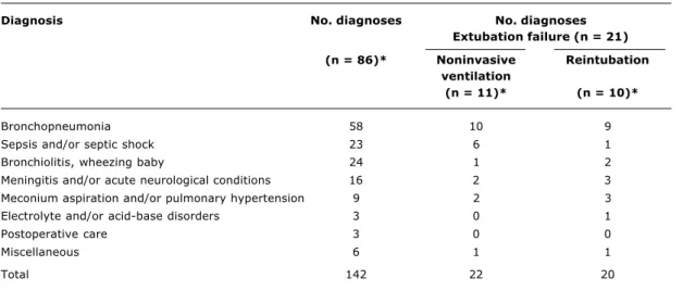
Table 1 lists the main diagnoses on admission for the sample as a whole, for extubation failure cases and for reintubated patients. The total number of diagnoses exceeds the number of patients, because many children presented with more than one condition on admission.
Diagnosis No. diagnoses No. diagnoses
Extubation failure (n = 21)
(n = 86)* Noninvasive Reintubation
ventilation
(n = 11)* (n = 10)*
Bronchopneumonia 58 10 9
Sepsis and/or septic shock 23 6 1
Bronchiolitis, wheezing baby 24 1 2
Meningitis and/or acute neurological conditions 16 2 3
Meconium aspiration and/or pulmonary hypertension 9 2 3
Electrolyte and/or acid-base disorders 3 0 1
Postoperative care 3 0 0
Miscellaneous 6 1 1
Total 142 22 20
Table 1 - Main diagnoses at admission for 86 patients studied
The mean VD/VT ratio for all cases was 0.62 (±0.18). Means and standard deviations of the VD/VT ratios for patients with successful extubation and for those whose extubation failed were 0.62 (±0.17) and 0.65 (±0.21), respectively (p = 0.472). Means and standard deviations for the VD/VT of patients who tolerated extubation (success-R), and for those who were reintubated were 0.62 (±0.18) and 0.64 (±0.21), respectively (p = 0.765).
The VD/VT ratios (mean and standard deviation) for patients given noninvasive support (n = 11) and reintubated patients (n = 10) were 0.65±0.22 and 0.64±0.21, respectively (p = 0.170).
With relation to the performance of the ratio in terms of sensitivity, specificity, positive predictive value (PPV) and negative predictive value (NPV), we observed that, for extubation failure, sensitivity was 72.3% (95%CI 60.0-81.7) and specificity was 61.9% (95%CI 40.8-79.2), with PPV of 85.4% (95%CI 73.8-92.4) and NPV of 41.9% (95%CI 26.4-59.2). For reintubation these figures were 65.8% (95%CI 54.6-75.4); 50.0% (95%CI 26.3-76.3);
90.9% (95%CI 80.4-96.0%) and 16.1% (95%CI 7.1-32.6), respectively.
The ROC curve for VD/VT ratio for extubation failure demonstrated that the ratio is a poor predictor of extubation, with an area under the curve (AUC) of 0.621.
The positive likelihood ratio for the VD/VT ratio, at a cutoff of 0.65, for extubation failure, was 1.89, while, in the event that the ratio is > 0.65, the chance of success is 0.45. In the case of those who were reintubated, the positive ratio was 1.31, and the negative, 0.68.
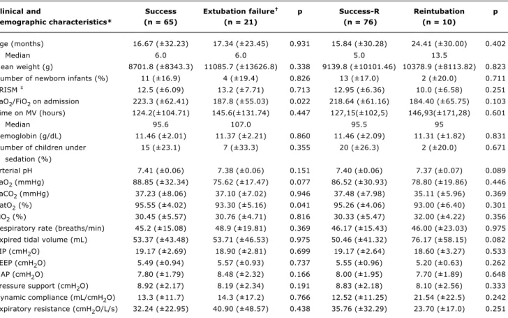
Table 2 lists demographic characteristics, together with clinical and ventilatory data, as predictive factors of extubation and reintubation. The univariate analysis revealed statistical significance for just pre-extubation O2 saturation and PaO2/FiO2 on admission, with p = 0.041 and p = 0.022, respectively.
None of the demographic characteristics or the pre-extubation clinical or ventilatory data exhibited any statistical differences for reintubated patients in the univariate analysis.
Table 2 - Pre-extubation demographic, clinical and ventilatory characteristics as predictive factors of extubation failure and reintubation
MAP = mean airway pressure; MV = mechanical ventilation; PEEP = positive end expiratory pressure; PIP = peak inspiratory pressure; PRISM = pediatric risk of mortality.
* Data expressed as means and standard deviations, except where specified otherwise. † Need for reintubation or noninvasive support.
‡ Not applicable to the 15 newborn infants.
Clinical and Success Extubation failure p Success-R Reintubation p
demographic characteristics* (n = 65) (n = 21) (n = 76) (n = 10)
Age (months) 16.67 (±32.23) 17.34 (±23.45) 0.931 15.84 (±30.28) 24.41 (±30.00) 0.402
Median 6.0 6.0 5.0 13.5
Mean weight (g) 8701.8 (±8343.3) 11085.7 (±13626.8) 0.338 9139.8 (±10101.46) 10378.9 (±8113.82) 0.823
Number of newborn infants (%) 11 (±16.9) 4 (±19.4) 0.826 13 (±17.0) 2 (±20.0) 0.711 PRISM 12.5 (±6.09) 13.2 (±7.71) 0.713 12.95 (±6.36) 10.0 (±6.58) 0.251
PaO2/FiO2 on admission 223.3 (±62.41) 187.8 (±55.03) 0.022 218.64 (±61.16) 184.40 (±65.75) 0.103
Time on MV (hours) 124.2(±104.71) 145.6(±131.74) 0.447 127,15(±102,5) 146,93(±171,28) 0.601
Median 95.6 107.0 95.5 95
Hemoglobin (g/dL) 11.46 (±2.01) 11.37 (±2.21) 0.860 11.46 (±2.09) 11.31 (±1.82) 0.831
Number of children under 15 (±23.1) 7 (±33.3) 0.355 20 (±26.3) 2 (±20.0) 0.671 sedation (%)
arterial pH 7.41 (±0.06) 7.38 (±0.06) 0.151 7.40 (±0.06) 7.37 (±0.07) 0.089
PaO2 (mmHg) 88.85 (±32.34) 75.62 (±17.47) 0.077 86.52 (±30.93) 78.80 (±19.86) 0.446
PaCO2 (mmHg) 37.23 (±8.06) 37.10 (±7.02) 0.946 37.48 (±7.98) 35.11 (±5.96) 0.369
SatO2 (%) 95.55 (±4.02) 93.30 (±5.16) 0.041 95.26 (±4.06) 93.00 (±6.40) 0.301
FiO2 (%) 30.45 (±5.57) 30.76 (±4.71) 0.816 30.33 (±5.47) 32.00 (±4.22) 0.356
Respiratory rate (breaths/min) 45.2 (±15.08) 48.9 (±19.81) 0.369 46.17 (±15.43) 46.00 (±23.03) 0.975 Expired tidal volume (mL) 53.37 (±43.48) 53.71 (±46.53) 0.975 50.46 (±41.32) 76.17 (±58.15) 0.082
PIP (cmH2O) 19.17 (±2.69) 18.90 (±2.81) 0.699 19.17 (±2.64) 18.60 (±3.27) 0.533
PEEP (cmH2O) 5.49 (±0.94) 5.57 (±0.93) 0.737 5.55 (±0.96) 5.20 (±0.63) 0.262
MAP (cmH2O) 7.80 (±1.79) 8.48 (±2.32) 0.166 8.00 (±1.95) 7.70 (±1.89) 0.648
Pressure support (cmH2O) 8.92 (±2.17) 8.19 (±2.34) 0.191 8.83 (±2.18) 8.10 (±2.56) 0.333
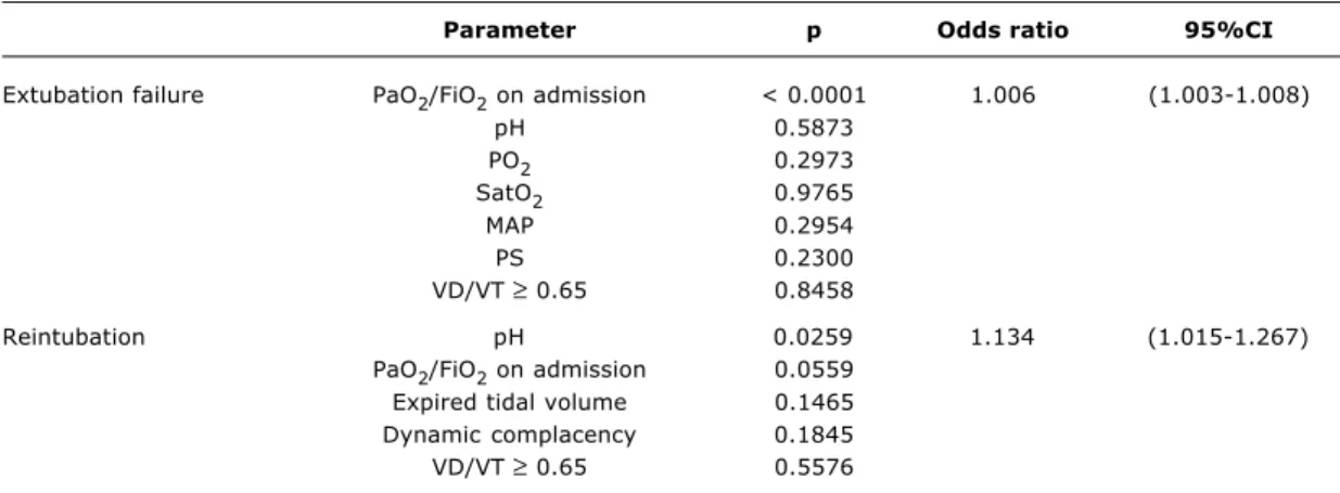
The predictive value of the VD/VT ratio for extubation failure and for reintubation was evaluated in association with other predictive demographic, clinical and ventilatory parameters, by means of logistic regression and multivariate analysis. All parameters that produced p < 0.25 in the univariate analysis were chosen for the multivariate analysis (Table 2). The results of the logistic regression are presented in Table 3. In this table it is possible to observe that the VD/VT ratio with a 0.65 cutoff does not exhibit a statistically significant correlation with success or failure of extubation (p = 0.8458), nor with the incidence of reintubation (p = 0.5576). The only parameter that exhibited a significant association with extubation failure was PaO2/FiO2 on admission. Here there was an inverse correlation between PaO2/FiO2 on admission and extubation failure (p < 0.0001). These data indicate that for each extra unit of PaO2/FiO2 on admission there was a 0.6% reduction in the risk of extubation failure.
The only predictive parameter associated with success-R was pre-extubation arterial pH. In this case, each 0.01 increase in pH above the mean determined a 13% increase in extubation success.
Discussion
As a predictor of extubation success, the VD/VT ratio was evaluated against two objectives to maintain the patient with no respiratory support whatsoever (success) and to avoid reintubation (success-R). A study in which Hubble et al.23 applied a similar analysis strategy reported
very different results from the ones observed here. Those
authors observed a significant difference in mean VD/VT between extubated patients failed extubation cases (0.44±0.17 vs.0.68±0.16; p = 0.0001).
It is important to point out, however, that the sample studied by Hubble et al.23 consisted of 45 patients aged 1
week to 18 years, with mean age of 43 (±62) months. Twenty-one of these were ventilated in postoperative care after elective surgery, 21 had acute lung injuries and three patients were trauma victims. Considering that the postoperative patients were in good general condition, with normal pulmonary parenchyma, it is possible that these factors favored successful extubation, even for patients whose VD/VT ratios were in the higher ranges. The research carried out by Hubble et al.23 included only
21 cases of lung disease. In contrast, in our study, the sample was essentially comprised of infants (mean age = 16.8±30.1 months; median = 5.5 months). Furthermore, they presented severe acute disease and, in general, some type of lung injury.
The ROC curve, which integrated sensitivity, specificity, PPV and NPV, demonstrates that the behavior of the VD/ VT ratio had very limited capacity to identify children at risk of extubation failure (AUC = 0.621). This finding once more diverges from data observed by Hubble et al.,23 who
described sensitivity of 72% and specificity of 92%.
The estimated likelihood ratios for the VD/VT ratio found in our study indicate limited prediction of success or failure, but suggest that there is some association with the clinical event of extubation. To date, however, there are no other studies that have evaluated VD/VT in terms of likelihood ratio.
Parameter p Odds ratio 95%CI
Extubation failure PaO2/FiO2 on admission < 0.0001 1.006 (1.003-1.008)
pH 0.5873
PO2 0.2973
SatO2 0.9765
MAP 0.2954
PS 0.2300
VD/VT ≥ 0.65 0.8458
Reintubation pH 0.0259 1.134 (1.015-1.267)
PaO2/FiO2 on admission 0.0559 Expired tidal volume 0.1465 Dynamic complacency 0.1845
VD/VT ≥ 0.65 0.5576
Table 3 - Multivariate analysis, with logistic regression, of indicators of extubation failure and of reintubation: VD/ VT ratio and clinical parameters*
95%CI = 95% confidence interval; MAP = mean airway pressure; SP = support pressure; VD/VT = ratio between dead space and tidal volume.
The multivariate analysis of extubation failure and reintubation indicators that had a p value of < 0.25 in the univariate analysis identified that the VD/VT ratio did not exhibit any significant association with these variables. Once more our data contradicts the results of Hubble et al.,23 whose logistic regression demonstrated a strong
association between VD/VT > 0.65 and extubation failure (p < 0.002). The clinical significance of the VD/VT ratio will be better understood after the completion of a multicenter study, that aims to enroll approximately 1,300 children, currently ongoing at 12 North-American PICUs and run by the Pediatric Acute Lung Injury Clinical Investigators (PALICI) network. The reasons for which the VD/VT ratio did not prove a useful parameter for predicting extubation success should be evaluated, especially because of the divergence from the pioneering study conducted by Hubble et al.23 Certain hypotheses can be formulated on the basis
of the study sample characteristics and the treatment given, in particular the use of mechanical respiratory assistance.
The study population was predominantly composed of young infants, which could have prejudiced the performance of the VD/VT ratio. There are certain pathophysiologic characteristics specific to young age: small infants exhibit less resistance to muscle fatigue,24 cough less efficiently,
have less capacity to eliminate respiratory secretion, and present proportionally narrower airways.25 These
characteristics are not evaluated by the VD/VT ratio. The patients studied by Hubble et al.23 were older, even
including adolescents of up to 18 years of age. In the absence of these influencing factors, the VD/VT ratio may have reflected the risk of extubation failure more appropriately.
A large number of other factors that could have influenced the performance of the VD/VT ratio26-28 were
studied and considered not to be significant in the study population. This was true of the number of newborn infants, time on mechanical ventilation, incidence of post-extubation laryngitis and presence of anemia.
Nevertheless, the presence and degree of muscular dysfunction after mechanical ventilation is difficult to assess. Muscular dysfunction may have had an effect on the capacity of those patients that failed to sustain their respiratory function unassisted.
It is recognized that patients with more intense pulmonary damage on admission have greater difficulty with removal of respiratory support.12 Nevertheless, if
patients are capable of tolerating the gradual reduction of ventilator parameters, why are they unable to sustain themselves after extubation? It is possible that the processes that damaged the lungs so intensely at the start of mechanical ventilation are still the cause of significant structural alterations that maintain the patients at the limit of viability when the respirator settings are
turned down. Once extubated, however, the patients are unable to sustain themselves due to these alterations affecting pulmonary function. Another possibility is that patients with worse PaO2/FiO2 ratios at admission would be subjected to more intense treatment regimes, with more aggressive parameters ventilator settings, thus resulting in greater dysfunction of the thoracic musculature after extubation. This hypothesis was also suggested by Manczur et al.29 who argue that intensity
of disease could be a factor influencing extubation success.
Pre-extubation arterial pH had a significant association with success-R (p = 0.0259) in the multivariate analysis, with acidosis favoring failure. In the sample studied, for each 0.01 increase in pH, there was a 13% increase in successful extubation. While the changes in arterial pH are apparently minor, it is possible that this finding with the reintubated patients is linked to the occurrence of hypoxemia and metabolic dysfunction by the production of organic acids. This assessment is not referred to in currently available studies and needs to be reproduced in studies with larger sample sizes.
We conclude that, in a pediatric population receiving mechanical ventilation due to a variety of etiologies, the VD/VT ratio, with a 0.65 cutoff, was not capable of predicting risk of extubation failure or reintubation.
References
1. Steinhorn DM, Green T. The treatment of acute respiratory failure in children: a historical examination of landmark advances. J Pediatr. 2001;139:604-8.
2. Gil B, Frutos-Vivar F, Esteban A. Deleterious effects of reintubation of mechanical ventilated patients. Clin Pulm Med. 2003;10: 226-30.
3. Rothaar RC, Epstein SK. Extubation failure: magnitude of the problem, impact on outcomes, and prevention. Curr Opin Crit Care. 2003;9:59-66.
4. Meade M, Guyatt G, Cook D, Griffith L, Sinuff T, Kergl C, et al. Predicting success in weaning from mechanical ventilation. Chest. 2001;120:400S-24S.
5. Teague WG. Noninvasive ventilation in the pediatric intensive care unit for children with acute respiratory failure. Pediatr Pulmonol. 2003;35:418-26.
6. Kurachek SC, Newth CJ, Quasney MW, Rice T, Sachdeva RC, Patel NR, et al. Extubation failure in pediatric intensive care: a multiple-center study of risk factors and outcomes. Crit Care Med. 2003;31:2657-64.
7. Hughes MR, Smith CD, Tecklenburg FW, Habib DM, Hulsey TC, Ebeling M. Effects of a weaning protocol on ventilated pediatric intensive care unit (PICU) patients. Top Health Inf Manage. 2001;22:35-43.
8. Epstein SK. Controversies in weaning from mechanical ventilation. J Intensive Care Med. 2001;16:270-86.
9. Farias JA, Alia I, Retta A, Olazarri F, Fernández A, Esteban A, et al. An evaluation of extubation failure predictors in mechanically ventilated infants and children. Intensive Care Med. 2002,28: 752-7.
12. Kline JA, Kubin AK, Patel MM, Easton EJ, Seupal RA. Alveolar dead space as a predictor of severity of pulmonary embolism. Acad Emerg Med. 2000;7:611-7.
13. Manthous CA, Goulding P. The effect of volume infusion on dead space in mechanically ventilated patients with severe asthma. Chest. 1997;112:843-6.
14. Nuckton TJ, Alonso JA, Kallet RH, Daniel BM, Pittet JF, Eisner MD, et al. Pulmonary dead-space fraction as a risk factor for death in the acute respiratory distress syndrome. New Engl J Med. 2002;346:1281-6.
15. Cala K, Pilas V, Vucic N. Vd/Vt test in the detection of pulmonary dysfunction in sepsis. Crit Care. 2004;8:P20.
16. Nozawa E, Kobayashi E, Matsumoto ME, Feltrim MI, Carmona MJ, Auler JJ. Assessment of factors that influence weaning from long-term mechanical ventilation after cardiac surgery. Arq Bras Cardiol. 2003;80:301-10.
17. Arnold JH, Bower LK, Thompson JE. Respiratory deadspace measurements in neonates with congenital diaphragmatic hernia. Crit Care Med. 1995;23:371-5.
18. Lum L, Saville A, Venkataraman ST. Accuracy of physiologic deadspace measurement in intubated pediatric patients using a metabolic monitor: comparison with the Douglas bag method. Crit Care Med. 1998;26:760-4.
19. Coss-Bu JA, Walding DL, David YB, Jefferson LS. Dead space ventilation in critically ill children with lung injury. Chest. 2003;123:2050-6.
20. Downes JJ, Raphaely RC. Pediatric intensive care. Anesthesiology. 1975;43:238-50.
21. Farias AMC, Guanaes A, Carvalho CRR, David CMN, Mena Barreto SS, Terzi RGG. II Consenso brasileiro de ventilação mecânica. J Pneumol. 2000;25:1-68.
22. Ball JA, Grounds RM. Calibration of three capnographs for use with helium and oxygen gas mixtures. Anaesthesia. 2003;58: 156-60.
23. Hubble CL, Gentile MA, Tripp DS, Craig DM, Meliones JN, Cheifetz IM. Deadspace to tidal volume ratio predicts successful extubation in infants and children. Crit Care Med. 2000;28: 2034-40.
24. Mulreany LT, Weiner DJ, McDonough JM, Panitch HB, Allen JL. Noninvasive measurement of the tension-time index in children with neuromuscular disease. J Appl Physiol. 2003;95:931-7. 25. Manczur TI, Greenough A, Pryor D, Rafferty GF. Comparison of
predictors of extubation from mechanical ventilation in children. Pediatr Crit Care Med. 2000;1:28-32.
26. Dimitriou G, Greenough A, Endo A, Cherian S, Rafferty GF. Prediction of extubation failure in preterm infants. Arch Dis Child Fetal Neonatal Ed. 2002;86:F32-5.
27. Edmunds S, Weiss I, Harrison R. Extubation failure in a large pediatric ICU population. Chest. 2001;119:897-900.
28. Randolph AG, Wypij D, Venkataraman ST, Hanson JH, Gedeit RG, Meert KL, et al. Effect of mechanical ventilator weaning protocols on respiratory outcomes in infants and children: a randomized controlled trial. JAMA. 2002;288:2561-8. 29. Manczur TI, Greenough A, Pryor D, Rafferty GF. Assessment of
respiratory drive and muscle function in the pediatric intensive care unit and prediction of extubation failure. Pediatr Crit Care Med. 2000;1:124-6.
Correspondence: Albert Bousso