Risk factors for post-extubation stridor in children:
the role of orotracheal cannula
Fatores de risco para estridor pós-extubação em crianças: o papel da cânula orotraqueal
Milena Siciliano Nascimento1, Cristiane Prado1, Eduardo Juan Troster1, Naiana Valério1,
Marcela Batan Alith2, João Fernando Lourenço de Almeida1
1 Hospital Israelita Albert Einstein, São Paulo, SP, Brazil.
2 Hospital Universitário, Universidade de São Paulo, São Paulo, SP, Brazil.
Corresponding author: Milena Siciliano Nascimento – Centro de Terapia Intensiva Pediátrica, Avenida Albert Einstein, 627/701 – Morumbi – Zip code: 05652-900 – São Paulo, SP, Brazil – Phone: (5511) 2151-8242 E-mail: milenasn@einstein.br
Received on: Sep 11, 2014 – Accepted on: Jan 20, 2015
Conflict of interest: none.
DOI: 10.1590/S1679-45082015AO3255 aBStract
objective: To determine the risk factors associated with stridor, with special attention to the role of the cuffed orotracheal cannula. Methods: Prospective analysis of all the intubated patients submitted to mechanical ventilator support from January 2008 to April 2011. The relevant factors for stridor collected were age, weight, size and type of airway tube, diagnosis, and duration of mechanical ventilation. The effects of variables on stridor were evaluated using uni- and multivariate logistic regression models. results: A total of 136 patients were included. Mean age was 1.4 year (3 days to 17 years). The mean duration of mechanical ventilation was 73.5 hours. Fifty-six patients (41.2%) presented with stridor after extubation. The total reintubation rate was 19.6% and 12.5 in patients with and without stridor, respectively. The duration of mechanical ventilation (>72 hours) was associated with a greater risk for stridor (odds ratio of 8.60; 95% confidence interval of 2.98-24.82; p<0.001). The presence of the cuffed orotracheal cannula was not associated with stridor (odds ratio of 98; 95% confidence interval of 0.46-2.06; p=0.953). conclusion: The main risk factor for stridor after extubation in our population was duration of mechanical ventilation. The presence of the cuffed orotracheal cannula was not associated with increased risk for stridor, reinforcing the use of the cuffed orotracheal cannula in children with respiratory distress.
Keywords: Respiratory sounds; Risk factors; Intubation, intratracheal/ instrumentation; Child
reSUMo
objetivo: Determinar os fatores de risco associados ao estridor, com especial atenção para o papel da cânula orotraqueal. Métodos: Análise prospectiva de todos os pacientes entubados submetidos à
ventilação mecânica no período de janeiro de 2008 a abril de 2011. Os fatores relevantes para estridor coletados foram idade, peso, tamanho e tipo da cânula orotraqueal, diagnóstico, e duração da ventilação mecânica. Os efeitos das variáveis sobre estridor foram avaliados utilizando modelos de regressão logística uni e multivariada. resultados: Foram incluídos 136 pacientes. A média de idade foi 1,4 ano (3 dias a 17 anos). O tempo médio de ventilação mecânica foi 73,5 horas. Apresentaram estridor após extubação 56 pacientes (41,2%). A taxa de reintubação foi de 19,6% e 12,5% em pacientes com ou sem estridor, respectivamente. A duração da ventilação mecânica (>72 horas) foi associada a um maior risco de estridor (odds ratio de 8,60; intervalo de confiança de 95% de 2,98-24,82; p<0,001). A presença da cânula orotraqueal não foi associada ao estridor (odds ratio de 0,98; intervalo de confiança de 95% de 0,46-2,06; p=0,953). conclusão: O principal fator de risco para estridor após extubação em nossa população foi o tempo de ventilação mecânica. A presença da cânula orotraqueal não foi associada a maior risco de estridor, reforçando o uso de cânulas com balonete em crianças com dificuldade respiratória.
Descritores: Sons respiratórios; Fatores de risco; Intubação intratraqueal/ instrumentação; Criança
introDUction
presence of atelectasis and post-extubation stridor accounts for 30% of all complications related to MV
in children.(1) The incidence of stridor after extubation in
children ranges from 3.5 to 30.2%.(1,2) This wide range of
incidence can be explained by the variability and lack of
objectivity of the definitions of stridor.(3) The presence
of post-extubation stridor in children may prolong length of stay in the PICU, particularly if reintubation is necessary.
Several factors such as age, weight, length of MV, size of orotracheal cannula (OTC), presence of a cuffed tube, and underlying indication for MV that have been associated with post-extubation stridor. Some methods have been used to predict post-extubation stridor, such as the air leak test, but the low sensitivity in young children makes the identification of patients at risk
troublesome.(4,5) As a result, pediatric intensivists must
be aware of all the main risk factors associated with stridor when attending to an intubated child.
The presence of a cuffed OTC has been traditionally associated with stridor in young children, leading to the classical indication of uncuffed OTC in children under
the age of 8 years.(6) However, in recent years, and after
the recommendation of cuffed OTC use in certain
circumstances (e.g., poor lung compliance, high airway
resistance, or a large glottic air leak) by the American
Heart Association,(7) there has been an increasing
interest in cuffed tubes in pediatric practice, in the operating room or in the PICU.
oBJectiVe
To identify the predictors of stridor after extubation in children admitted to a pediatric intensive care unit of a private hospital with special attention regarding the relation between cuffed and uncuffed orotracheal cannula.
MetHoDS
This prospective cohort study was performed in a
multidisciplinary, 14-bed PICU of Hospital Israelita Albert
Einstein, a private organization, from January 2008 to May 2011. The study was approved by the hospital Ethics Committee, under the registration number 10/1450, and the need for informed consent was waived because no intervention was attempted and the confidentiality of the patients’ data was preserved.
All patients that met the following inclusion criteria were included: age less than 18 years; and requirement of OTC and MV for more than 24 hours. Patients were excluded for death after MV without extubation attempts;
receiving prophylactic corticosteroids; known vocal cord paralysis or malacia; and limitations of medical care in place. Assigned patients were accompanied for 72 hours after extubation. We used the definition of stridor as any noisy breathing, specifically a high-pitched crowing sound associated with airway obstruction after intubation. Although there are some scores in the literature, no standardized score for stridor was used. The daily evaluation was performed by the pediatric intensivist and the respiratory physical therapist. Along with presence of stridor, signs of respiratory distress,
(SpO2) <92%, and level of consciousness were also
observed. The patient data collected regarding the risk factors associated with stridor were age, weight, size and type of airway tube (no cuff, de-inflated cuff or inflated cuff), admission diagnosis, and duration of MV. The list of possible risk factors was selected before the initiation of the study. The selection was made by the authors based on pediatric and adult literature in addition to personal experience using the Delphi
method for consensus.(8)
The patients submitted to VM used plastic OTC (Mallinckrodt Medical, Ireland; SIMS Portex Ltd, United Kingdom; Rüsch GmbH, Germany). The OTC choice was standardized using the Pediatric Advanced Life Support formula: OTC internal diameter (in mm) = 4 + age (in years)/4 for uncuffed OTC; and OTC internal diameter (in mm) = 3.5 + age (in years)/4 for cuffed OTC. The choice for cuffed or uncuffed OTC was determined by the option of the attending clinician. The group of patients that used cuffed OTC had the endotracheal cuff balloon pressure controlled. The
cuff balloon pressure was maintained below 20cmH2O
through all the MV length of utilization. The time of extubation during the study was determined by the care team based on clinical assessment and the use of spontaneous breathing trial.
Statistical analysis
The observed characteristics possibly associated with stridor were described in absolute frequencies and percentages, if categorical, or as mean and standard deviation, if quantitative, among patients with or without obstruction. The effects of variables on stridor were evaluated using uni-and multivariate logistic regression models. Variables with p value <0.10 in the univariate approach were included in the multivariate model.
Effect sizes were presented as odds ratios (OR) and
reSUltS
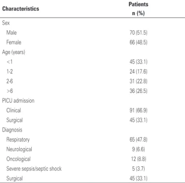
A total of 148 patients used MV for more than 24 hours and were eligible for the study. Twelve of them were excluded (eight for receiving prophylactic corticosteroids and four for death before extubation attempt). Their mean age was 1.4 year (range: 3 days to 17 years). Thirty-three percent of patients were less than 1 year old. The mean weight was 16.6kg. Table 1 (136 patients) shows that PICU received predominantly clinical patients with respiratory disease (71% of the non-surgical patients).
Figure 1 shows that the size of OTC most frequently used was 3.5mm. Seventy-four patients (54%) used the cuffed OTC; 19 (14%) patients used the deflated cuffed OTC. The uncuffed OTC was preferred in
infants (76% uncuffed OTC versus 24% cuffed OTC).
Moreover, among children aged over 1 year, uncuffed OTC was used in 22% and cuffed OTC in 78% of them (Figure 2).
table 1. Demographics and characteristics of patients submitted to mechanical ventilation (>24 hours)
characteristics Patients
n (%)
Sex
Male 70 (51.5)
Female 66 (48.5)
Age (years)
<1 45 (33.1)
1-2 24 (17.6)
2-6 31 (22.8)
>6 36 (26.5)
PICU admission
Clinical 91 (66.9)
Surgical 45 (33.1)
Diagnosis
Respiratory 65 (47.8)
Neurological 9 (6.6)
Oncological 12 (8.8)
Severe sepsis/septic shock 5 (3.7)
Surgical 45 (33.1)
PICU: pediatric intensive care unit.
OTC size: orotracheal cannula size.
Figure 1. Size of orotracheal cannula, in millimeters, and number of patients
Cuffed OTC: cuffed of orotracheal cannula; Uncuffed OTC: uncuffed of orotracheal cannula.
Figure 2. Age distribution of cuffed orotracheal cannula and uncuffed orotracheal cannula (%)
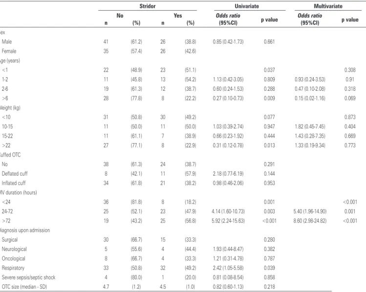
In the univariate analysis, age of less than 12 months (p<0.037), weight under 10kg (p<0.77), and duration of MV between 24 and 72 hours (OR: 4.14; 95%CI: 1.60-10.73; p=0.001) and higher than 72 hours (OR: 5.92; 95%CI: 2.24-15.63; p<0.01) were statistically associated with stridor.
Regarding the multivariate analysis, the risk factors associated with stridor were duration of MV between 24 to 72 hours (OR: 5.40; 95%CI: 1.96-14.90; p<0.001). This risk increased if the duration of MV was greater than 72 hours (OR: 8.60; 95%CI: 2.98-24.82; p<0.001). With this analysis, the only risk factor associated with stridor in our population was the duration of MV.
Two known risk factors with an increased likelihood of determining post-extubation stridor were evaluated with special attention. The presence of cuffed OTC (regardless of being inflated or deflated) and children with less than 12 months and 10kg (infants) were not associated with increased risk of stridor in our population.
table 2. Effects of variables on stridor using univariate and multivariate logistic regression models
Stridor Univariate Multivariate
no Yes Odds ratio
p value Odds ratio p value
n (%) n (%) (95%ci) (95%ci)
Sex
Male 41 (61.2) 26 (38.8) 0.85 (0.42-1.73) 0.661
Female 35 (57.4) 26 (42.6)
Age (years)
<1 22 (48.9) 23 (51.1) 0.037 0.308
1-2 11 (45.8) 13 (54.2) 1.13 (0.42-3.05) 0.809 0.93 (0.24-3.53) 0.91
2-6 19 (61.3) 12 (38.7) 0.60 (0.24-1.53) 0.288 0.47 (0.10-2.08) 0.318
>6 28 (77.8) 8 (22.2) 0.27 (0.10-0.73) 0.009 0.15 (0.02-1.16) 0.069
Weight (kg)
<10 31 (50.8) 30 (49.2) 0.077 0.873
10-15 11 (50.0) 11 (50.0) 1.03 (0.39-2.74) 0.947 1.82 (0.45-7.45) 0.404
15-22 11 (61.1) 7 (38.9) 0.66 (0.23-1.92) 0.444 1.43 (0.28-7.35) 0.669
>22 27 (77.1) 8 (22.9) 0.31 (0.12-0.78) 0.013 1.33 (0.19-9.34) 0.773
Cuffed OTC
No 38 (61.3) 24 (38.7) 0.291
Deflated cuff 8 (42.1) 11 (57.9) 2.18 (0.77-6.19) 0.144
Inflated cuff 34 (61.8) 21 (38.2) 0.98 (0.46-2.06) 0.953 MV duration (hours)
<24 36 (81.8) 8 (18.2) 0.001 <0.001
24-72 25 (52.1) 23 (47.9) 4.14 (1.60-10.73) 0.003 5.40 (1.96-14.90) 0.001 >72 19 (43.2) 25 (56.8) 5.92 (2.24-15.63) <0.001 8.60 (2.98-24.82) <0.001 Diagnosis upon admission
Surgical 30 (66.7) 15 (33.3) 0.280
Neurological 5 (55.6) 4 (44.4) 1.93 (0.44-8.47) 0.382
Oncological 8 (66.7) 4 (33.3) 1.21 (0.31-4.78) 0.787
Respiratory 33 (50.8) 32 (49.2) 2.42 (1.05-5.58) 0.039
Severe sepsis/septic shock 4 (80.0) 1 (20.0) 0.81 (0.08-8.54) 0.858 OTC size (median - SD) 4.7 (1.2) 4.5 (1.0) 0.82 (0.60-1.13) 0.218
95%CI: 95% confidence interval; OTC: orotracheal cannula; MV: mechanical ventilation; SD: standard deviation.
DiScUSSion
Our findings show that the duration of MV and consequently the duration of OTC utilization were the only risk factors associated with stridor after extubation in our population. Although it could be considered an obvious conclusion, the literature remains controversial about the duration of intubation and the subsequent risk
of developing complications.(2) Analyzing specifically
the presence of post-extubation stridor, the duration of MV for more than 3 days was associated with increased
risk for stridor in the adult literature.(9,10) Nevertheless,
other authors fail to show this association, maintaining
the subject in debate.(5,11,12) These considerations
are important because extubation failure results in prolonged utilization of MV and is independently associated with a five-fold increased risk of death in pediatric patients.(13)
When the duration of MV is associated with stridor and used for more than 72 hours, it is often associated
with increased risk.(14) In other studies, the risk is
increased only after 5 to 6 days.(9,15) It is interesting to
note that in our study, a duration of MV as low as 24 hours was sufficient to demonstrate significance in the presence of stridor.
Our incidence of stridor after extubation (42%)
is greater than other pediatric studies.(1,2) This could
contribute to inconsistent findings in assessment of risk factors and in incidence of stridor.(3,16)
Regarding gender, the adult literature indicates that females are at increased risk of developing stridor after
extubation.(9,12) The smaller size of larynx of females
explains this increased risk when compared to males.(17)
On the other hand, in children, the male gender is
associated with post-extubation airway compromise.(5)
In our population, neither gender was associated with greater risk.
According to previous studies, non-surgical patients (medical patients) are at risk of developing stridor after
extubation.(10) Our PICU received the majority of children
with non-surgical diseases (66%). In our population, there was a clear prevalence of respiratory diseases, with more than 70% of the non-surgical patients. Nevertheless, the admission diagnosis was not associated with a higher risk of stridor.
It was expected to find in literature that children under 24 months of age would be at risk for post-extubation
stridor, due to the smaller caliber of the airways and(5)
we opted to also include weight as a risk factor, and both revealed that children under 12 months old and with less than 10kg had increased risk of stridor in univariate
analysis. Otherwise, in the multivariate analysis each
predictor would fail to show its association with stridor. It is possible that these predictors associated with infants (age <12 months and weight <10kg) could show significance if we had a larger sample.
The published studies on utilization of cuffed OTC in small children have demonstrated different and better results when compared with the standard and classical affirmation that cuffed OTC is associated with airway mucosa injury and consequently post-extubation stridor. These studies show that the use of cuffed OTC is not related with airway damage when adequate OTC diameter is used and the cuff pressure is sustained
below 20mmHg.(4,18-20)
Therefore, the traditional clinical practice for the last half century that children under 8 to 10 years should use uncuffed OTC was based on experienced medical opinion and empiricism rather than scientific evidence. The assumption was that the cricoid cartilage would always be a perfectly circular structure and therefore would provide a physiologic seal with airway pressures
fewer than <25cmH2O with uncuffed OTCs.(21)
Other studies show that cuffed OTC in small children could lead to laryngeal injury and subsequent post-extubation stridor and other airway morbidity. Nevertheless, it is important to mention that some of these studies are case reports and/or studies related with wrong OTC utilization, such as oversized OTC
diameters, inadequately designed cuffs, inadequate tube position, cuff overinflation, or absence of cuff pressure control.(22-26)
Regardless of the controversy about airway injury and the fear of changing traditional concepts, cuffed OTC has determinant advantages in anesthetic and pediatric intensive care use. For anesthesia, the cuffed OTC allows the use of lower flow of fresh gas and therefore, decreases atmospheric pollution by anesthetic gases, lowering the health risk for operating room personnel and decreasing the consumption of these gases, with economic implications. For anesthesia and pediatric intensive care, it decreases the risk of aspiration and improves the ventilation and end-tidal carbon dioxide
monitoring with better control of air leakage.(27,28) In
children with severe lung disease requiring higher mean airway pressures for lung recruitment, like in protective lung strategies of ventilation (low tidal volume and high positive end-expiratory pressure − PEEP) or high frequency oscillatory ventilation, the use of these strategies
would be impossible without a cuffed OTC.(29)
One limitation of our study was not evaluating the number of intubation attempts and the location of
the procedure, as it could be associated with stridor.(5)
Another limitation is the design of the study, because a clinical trial would provide stronger evidence when compared with a cohort.
In contrast, our tertiary PICU has high-complexity patients submitted to liver and bone marrow transplants and cardiac surgery, but also admitting less severe patients, with a great number of respiratory diseases in previously healthy children, like bronchiolitis, laryngitis and pneumonia. Therefore, one of the strengths of this study is the analysis of the risk factors of stridor in this heterogeneous group, with the particular issue of being a private PICU, with a large sample for a single pediatric center. Another advantage is that it emphasizes the concept that days of ventilation correlate with post-extubation outcome, as previously demonstrated in the literature.(9,10,13,15)
As to OTC, our study increases the body of evidence that cuffed OTC is not associated with a higher risk of stridor in the PICU setting. Unfortunately, this study does not have sufficient statistical power to enlighten the affirmation of previous authors to support pediatric intensive care providers and anesthesiologists in the use cuffed OTC in all pediatric situations. There is a trend in the literature, with increasing number of published articles, showing that cuffed OTC should be used in all pediatric patients, but this should not be extrapolated to
conclUSion
The main risk factor for stridor after extubation in our population was duration of mechanical ventilation greater than 24 hours. The risk increased if the duration of mechanical ventilation were greater than 72 hours. The presence of cuffed orotracheal cannula was not associated with increased risk for stridor in children, but lacked sufficient statistical power to make a recommendation in favor of or against the routine use of cuffed orotracheal cannula.
acKnoWleDgMentS
We would like to thank Elivane da Silva Victor for her help with the statistics issues.
reFerenceS
1. Principi T, Fraser DD, Morrison GC, Farsi SA, Carrelas JF, Maurice EA, et al. Complications of mechanical ventilation in the pediatric population. Pediatr Pulmonol. 2011;46(5):452-7.
2. Wittekamp BH, van MooK WN, Tjan DH, Zwaveling JH, Bergmans DC. Clinical review: post-extubation laryngeal edema and extubation failure in critically ill adult patients. Crit Care. 2009;13(6):233. Review.
3. Khemani RG. Post extubation stridor the call for objectivity. Indian Pediatr. 2010;47(4):307-8.
4. Mhanna MJ, Zamel YB, Tichy CM, Super DM. The “air leak” test around the endotracheal tube, as a predictor of post-extubation stridor, is age-dependent in children. Crit Care Med. 2002;30(12):2639-43.
5. Wratney AT, Benjamin DK Jr, Slonim AD, He J, Hamel DS, Cheifetz IM. The endotracheal tube air leak test does not predict extubation outcome in critically ill pediatric patients. Pediatr Crit Care Med. 2008;9(5):490-6. 6. Flynn PE, Black AE, Mitchell V. The use of cuffed tracheal tubes for paediatric
tracheal intubation, a survey of specialist practice in the United Kingdom. Eur J Anaesthesiol. 2008;25(8):685-8.
7. Kleinman ME, Chameides L, Schexnayder SM, Samson RA, Hazinski MF, Atkins DL, et al. Part 14: pediatric advanced life support: 2010 American Heart Association Guidelines for Cardiopulmonary Resuscitation and Emergency Cardiovascular Care. Circulation. 2010;122(18 Suppl 3):S876-908. Review. 8. Jones J, Hunter D. Consensus methods for medical and health services
research. BMJ. 1995;311(7001):376-80. Review.
9. Kriner EJ, Shafazand S, Colice GL. The endotracheal tube cuff-leak test as a predictor for post-extubation stridor. Respir Care. 2005;50(12):1632-8. 10. Jaber S, Chanques G, Matecki S, Ramonatxo M, Vergne C, Souche B, et al.
Post-extubation stridor in intensive care unit patients. Risk factors evaluation and importance of the cuff-leak test. Intensive Care Med. 2003;29(1):69-74. 11. Cheng KC, Hou CC, Huang HC, Lin SC, Zhang H. Intravenous injection of
methylprednisolone reduces the incidence of postextubation stridor in intensive care unit patients. Crit Care Med. 2006;34(5):1345-50. Erratum in: Crit Care Med. 2007;35(5):1454.
12. Wang CL, Tsai YH, Huang CC, Wu YK, Ye MZ, Chou HM, et al. The role of the cuff leak test in predicting the effects of corticosteroid treatment on postextubation stridor. Chang Gung Med J. 2007;30(1):53-61.
13. Harkel AD, van der Vorst MM, Hazekamp MG, Ottenkamp J. High mortality rate after extubation failure after pediatric cardiac surgery. Pediatric Cardiol. 2005;26(6):756-61.
14. Sandhu RS, Pasquale MD, Miller K, Wasser TE. Measurement of endotracheal tube cuff leak to predict postextubation stridor and need for reintubation. J Am Coll Surg. 2000;190(6):682-7.
15. Erginel S, Ucgun I, Yildirim H, Mentintas M, Parspour S. High body mass index and long duration of intubation increase post-extubation stridor in patients with mechanical ventilation. Tohoku J Exp Med. 2005;207(2):125-32. 16. Khemani RG, Schneider JB, Morzov R, Markovitz B, Newth CJ. Pediatric
upper airway obstruction: interobserver variability is the road to perdition. J Crit Care. 2013;28(4):490-7.
17. Malhotra D, Gurcoo S, Qazi S, Gupta S. Randomized comparative efficacy of dexamethasone to prevent post-extubation upper airway complications in children and adults in ICU. Indian J Anaesth. 2009;53(4):442-9.
18. Deakers TW, Reynolds G, Stretton M, Newth CJ. Cuffed endotracheal tubes in pediatric intensive care. J Pediatr. 1994;125(1):57-62.
19. Khine HH, Corddry DH, Kettrick RG, Martin TM, McCloskey JJ, Rose JB, et al. Comparison of cuffed and uncuffed endotracheal tubes in young children during general anesthesia. Anesthesiology. 1997;86(3):627-31; discussion 27A. 20. Weiss M, Dullenkopf A, Fischer JE, Keller C, Gerber AC; European Paediatric
Endotracheal Intubation Study Group. Prospective randomized controlled multi-centre trial of cuffed or uncuffed endotracheal tubes in small children. Br J Anaesth. 2009;103(6):867-73.
21. Fisher DM. Anesthesia equipment for pediatrics. In: Gregory GA, editor. Pediatric anesthesia. 4th ed. New York: Churchill Livingstone; 2003. p. 214-6. 22. Dillier CM, Trachsel D, Baulig W, Gysin C, Gerber AC, Weiss M. Laryngeal damage due to an unexpectedly large and inappropriately designed cuffed paediatric tracheal tube in a 13-month-old child. Can J Anaesth. 2004;51(1):72-5. 23. Holzki J. Laryngeal damage from tracheal intubation. Paediatr Anaesth.
1997;7(6):435-37.
24. Holzki J. [Tubes with cuffs in newborn and young children are a risk! Remarks on the paper by T. Erb and F. J. Frei (Anaesthesist (2001) 50:395-400]. Anaesthesist. 2002;51(4):321-3; author reply 325-6. German.
25. Silva MJ, Aparício J, Mota T, Spratley J, Ribeiro A. Ischemic subglottic damage following a short-time intubation. Eur J Emerg Med. 2008;15(6):351-3. 26. Weiss M, Dullenkopf A, Gysin C, Dillier CM, Gerber AC. Shortcomings of
cuffed paediatric tracheal tubes. Br J Anaesth. 2004;92(1):78-88.
27. Bhardwaj N. Pediatric cuffed endotracheal tubes. J Anaesthesiol Clin Pharmacol. 2013;29(1):13-8.
28. Fine GF, Borland LM. The future of the cuffed endotracheal tube. Paediatr Anaesth. 2004;14(1):38-42. Review.