Outcome of children and adolescents with lymphoblastic
lymphoma
MARIA CHRISTINA LOPES ARAÚJO OLIVEIRA1*, KEYLA CHRISTY SAMPAIO2, ALINE CARNEIRO OLIVEIRA3, AIESKA DANTAS SANTOS4,
LÚCIA PORTO CASTRO5, MARCOS BORATO VIANA6
1PhD – Associate Professor, Department of Pediatrics, Universidade Federal de Minas Gerais, Belo Horizonte, MG, Brazil 2PhD – Adjunct Professor – Department of Pediatrics, Universidade Federal de Minas Gerais, Belo Horizonte, MG, Brazil 3Physician, Hospital das Clínicas, UFMG, Belo Horizonte, MG, Brazil
4Physician, HC-UFMG, Belo Horizonte, MG, Brazil
5MSc – Assistant Professor, Pathology Department, UFMG, Belo Horizonte, MG, Brazil 6PhD – Full Professor, Pediatrics Department, UFMG, Belo Horizonte, MG, Brazil
S
UMMARYStudy conducted at Pediatrics Department, Faculdade de Medicina, Universidade Federal de Minas Gerais, Belo Horizonte, MG Brazil
Article received: 1/19/2015
Accepted for publication: 5/4/2015
*Correspondence:
Address: Av. Professor Alfredo Balena, 100 Belo Horizonte, MG – Brazil Postal code: 30130100 [email protected]
http://dx.doi.org/10.1590/1806-9282.62.01.59
Financial support: CAPES
Introduction: lymphoblastic lymphoma (LBL) is the second most common
sub-type of non-Hodgkin lymphoma in children. The aim of this study was to char-acterize the clinical course of children and adolescents with LBL treated at a ter-tiary center.
Methods: this is a retrospective cohort study of 27 patients aged 16 years or less with LBL admitted between January 1981 and December 2013. Patients received intensive chemotherapy regimen derived from acute lymphoblastic leukemia (ALL) therapy. Diagnosis was based on biopsy of tumor and/or cytological ex-amination of pleural effusions. The overall survival was analyzed using the Ka-plan-Meier method.
Results: the median age at diagnosis was 11.6 years (interquartile range,
4.6-13.8). LBL had T cell origin in 16 patients (59%). The most common primary manifestation in T-cell LBL was mediastinum involvement in 9 patients (56%). Intra-abdominal tumor was the major site of involvement in patients with pB-LBL. Most patients had advanced disease (18 patients – 67%) at diagnosis. Twen-ty-four patients (89%) achieved complete clinical remission. After a median fol-low-up of 43 months (interquartile range, 6.4-95), 22 patients (81%) were alive in irst complete remission. Five children (18.5%) died, three of them soon after admission and two after relapsing. The probability of survival at ive years for 20 patients with de novo LBL was 78% (SD 9.4).
Conclusion: our indings conirm the favorable prognosis of children with LBL
with an intensive chemotherapy regimen derived from ALL therapy.
Keywords: precursor cell lymphoblastic leukemia-lymphoma, lymphoma,
non--Hodgkin, pediatrics, survival.
I
NTRODUCTIONLymphoblastic lymphomas (LBL) are lymphoid malignan-cies from immature or precursor cells representing one third of the cases of non-Hodgkin lymphoma (NHL) in children and adolescents.1-3 The current World Health Or-ganization (WHO) classiication assigns tumours of he-matopoietic and lymphoid tissues in T lymphoblastic leu-kemia/lymphoma and B lymphoblastic leukemia/ lymphoma.4,5 Of note, only approximately 10% of LBL ex-press B-cell markers in contrast to approximately 85% of
acute lymphoblastic leukemia (ALL).6 The distinction be-tween leukemia and lymphoma is arbitrary and based on the extent of involvement of bone marrow (BM). It is usu-al to diagnose patients with ≥ 25% lymphoblasts in BM as having ALL, whereas patients with extra-medullary disease and less than 25% blasts in BM are diagnosed as LBL.7,8
lym-phomas (pB-LBL) are usually localized in peripheral lymph nodes and extranodal sites, such as skin, soft tissues, and bone, with a preference for the head and neck regions.10,11
Whether LBL and ALL in childhood are biologically identical or rather distinct disorders is not entirely clear.12,13 To date, the pathogenesis and genetic changes of LBL is poorly understood.3 LBLs are most effectively treated us-ing ALL-based therapies.14 Nowadays, event-free survival (EFS) can be reached by 75-90% of children and adoles-cents.3,15,16
The objective of this study was to contribute to the knowledge of the clinical course and treatment outcome of 27 children and adolescents with LBL followed up at a single tertiary center.
M
ETHODSThis is a longitudinal retrospective observational cohort study that evaluated 27 children and adolescents aged 16 years or less with LBL. The patients were admitted to the Pediatric Hematology Unit, University Hospital, Univer-sidade Federal de Minas Gerais (UFMG), between Janu-ary 1981 and December 2013. Seven patients were exclud-ed from the Kaplan-Meier survival analysis (see statistical session) because of previous treatment at other institu-tions (n=3), severe concomitant immunodeiciency (one with primary and two with secondary immunodeicien-cy), and one with bone marrow involvement with atypi-cal cells that were not considered lymphoblasts.
Medical records were reviewed to collect demographic data (age, gender), clinical data (medical history, physical examination, clinical presentation), diagnostic procedures (imaging studies, bone marrow aspiration, cerebrospinal luid [CSF] analysis), staging, laboratory data (lactate de-hydrogenase [LDH] levels, blood counts, serum electrolytes, liver and kidney tests), treatment, and outcome. Diagnosis was made by incisional or excisional biopsy, or cytological examination of pleural or abdominal effusions. Karyotype studies were not available at the time. All the diagnoses were conirmed by morphological and immunohistochem-istry criteria deined by the World Health Organization (WHO) classiication.17 Immunohistochemistry was per-formed using monoclonal antibodies CD20, CD10, CD79a, CD30, CD3, CD15, TdT, CD45, and CD45RO for the de-tection of B and T cells. Cell lineage assignment required 50% or more of positive neoplastic B or T cells. Pleural or abdominal effusions were examined by low cytometry.
Clinical staging was based on the St. Jude Children’s Research Hospital staging system.18 Central nervous sys-tem (CNS) disease was diagnosed by the presence of mor-phologically identiiable lymphoma cells (regardless of
quantity) in CSF, an intracranial mass or cranial nerve palsy not caused by an extradural mass.
Treatment
Patients with LBL were treated according to protocols based on an ALL-type strategy. Patients admitted between 1981 and 1987 were treated according to the modiied LSA2L2 protocol of the Memorial Sloan-Kettering Can-cer Center.19 After 1987, patients were treated with a BFM-83-based protocol (Berlin-Frankfurt-Münster).20
Response criteria
Complete remission (CR) was deined as the disappear-ance of all tumor masses conirmed by clinical examina-tion and imaging investigaexamina-tions, one month after thera-py. After the end of treatment, the patients were followed at 30-day intervals during the irst year, at 60-day inter-vals during the second year and at 3- to 6-month interinter-vals up to ive years. Progression of the local tumor was deined if the tumor site showed no decrease in size after starting chemotherapy. Relapse was deined as the recurrence of lymphoma with the same histological or immunopheno-typic features as the initial one at any site after CR was achieved. Local relapse was diagnosed when it involved a previously involved site (except bone marrow and CSF).
Statistical analysis
The time limit for the current study was the end of De-cember 2013. Data are reported as medians and interquar-tile range (IQ) or means and standard deviation (SD), when appropriate. Mann-Whitney or Kruskal-Wallis tests were used to compare nonparametric continuous variables. Di-chotomous variables were compared using two-tailed chi-square test or Fisher exact test. Overall survival (OS) was deined as the time from diagnosis to date of death due to any cause or date of last follow-up contact for patients who were alive. The OS was analyzed using the Kaplan– Meier method for 20 patients, as mentioned before.
Ethical issues
The study was approved by the Research Ethics Commit-tee of UFMG. Written informed consent was obtained from the guardians of the patients and, when appropri-ate, from the patients themselves, according to the Hel-sinki Declaration.
R
ESULTSPatient characteristics
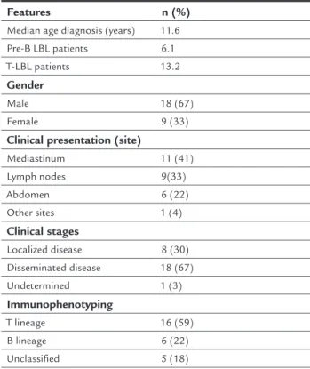
pB-LBL and 13.2 for those with T-pB-LBL. There was a predom-inance of males (1.6:1). Diagnosis was based on cytological examination of pleural effusions in two pa-tients (7%), and on tumor biopsies for the remaining cas-es. Immunohistochemistry was performed in 21 patients and immunophenotyping in two patients. Immunohis-tochemistry was inconclusive in six patients due to tech-nical problems with the ixing of samples. The clitech-nical and demographic characteristics of the patients are shown in Table 1. LBLs were of T-precursor cell origin in the ma-jority of cases (16 patients- 59%). The most common man-ifestation at diagnosis in T-cell cases was mediastinal enlargement, observed in nine patients, followed by lymph nodes in six. Intra-abdominal tumor was the major site of involvement in patients with pB-LBL, observed in three cases followed nodal disease in two cases. A para-vertebral tumor was the initial manifestation in one tient who had a secondary immunodeiciency. Most pa-tients had advanced disease (67%). Serum LDH concentration immediately at diagnosis was available only in 16 cases and the mean concentration was 831.63 IU/L (range: 152 to 3,897 IU/L). Serum uric acid was available for 22 patients with a mean value of 4.3 mg/dL (range: 2.0 to 8.5 mg/dL).
TABLE 1 Baseline clinical characteristics of 27 children with lymphoblastic lymphoma.
Features n (%)
Median age diagnosis (years) 11.6
Pre-B LBL patients 6.1
T-LBL patients 13.2
Gender
Male 18 (67)
Female 9 (33)
Clinical presentation (site)
Mediastinum 11 (41)
Lymph nodes 9(33)
Abdomen 6 (22)
Other sites 1 (4)
Clinical stages
Localized disease 8 (30)
Disseminated disease 18 (67)
Undetermined 1 (3)
Immunophenotyping
T lineage 16 (59)
B lineage 6 (22)
Unclassified 5 (18)
(continue)
TABLE 1 (Cont.) Baseline clinical characteristics of 27 children with lymphoblastic lymphoma.
Features n (%)
Characteristics
De novo LBL 24 (89)
Primary immunodeficiency 1 (4)
Secondary immunodeficiency 2 (7)
Outcome
Death before remission 3 (11)
Death after relapse 2 (7)
Alive in first remission 22 (81.5)
Outcome
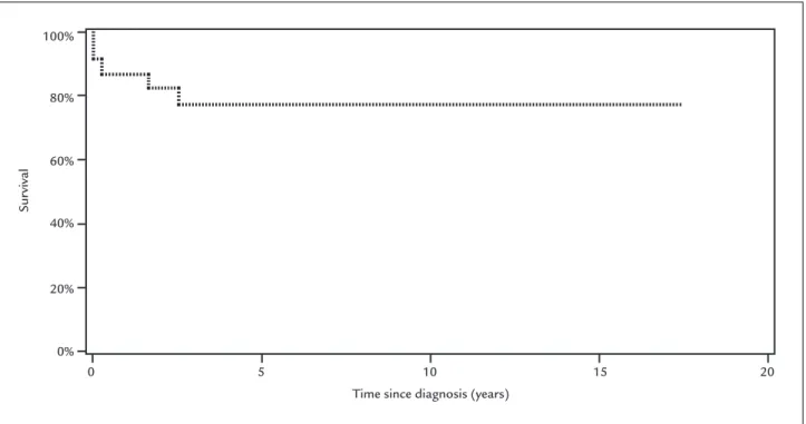
24 patients (9%) reached complete remission. After a me-dian follow-up of 43 months (interquartile range, 6.4-95), 22 patients (81%) were alive in irst CR. Five children (18.5%) died, three of them soon after admission and two (7%) after relapsing. Among the patients with immuno-deiciency, one died and the other two are alive in irst re-mission. The probability of survival at ive years for 20 patients with de novo LBL and similarly treated was 78%
(SD 9.4) (Figure 1).
D
ISCUSSIONLBLs comprise approximately 30% of the NHLs that oc-cur in children and young adults. The vast majority of them arise from immature T-cells, corresponding to de-ined stages of thymocyte differentiation, and approxi-mately 10 to 15% are precursor B-cell lymphomas.2,15,21 LBL accounted for 23% of cases of NHL enrolled in our institution in the period of 26 years. Burkitt’s lymphoma is the leading subtype in our institution.22
In agreement with the literature, the most frequent phenotype among our patients had T-cell origin. Approx-imately 20% were of pB-cell origin, and the remaining cas-es were unclassiied. In a Brazilian epidemiologic study, LBL represented 36% of all NHLs (272 cases). T-cell phe-notype was the most prevalent representing 60% of cas-es; 25% were of the pB-cell phenotype, and the remaining 15% were unclassiied.23
Patients with pB-LBL were younger than those with T-LBL, as reported by others (15, 24). However, the medi-an age at diagnosis for our patients with T-LBL was a lit-tle higher than reported before (around 8.8 years old).24 As also reported in previous studies, there was a clear pre-dominance of males (2:1).
pared with boys might have contributed to worst treat-ment results for females with increasing age.24
In general, the outcome is worst for CNS-positive as compared with CNS-negative patients, although not for all NHL subgroups. CNS involvement occurs more fre-quently in patients with advanced disease. In contrast to patients with Burkitt’s lymphoma, CNS involvement does not negatively impact treatment results for LBL patients.29
Over 80% of patients with T-LBL do not have mar-row involvement at diagnosis by morphologic examina-tion of bilateral marrow aspirates and biopsies.2 Howev-er, in a series of 99 children with T-LBL, more than two thirds of them had neoplastic lymphoblasts detected by low cytometry. This inding implies that a more sensi-tive method for investigating neoplastic cells in the BM at diagnosis may contribute to a more appropriate risk classiication.30 Persistent minimal disease during and/or at the end of induction therapy may also identify a poor risk group of patients.30
In relation to stage, patients with localized disease have a good prognosis.31 Disease stage was a major prog-nostic factor for children with pB-LBL, with signiicant better overall survival and event-free survival rates ob-served in patients with stage I to III as compared to those with stage IV.21 In the present study, out of ive patients who died, four had disseminated disease. The ifth pa-tient had localized disease and primary immunodei-ciency.
predominant in T-LBL.25 In the present study, mediasti-nal enlargement was present in more than half of the cas-es. Two patients had BM invasion, but CNS disease was not observed. The clinical presentation of children with pB-LBL varies considerably. Bone, soft tissue, and viscer-al organs can viscer-all be involved. In a series of twenty-seven patients with pB-LBL, the majority of them had nodal disease.15 In the present study, although there were only six patients, ive had intra-abdominal or lymph nodal dis-ease as the presenting clinical manifestation.
T-LBL stage is often advanced at diagnosis (stage III--IV), unlike pB-cell LBL, which primarily is a localized dis-ease.26,27 These results are coincident with those observed in our series. The majority of patients were diagnosed with advanced stages of disease.
Prognostic markers in LBL are yet to be clearly de-ined. Age, gender, response to treatment, stage, molec-ular prognostic markers, and marrow and CNS involve-ment remain unclear as prognostic factors in LBL. For instance, in a series of 85 children and adolescents with advanced LBL the multivariate analysis failed to dem-onstrate age, gender, lactate dehydrogenase level, and marrow or central nervous system disease to have inde-pendent prognostic value.28 In a recent study by Bur-khardt, adolescent females with T-LBL had a worse out-come. The worst outcome of girls compared with boys observed only in patients older than 9 years arose the is-sue whether the advanced pubertal changes of girls
com-Sur
vival
Time since diagnosis (years) 100%
80%
60%
40%
20%
0%
0 5 10 15 20
No single recurrent chromosome abnormality has been identiied as characteristic of LBL.31 Recently, activating
NOTCH1 mutations (chromosome 9) were reported to be
associated with favorable prognosis. Loss of heterozygos-ity at chromosome 6q (LOH6q) was reported to be associ-ated with increased relapse risk for patients with T-LBL.26
Response to treatment has been considered an im-portant prognostic factor. In a series of 121 children with T-LBL, the ones who had reached CR on the seventh day of chemotherapy (prephase) had a better prognosis than those who had not reached CR.9
Because of its biological relationship to ALL, patients with LBL have been treated with therapeutic protocols used for ALL, which are based on the principle of contin-ual exposure to cytostatics over a long period of time.32 Among our patients this strategy also proved highly
efi-cacious. Only two patients relapsed (7.4%), and both died. As reported in modern treatment protocols for LBL, 10%
of patients with progressive disease or relapse have in-deed an extremely poor prognosis.1 The probability of 5-year survival for 20 patients with de novo LBL and
sim-ilarly treated was almost 80%, similar to those achieved in major treatment centers (75 to 90%).3
C
ONCLUSIONOur indings conirm a favorable prognosis for children with LBL treated with ALL-based therapies. The subtle differences between LBL and ALL are elusive, and have raised questions as to whether identical therapeutic ap-proaches are warranted for each immunophenotypic cat-egory. Prognostic markers in LBL have also yet to be clear-ly deined. The outcome for patients with recurrent disease is poor. Molecular and cellular pathogenesis of malig-nant transformation to LBL is still a challenge to be tack-led in order to develop strategies for prevention, early identiication, and targeted therapies.
A
CKNOWLEDGMENTSThis study was supported by CAPES. Dr. M. C. Oliveira received a research grant from CAPES (2767-15-5).
R
ESUMOEvolução de crianças e adolescentes com linfoma linfo-blástico
Objetivos: linfoma linfoblástico (LL) é o segundo subti-po mais comum de linfoma não Hodgkin em crianças. O objetivo deste estudo foi caracterizar a evolução clínica de crianças e adolescentes com LL em um centro terciário.
Métodos: estudo de coorte retrospectivo de 27 pacientes com idade de até 16 anos com LL admitidos entre janei-ro de 1981 e dezembjanei-ro de 2013. Os pacientes foram tra-tados de acordo com o protocolo de tratamento para leu-cemia linfoblástica aguda (LLA). O diagnóstico foi baseado em biópsia do tumor e/ou no exame citológico de derrame pleural. A sobrevida global foi analisada pelo método de Kaplan-Meier.
Resultados: a média de idade ao diagnóstico foi de 11,6 anos (variação interquartil, 4,6-13,8). LL de células T foi identiicado em 16 pacientes (59%) e a manifestação pri-mária mais comum foi o acometimento mediastinal em 9 pacientes (56%). Tumor intra-abdominal foi a manifes-tação clínica principal nos pacientes com LL de células pré-B. A maioria dos pacientes apresentava doença avan-çada (18 pacientes – 67%) ao diagnóstico. Vinte e quatro pacientes (89%) alcançaram remissão clínica completa. Após um período de acompanhamento médio de 43 me-ses (intervalo interquartil, 6,4-95), 22 pacientes (81%) con-tinuam vivos em primeira remissão clínica completa. Cin-co crianças (18,5%) morreram, três delas logo após a admissão e duas após recidiva. A probabilidade de sobre-vida em cinco anos para 20 pacientes com LL de novo foi de 78% (SD 9.4).
Conclusão: nossos resultados conirmam o prognóstico
favorável de crianças com LL tratadas com regime de qui-mioterapia intensiva derivado da terapia de LLA.
Palavras-chave: leucemia-linfoma linfoblástico de células T precursoras, leucemia-linfoma linfoblástico de células precursoras B, linfoma não Hodgkin, pediatria, sobrevida.
R
EFERENCES1. Burkhardt B, Reiter A, Landmann E, Lang P, Lassay L, Dickerhoff R, et al. Poor outcome for children and adolescents with progressive disease or relapse of lymphoblastic lymphoma: a report from the Berlin-Frankfurt-Muenster Group. J Clin Oncol. 2009; 27(20):3363-9.
2. Cairo MS, Raetz E, Lim MS, Davenport V, Perkins SL. Childhood and adolescent non-Hodgkin lymphoma: new insights in biology and critical challenges for the future. Pediatr Blood Cancer. 2005; 45(6):753-69. 3. Schmidt E, Burkhardt B. Lymphoblastic lymphoma in childhood and
adolescence. Pediatr Hematol Oncol. 2013; 30(6):484-508.
4. Borowitz MJ, Chan J. B lymphoblastic leukaemia/lymphoma, not otherwise speciied. In: Swerdlow SH, Campo E, Harris NL (eds.). WHO classiication of tumours of haematopoietic and lymphoid tissues. Lyon: WHO, 2008. p.168-70. 5. Borowitz MJ, Chan JK. T lymphoblastic leukaemia/lymphoma. In: Swerdlow SH, Campo E, Harris NL (eds.). WHO classification of tumors of haematopoietic and lymphoid tissues. Lyon: WHO, 2008. p.176-8. 6. Maitra A, McKenna RW, Weinberg AG, Schneider NR, Kroft SH. Precursor
B-cell lymphoblastic lymphoma. A study of nine cases lacking blood and bone marrow involvement and review of the literature. Am J Clin Pathol. 2001; 115(6):868-75.
8. Reiter A, Tiemann M, Ludwig WD, Wacker HH, Yakisan E, Schrappe M, et al. [NHL-BFM 90 therapy study in treatment of malignant non-Hodgkin’s lymphomas in children and adolescents. Part 1: Classiication and allocation to strategic therapy groups. BIF study group]. Klin Padiatr. 1994; 206(4):222-33. 9. Uyttebroeck A, Suciu S, Laureys G, Robert A, Pacquement H, Ferster A, et al.
Treatment of childhood T-cell lymphoblastic lymphoma according to the strategy for acute lymphoblastic leukaemia, without radiotherapy: long term results of the EORTC CLG 58881 trial. Eur J Cancer. 2008; 44(6):840-6. 10. Muljono A, Graf NS, Arbuckle S. Primary cutaneous lymphoblastic lymphoma
in children: series of eight cases with review of the literature. Pathology. 2009; 41(3):223-8.
11. Ozdemirli M, Fanburg-Smith JC, Hartmann DP, Shad AT, Lage JM, Magrath IT, et al. Precursor B-lymphoblastic lymphoma presenting as a solitary bone tumor and mimicking Ewing’s sarcoma: a report of four cases and review of the literature. Am J Surg Pathol. 1998; 22(7):795-804.
12. Burkhardt B. Paediatric lymphoblastic T-cell leukaemia and lymphoma: one or two diseases? Br J Haematol. 2010; 149(5):653-68.
13. Reiter A, Schrappe M, Ludwig WD, Tiemann M, Parwaresch R, Zimmermann M, et al. Intensive ALL-type therapy without local radiotherapy provides a 90% event-free survival for children with T-cell lymphoblastic lymphoma: a BFM group report. Blood. 2000; 95(2):416-21.
14. Anderso JR, Jenkin RD, Wilson JF, Kjeldsberg CR, Sposto R, Chilcote RR, et al. Long-term follow-up of patients treated with COMP or LSA2L2 therapy for childhood non-Hodgkin’s lymphoma: a report of CCG-551 from the Children’s Cancer Group. J Clin Oncol. 1993; 11(6):1024-32.
15. Neth O, Seidemann K, Jansen P, Mann G, Tiemann M, Ludwig WD, et al. Precursor B-cell lymphoblastic lymphoma in childhood and adolescence: clinical features, treatment, and results in trials NHL-BFM 86 and 90. Med Pediatr Oncol. 2000; 35(1):20-7.
16. Termuhlen AM, Smith LM, Perkins SL, Lones M, Finlay JL, Weinstein H, et al. Disseminated lymphoblastic lymphoma in children and adolescents: results of the COG A5971 trial: a report from the Children’s Oncology Group. Br J Haematol. 2013; 162(6):792-801.
17. Swerdlow SH, Campo E, Harris NL, Jaffe E, Pieleri SA, Stein H (eds.). WHO classiication of tumors of haematopoietic and lymphoide tissues. IARC WHO Classiication of tumors. 2.ed. Lyon: IARC, 2008.
18. Murphy SB. Classiication, staging and end results of treatment of childhood non- Hodgkin’s lymphomas: dissimilarities from lymphomas in adults. Semin Oncol. 1980; 7(3):332-9.
19. Wollner N, Wachtel AE, Exelby PR, Centore D. Improved prognosis in children with intra-abdominal non-Hodgkin’s lymphoma following LSA2L2 protocol chemotherapy. Cancer. 1980; 45(12):3034-9.
20. Reiter A, Schrappe M, Ludwig WD, Lampert F, Harbott J, Henze G, et al. Favorable outcome of B-cell acute lymphoblastic leukemia in childhood: a report of three consecutive studies of the BFM group. Blood. 1992; 80(10):2471-8.
21. Ducassou S, Ferlay C, Bergeron C, Girard S, Laureys G, Pacquement H, et al. Clinical presentation, evolution, and prognosis of precursor B-cell lymphoblastic lymphoma in trials LMT96, EORTC 58881, and EORTC 58951. Br J Haematol. 2011; 152(4):441-51.
22. Cunha KC, Oliveira MC, Gomes AC, de Castro LP, Viana MB. Clinical course and prognostic factors of children with Burkitt’s lymphoma in a developing country: the experience of a single centre in Brazil. Rev Bras Hematol Hemoter. 2012; 34(5):361-6.
23. Gualco G, Klumb CE, Barber GN, Weiss LM, Bacchi CE. Pediatric lymphomas in Brazil. Clinics. 2010; 65(12):1267-77.
24. Burkhardt B, Zimmermann M, Oschlies I, Niggli F, Mann G, Parwaresch R, et al. The impact of age and gender on biology, clinical features and treatment outcome of non-Hodgkin lymphoma in childhood and adolescence. Br J Haematol. 2005; 131(1):39-49.
25. Sheibani K, Nathwani BN, Winberg CD, Burke JS, Swartz WG, Blayney D, et al. Antigenically defined subgroups of lymphoblastic lymphoma. Relationship to clinical presentation and biologic behavior. Cancer. 1987; 60(2):183-90.
26. Bonn BR, Rohde M, Zimmermann M, Krieger D, Oschlies I, Niggli F, et al. Incidence and prognostic relevance of genetic variations in T-cell lymphoblastic lymphoma in childhood and adolescence. Blood. 2013; 121(16):3153-60.
27. El-Mallawany NK, Frazer JK, Van Vlierberghe P, Ferrando AA, Perkins S, Lim M, et al. Pediatric T- and NK-cell lymphomas: new biologic insights and treatment strategies. Blood Cancer J. 2012; 2(4):e65.
28. Abromowitch M, Sposto R, Perkins S, Zwick D, Siegel S, Finlay J, et al. Shortened intensiied multi-agent chemotherapy and non-cross resistant maintenance therapy for advanced lymphoblastic lymphoma in children and adolescents: report from the Children’s Oncology Group. Br J Haematol. 2008; 143(2):261-7.
29. Salzburg J, Burkhardt B, Zimmermann M, Wachowski O, Woessmann W, Oschlies I, et al. Prevalence, clinical pattern, and outcome of CNS involvement in childhood and adolescent Hodgkin’s lymphoma differ by non-Hodgkin’s lymphoma subtype: a Berlin-Frankfurt-Munster Group Report. J Clin Oncol. 2007; 25(25):3915-22.
30. Coustan-Smith E, Sandlund JT, Perkins SL, Chen H, Chang M, Abromowitch M, et al. Minimal disseminated disease in childhood T-cell lymphoblastic lymphoma: a report from the Children’s Oncology Group. J Clin Oncol. 2009; 27(21):3533-9.
31. Lones MA, Heerema NA, Le Beau MM, Sposto R, Perkins SL, Kadin ME, et al. Chromosome abnormalities in advanced stage lymphoblastic lymphoma of children and adolescents: a report from CCG-E08. Cancer Genet Cytogenet. 2007; 172(1):1-11.