Outcome of children and adolescents with lymphoblastic
lymphoma
MARIA CHRISTINA LOPES ARAÚJO OLIVEIRA1*, KEYLA CHRISTY SAMPAIO2, ALINE CARNEIRO OLIVEIRA3, AIESKA DANTAS SANTOS3,
LÚCIA PORTO CASTRO4, MARCOS BORATO VIANA5
1 PhD – Associate Professor, Hematology Division, Pediatrics Department, Universidade Federal de Minas Gerais (UFMG), Belo Horizonte, MG, Brazil 2PhD – Adjunct Professor – Pediatrics Department – UFMG, Belo Horizonte, MG, Brazil
3Physician – Hospital das Clínicas, UFMG, Belo Horizonte, MG, Brazil
4MSc – Assistant Professor – Pathology Department – UFMG, Belo Horizonte, MG, Brazil 5PhD – Full Professor – Pediatrics Department – UFMG, Belo Horizonte, MG, Brazil
S
UMMARYStudy conducted at Pediatrics Department – Faculdade de Medicina, Universidade Federal de Minas Gerais (UFMG),
Belo Horizonte, MG, Brazil
Article received: 1/19/2015
Accepted for publication: 5/4/2015
*Correspondence:
Address: Av. Professor Alfredo Balena, 100 Belo Horizonte, MG – Brazil
Postal code: 30130-100 [email protected]
http://dx.doi.org/10.1590/1806-9282.61.05.417
Introduction: lymphoblastic lymphoma (LBL) is the second most common
sub-type of non-Hodgkin lymphoma in children. The aim of this study was to char-acterize the clinical course of children and adolescents with LBL treated at a ter-tiary center.
Methods: this is a retrospective cohort study of 27 patients aged 16 years or young-er with LBL admitted between January 1981 and Decembyoung-er 2013. Patients wyoung-ere treated according to the therapy protocol used for acute lymphoblastic leucemia. Diagnosis was based on biopsy of tumor and/or cytological examination of pleu-ral effusions. The ovepleu-rall survival was analyzed using the Kaplan-Meier method.
Results: the median age at diagnosis was 11.6 years (interquartile range, 4.6-13.8). LBL had T-cell origin in 16 patients (59%). The most common primary manifestation in T-cell LBL was mediastinal involvement, in 9 patients (56%). Intra-abdominal tumor was the major site of involvement in patients with pre-cursor B-LBL. Most patients had advanced disease (18 patients – 67%) at diag-nosis. Twenty-four patients (89%) achieved complete clinical remission. After a median follow-up of 43 months (interquartile range, 6.4-95), 22 patients (81%) were alive in first complete remission. Five children (18.5%) died, three of them soon after admission and two after relapsing. The probability of survival at five years for 20 patients with de novo LBL was 78% (SD 9.4).
Conclusion: our findings confirm the favorable prognosis of children with LBL with an intensive chemotherapy regimen derived from ALL therapy.
Keywords: precursor T-cell lymphoblastic-leukemia lymphoma, precursor B-cell
lymphoblastic-leukemia lymphoma, non-Hodgkin lymphoma, pediatrics, survival.
INTRODUCTION
Lymphoblastic lymphomas (LBL) are lymphoid malignan-cies of immature or precursor cells representing one third of the cases of non-Hodgkin lymphoma in children and adolescents.1-3 The current World Health Organization (WHO) classification assigns tumours of hematopoietic and lymphoid tissues in T lymphoblastic leukemia/lym-phoma and B lymphoblastic leukemia/lymleukemia/lym-phoma.4,5 Of note, only approximately 10% of LBL express B-cell mark-ers in contrast to approximately 85% of acute lymphoblas-tic leukemia (ALL).6 The distinction between leukemia and lymphoma is arbitrary and based on the extent of
in-volvement of bone marrow (BM). It is usual to diagnose patients with ≥ 25% lymphoblasts in BM as having ALL, whereas patients with extra-medullary disease and less than 25% blasts in BM are diagnosed as LBL.7,8
Whether LBL and ALL in childhood are biologically identical or rather distinct disorders is not entirely clear.12,13 To date, the pathogenesis and genetic changes of LBL is poorly understood.3 LBLs are most effectively treated us-ing ALL-based therapies.14 Nowadays event-free survival (EFS) can be reached by 75-90% of children and adoles-cents.3,15,16
The objective of this study was to contribute to the knowledge of the clinical course and treatment present-ing the outcome of 27 children and adolescents with LBL followed up in a single tertiary center.
METHODS
This is a longitudinal retrospective observational cohort study that evaluated 27 children and adolescents aged 16 years or younger with LBL. The patients were admitted to the Pediatric Hematology Unit, University Hospital, Universidade Federal de Minas Gerais (UFMG), between January 1981 and December 2013. Seven patients were excluded from the Kaplan-Meier survival analysis (see sta-tistical session) because of previous treatment at other institutions (n=3), severe concomitant immunodeficien-cy (one with primary and two with secondary immuno-deficiency), and one with bone marrow involvement with atypical cells that were not considered lymphoblasts.
Medical records were reviewed to collect demograph-ic data (age, gender), clindemograph-ical data (meddemograph-ical history, phys-ical examination, clinphys-ical presentation), diagnostic pro-cedures (imaging studies, bone marrow aspiration, cerebrospinal fluid – CSF analysis), staging, laboratory data (lactate dehydrogenase – LDH levels, blood counts, serum electrolytes, liver and kidney tests), treatment, and outcome. Diagnosis was made by incisional or excision-al biopsy, or cytologicexcision-al examination of pleurexcision-al or abdom-inal effusions. Karyotype studies were not available at the time. All the diagnoses were confirmed by morphologi-cal and immunohistochemistry criteria defined by the World Health Organization (WHO) classification.17 Im-munohistochemistry was performed using monoclonal antibodies CD20, CD10, CD79a, CD30, CD3, CD15, TdT, CD45, and CD45RO for the detection of B and T cells. Cell lineage assignment required 50% or more of positive neoplastic B or T cells. Pleural or abdominal effusions were examined by flow cytometry.
Clinical staging was based on the St. Jude Children’s Research Hospital staging system.18 Central nervous sys-tem (CNS) disease was diagnosed by the presence of mor-phologically identifiable lymphoma cells (regardless of quantity) in CSF, an intracranial mass or cranial nerve palsy not caused by an extradural mass.
• Treatment: patients with LBL were treated accor-ding to protocols based on an ALL-type strategy. Patients admitted between 1981 and 1987 were trea-ted according to the modified LSA2L2 protocol of the Memorial Sloan-Kettering Cancer Center.19 Af-ter 1987, patients were treated with a BFM-83-ba-sed protocol (Berlin-Frankfurt-Münster).20
• Response criteria: complete remission (CR) was defined as the disappearance of all tumor masses confirmed by clinical examination and imaging investigations, one month after therapy. After the end of treatment, the patients were followed at 30-day intervals during the first year, at 60-day intervals during the second year and at 3- to 6-month intervals up to five years. Progression of the local tumor was defined if the tumor site showed no decrease in size after starting chemotherapy. Relap-se was defined as the recurrence of lymphoma with the same histological or immunophenotypic features as the initial one at any site after CR was achieved. Local relapse was diagnosed when it involved a previously in-volved site (except bone marrow and CSF).
• Statistical analysis: the time limit for the current study was the end of December 2013. Data are reported as medians and interquartile range (IQ) or means and standard deviation (SD), when appropriate. Mann--Whitney or Kruskal-Wallis tests were used to compa-re nonparametric continuous variables. Dichotomous variables were compared by the two-tailed chi-squa-re test or Fisher’s exact test. Overall survival (OS) was defined as the time from diagnosis to date of death due to any cause or date of last follow-up contact for patients who were alive. The OS was analyzed using the Kaplan–Meier method for 20 patients, as men-tioned before.
• Ethical issues: the study was approved by the Research Ethics Committee of UFMG. Written informed con-sent was obtained from the guardians of the patients and, when appropriate, from the patients themsel-ves, according to the Helsinki Declaration.
RESULTS
Patient characteristics
inconclusive in six patients because of technical problems with the fixing of samples. The clinical and demograph-ic characteristdemograph-ics of the patients are shown in Table 1. LBLs were of T-precursor cell origin in the majority of cases (16 patients – 59%). The most common manifesta-tion at diagnosis in T-cell cases was mediastinal enlarge-ment, observed in nine patients, followed by lymph nodes in six. Intra-abdominal tumor was the major site of in-volvement in patients with pB-LBL, observed in three cas-es followed nodal disease in two cascas-es. A paravertebral tu-mor was the initial manifestation in one patient who had a secondary immunodeficiency. Most patients had ad-vanced disease (67%). Serum LDH concentration imme-diately at diagnosis was available only in 16 and the mean concentration was 831.63 IU/L (range: 152 to 3,897 IU/L). Serum uric acid was available for 22 patients with a mean value of 4.3 mg/dL (range: 2.0 to 8.5 mg/dL).
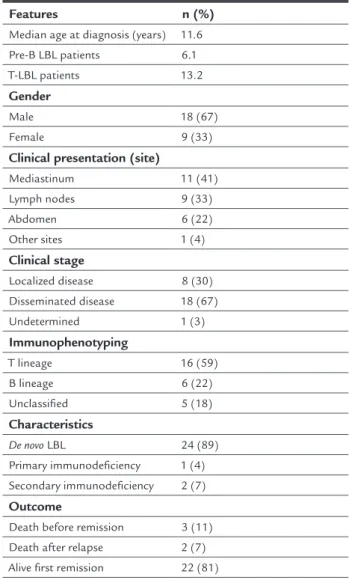
TABLE 1 Baseline clinical characteristics of 27 children
with lymphoblastic lymphoma.
Features n (%)
Median age at diagnosis (years) 11.6
Pre-B LBL patients 6.1
T-LBL patients 13.2
Gender
Male 18 (67)
Female 9 (33)
Clinical presentation (site)
Mediastinum 11 (41)
Lymph nodes 9 (33)
Abdomen 6 (22)
Other sites 1 (4)
Clinical stage
Localized disease 8 (30)
Disseminated disease 18 (67)
Undetermined 1 (3)
Immunophenotyping
T lineage 16 (59)
B lineage 6 (22)
Unclassified 5 (18)
Characteristics
De novo LBL 24 (89)
Primary immunodeficiency 1 (4) Secondary immunodeficiency 2 (7)
Outcome
Death before remission 3 (11)
Death after relapse 2 (7)
Alive first remission 22 (81)
Outcome
24 patients (89%) reached complete remission. After a me-dian follow-up of 43 months (interquartile range, 6.4-95), 22 patients (81%) were alive in first CR. Five children (18.5%) died, three of them soon after admission and two (7%) after relapsing. Among the patients with immuno-deficiency, one died and the other two are alive in first re-mission. The probability of survival at five years for 20 patients with de novo LBL and similarly treated was 78% (SD 9.4) (Figure 1).
DISCUSSION
LBLs comprise approximately 30% of the NHLs that occur in children and young adults. The vast majority of them arise from immature T-cells corresponding to defined stag-es of thymocyte differentiation, and approximately 10 to 15% are precursor B-cell lymphomas.2,15,21 LBL accounted for 23% of cases of non-Hodgkin’s lymphoma (NHL) en-rolled in our institution in the period of 26 years. Burkitt’s lymphoma is the leading subtype in our institution.22
In agreement with the literature, the most frequent phenotype among our patients had T-cell origin. Approx-imately 20% were of pB-cell origin, and the remaining were unclassified. In a Brazilian epidemiologic study, LBL represented 36% of all NHLs (272 cases). T-cell pheno-type was the most prevalent representing 60% of cases; 25% were of the pB-cell phenotype, and the remaining 15% were unclassified.23
Patients with pB-LBL were younger than those with T-LBL, as reported by others.15,24 However, the median age at diagnosis for our patients with T-LBL was a little high-er than reported before (around 8.8 years old).24 As also reported in previous studies, there was a clear predomi-nance of males (2:1).
Clinical features presented in T-LBL are quite distinct from those of pB- LBL. Mediastinal, bone marrow (BM) and central nervous system (CNS) involvement predom-inate in T-LBL.25 In the present study, mediastinal en-largement was seen in more than half of the cases. Two patients had BM invasion, but CNS disease was not ob-served. The clinical presentation in children with pB-LBL varies considerably. Bone, soft tissue, and visceral organs can all be involved. In a series of twenty-seven patients with pB-LBL the majority of them had nodal disease.15 In the present study, although there were only six patients, five had intra-abdominal or lymph nodal disease as the presenting clinical manifestation.
ob-classification.30 Persistent minimal disease during and/ or at the end of induction therapy may also identify a poor-risk group of patients.30
As for staging, patients with localized disease have a good prognosis.31 Disease stage was a major prognostic factor for children with pB-LBL, with significant better overall survival and event-free survival rates observed in patients with stage I to III, as compared to those with stage IV.21 In the present study, out of five patients who died, four had disseminated disease. The fifth patient had localized disease and primary immunodeficiency.
No single recurrent chromosome abnormality has been identified as characteristic of LBL.31 Recently, acti-vating NOTCH1 mutations (chromosome 9) were report-ed to be associatreport-ed with favorable prognosis. Loss of het-erozygosity at chromosome 6q (LOH6q) was reported to be associated with increased relapse risk for patients with T-LBL.26
Response to treatment has been considered an im-portant prognostic factor. In a series of 121 children with T-LBL who had reached CR on the seventh day of chemo-therapy (prephase) had a better prognosis than those who had not reached CR.9
Because of its biological relationship to ALL, patients with LBL have been treated with therapeutic protocols used for ALL, which are based on the principle of contin-ual exposure to cytostatic drugs over a long period of time.32 Among our patients this strategy also proved high-ly efficacious. Onhigh-ly two patients relapsed (7.4%), and both died. As reported in modern treatment protocols for LBL, 10% of patients with progressive disease or relapse have served in our series. Most patients were diagnosed with
advanced stages of disease.
Prognostic markers in LBL are yet to be clearly de-fined. Age, gender, response to treatment, stage, molecu-lar prognostic markers, and marrow and CNS involve-ment remain unclear as prognostic factors in LBL. For instance, in a series of 85 children and adolescents with advanced LBL, the multivariate analysis failed to demon-strate age, gender, lactate dehydrogenase level, and mar-row or central nervous system disease to have indepen-dent prognostic value.28 In a recent study by Burkhardt, adolescent females with T-LBL had a worse outcome. The worst outcome of girls compared with boys observed only in patients older than 9 years has given rise to the hypoth-esis that advanced pubertal changes in girls compared with boys might contribute to inferior treatment results for females with increasing age.24
In general, the outcome is inferior for CNS positive as compared with CNS-negative patients, although not for all NHL subgroups. CNS involvement occurs more frequently in patients with advanced disease. Unlike pa-tients with Burkitt’s lymphoma, CNS involvement does not negatively impact treatment results for LBL patients.29
Over 80% of patients with T-LBL do not have mar-row involvement at diagnosis by morphologic examina-tion of bilateral marrow aspirates and biopsies.2 Howev-er, in a series of 99 children with T-LBL, more than two thirds of them had neoplastic lymphoblasts detected by flow cytometry. This finding implies that a more sensi-tive method for investigating neoplastic cells in the BM at diagnosis may contribute to a more appropriate risk FIGURE 1 Overall survival of 20 patients with lymphoblastic lymphoma.
Sur
vival
100%
80%
60%
0 5 10 15 20
40%
20%
0
indeed an extremely poor prognosis.1 The probability of 5-year survival of 20 patients with de novo LBL treated in the same manner was almost 80%, similar to that achieved in major treatment centers (75 to 90%).3
CONCLUSION
Our findings confirm a favorable prognosis for children with LBL treated with ALL-based therapies. The subtle differences between LBL and ALL are elusive, and have raised questions as to whether identical therapeutic ap-proaches are warranted for each immunophenotypic cat-egory. Prognostic markers in LBL have also yet to be clear-ly defined. The outcome for patients with recurrent disease is poor. Molecular and cellular pathogenesis of malig-nant transformation to LBL is still a challenge to be tack-led in order to develop strategies for prevention, early identification, and targeted therapies.
RESUMO
Evolução de crianças e adolescentes com linfoma linfo-blástico
Objetivos: linfoma linfoblástico (LL) é o segundo subti-po mais comum de linfoma não Hodgkin em crianças. O objetivo deste estudo foi caracterizar a evolução clínica de crianças e adolescentes com LL em um centro terciário.
Métodos: estudo de coorte retrospectivo de 27 pacientes com idade de até 16 anos com LL admitidos entre janeiro de 1981 e dezembro de 2013. Os pacientes foram tratados de acordo com o protocolo de tratamento para leucemia linfoblástica aguda (LLA). O diagnóstico foi baseado em biópsia do tumor e/ou no exame citológico de derrame pleural. A sobrevida global foi analisada pelo método de Kaplan-Meier.
Resultados: a média de idade ao diagnóstico foi de 11,6 anos (variação interquartil, 4,6-13,8). Linfoma linfoblásti-co de células T foi identificado em 16 pacientes (59%) e a manifestação primária mais comum foi o acometimento mediastinal (56%). Tumor intra-abdominal foi a manifes-tação clínica principal nos pacientes com LL de células pré--B. A maioria dos pacientes apresentava doença avançada (18 pacientes, 67%) ao diagnóstico. Vinte e quatro pacien-tes (89%) alcançaram remissão clínica completa. Após um período de acompanhamento médio de 43 meses (interva-lo interquartil, 6,4-95), 22 pacientes (81%) continuam vi-vos em primeira remissão clínica completa. Cinco crianças (18,5%) morreram, três delas logo após a admissão e duas após recidiva. A probabilidade de sobrevida em cinco anos para 20 pacientes com LL de novo foi de 78% (DP 9,4).
Conclusão: os resultados confirmam o prognóstico
favo-rável de crianças com LL tratadas com regime de quimio-terapia intensiva derivado da quimio-terapia de LLA.
Palavras-chave: leucemia-linfoma linfoblástico de células T precursoras, leucemia-linfoma linfoblástico de células precursoras B, linfoma não Hodgkin, pediatria, sobrevida.
REFERENCES
1. Burkhardt B, Reiter A, Landmann E, Lang P, Lassay L, Dickerhoff R, et al. Poor outcome for children and adolescents with progressive disease or relapse of lymphoblastic lymphoma: a report from the Berlin-Frankfurt-Muenster Group. J Clin Oncol. 2009; 27(20):3363-9.
2. Cairo MS, Raetz E, Lim MS, Davenport V, Perkins SL. Childhood and adolescent non-Hodgkin lymphoma: new insights in biology and critical challenges for the future. Pediatr Blood Cancer. 2005; 45(6):753-69. 3. Schmidt E, Burkhardt B. Lymphoblastic lymphoma in childhood and
adolescence. Pediatr Hematol Oncol. 2013; 30(6):484-508.
4. Borowitz MJ, Chan J. B lymphoblastic leukaemia/lymphoma, not otherwise specified. In: Swerdlow SH, Campo E, Harris NL (eds.). WHO Classification of Tumours of Haematopoietic and Lymphoid Tissues. Lyon: WHO, 2008. p.168-70.
5. Borowitz MJ, Chan JK. T lymphoblastic leukaemia/lymphoma. In: Swerdlow SH, Campo E, Harris NL (eds.). WHO classification of tumors of haematopoietic and lymphoid tissues. Lyon: WHO, 2008. p.176-8. 6. Maitra A, McKenna RW, Weinberg AG, Schneider NR, Kroft SH. Precursor
B-cell lymphoblastic lymphoma. A study of nine cases lacking blood and bone marrow involvement and review of the literature. Am J Clin Pathol. 2001; 115(6):868-75.
7. Reiter A, Schrappe M, Parwaresch R, Henze G, Muller-Weihrich S, Sauter S, et al. Non-Hodgkin’s lymphomas of childhood and adolescence: results of a treatment stratified for biologic subtypes and stage – a report of the Berlin-Frankfurt-Munster Group. J Clin Oncol. 1995; 13(2):359-72.
8. Reiter A, Tiemann M, Ludwig WD, Wacker HH, Yakisan E, Schrappe M, et al. [NHL-BFM 90 therapy study in treatment of malignant non-Hodgkin’s lymphomas in children and adolescents. Part 1: Classification and allocation to strategic therapy groups. BIF study group]. Klin Padiatr. 1994; 206(4):222-33. 9. Uyttebroeck A, Suciu S, Laureys G, Robert A, Pacquement H, Ferster A, et
al. Treatment of childhood T-cell lymphoblastic lymphoma according to the strategy for acute lymphoblastic leukaemia, without radiotherapy: long term results of the EORTC CLG 58881 trial. Eur J Cancer. 2008; 44(6):840-6.
10. Muljono A, Graf NS, Arbuckle S. Primary cutaneous lymphoblastic lymphoma in children: series of eight cases with review of the literature. Pathology. 2009; 41(3):223-8.
11. Ozdemirli M, Fanburg-Smith JC, Hartmann DP, Shad AT, Lage JM, Magrath IT, et al. Precursor B-Lymphoblastic lymphoma presenting as a solitary bone tumor and mimicking Ewing’s sarcoma: a report of four cases and review of the literature. Am J Surg Pathol. 1998; 22(7):795-804.
12. Burkhardt B. Paediatric lymphoblastic T-cell leukaemia and lymphoma: one or two diseases? Br J Haematol. 2010; 149(5):653-68.
13. Reiter A, Schrappe M, Ludwig WD, Tiemann M, Parwaresch R, Zimmermann M, et al. Intensive ALL-type therapy without local radiotherapy provides a 90% event-free survival for children with T-cell lymphoblastic lymphoma: a BFM group report. Blood. 2000; 95(2):416-21.
14. Anderson JR, Jenkin RD, Wilson JF, Kjeldsberg CR, Sposto R, Chilcote RR, et al. Long-term follow-up of patients treated with COMP or LSA2L2 therapy for childhood non-Hodgkin’s lymphoma: a report of CCG-551 from the Children’s Cancer Group. J Clin Oncol. 1993; 11(6):1024-32.
15. Neth O, Seidemann K, Jansen P, Mann G, Tiemann M, Ludwig WD, et al. Precursor B-cell lymphoblastic lymphoma in childhood and adolescence: clinical features, treatment, and results in trials NHL-BFM 86 and 90. Med Pediatr Oncol. 2000; 35(1):20-7.
17. Swerdlow SH, Campo E, Harris NL, Jaffe E, Pieleri SA, Stein H. WHO classification of tumors of haematopoietic and lymphoide tissues. In: IARC W (ed.). 2.ed. 2008.
18. Murphy SB. Classification, staging and end results of treatment of childhood non- Hodgkin’s lymphomas: dissimilarities from lymphomas in adults. Semin Oncol. 1980; 7(3):332-9.
19. Wollner N, Wachtel AE, Exelby PR, Centore D. Improved prognosis in children with intra-abdominal non-Hodgkin’s lymphoma following LSA2L2 protocol chemotherapy. Cancer. 1980; 45(12):3034-9.
20. Reiter A, Schrappe M, Ludwig WD, Lampert F, Harbott J, Henze G, et al. Favorable outcome of B-cell acute lymphoblastic leukemia in childhood: a report of three consecutive studies of the BFM group. Blood. 1992; 80(10):2471-8.
21. Ducassou S, Ferlay C, Bergeron C, Girard S, Laureys G, Pacquement H, et al. Clinical presentation, evolution, and prognosis of precursor B-cell lymphoblastic lymphoma in trials LMT96, EORTC 58881, and EORTC 58951. Br J Haematol. 2011; 152(4):441-51.
22. Cunha KC, Oliveira MC, Gomes AC, de Castro LP, Viana MB. Clinical course and prognostic factors of children with Burkitt’s lymphoma in a developing country: the experience of a single centre in Brazil. Rev Bras Hematol Hemoter. 2012; 34(5):361-6.
23. Gualco G, Klumb CE, Barber GN, Weiss LM, Bacchi CE. Pediatric lymphomas in Brazil. Clinics (Sao Paulo). 2010; 65(12):1267-77.
24. Burkhardt B, Zimmermann M, Oschlies I, Niggli F, Mann G, Parwaresch R, et al. The impact of age and gender on biology, clinical features and treatment outcome of non-Hodgkin lymphoma in childhood and adolescence. Br J Haematol. 2005; 131(1):39-49.
25. Sheibani K, Nathwani BN, Winberg CD, Burke JS, Swartz WG, Blayney D, et al. Antigenically defined subgroups of lymphoblastic lymphoma.
Relationship to clinical presentation and biologic behavior. Cancer. 1987; 60(2):183-90.
26. Bonn BR, Rohde M, Zimmermann M, Krieger D, Oschlies I, Niggli F, et al. Incidence and prognostic relevance of genetic variations in T-cell lymphoblastic lymphoma in childhood and adolescence. Blood. 2013; 121(16):3153-60.
27. El-Mallawany NK, Frazer JK, Van Vlierberghe P, Ferrando AA, Perkins S, Lim M, et al. Pediatric T- and NK-cell lymphomas: new biologic insights and treatment strategies. Blood Cancer J. 2012; 2(4):e65.
28. Abromowitch M, Sposto R, Perkins S, Zwick D, Siegel S, Finlay J, et al. Shortened intensified multi-agent chemotherapy and non-cross resistant maintenance therapy for advanced lymphoblastic lymphoma in children and adolescents: report from the Children’s Oncology Group. Br J Haematol. 2008; 143(2):261-7.
29. Salzburg J, Burkhardt B, Zimmermann M, Wachowski O, Woessmann W, Oschlies I, et al. Prevalence, clinical pattern, and outcome of CNS involvement in childhood and adolescent Hodgkin’s lymphoma differ by non-Hodgkin’s lymphoma subtype: a Berlin-Frankfurt-Munster Group Report. J Clin Oncol. 2007; 25(25):3915-22.
30. Coustan-Smith E, Sandlund JT, Perkins SL, Chen H, Chang M, Abromowitch M, et al. Minimal disseminated disease in childhood T-cell lymphoblastic lymphoma: a report from the children’s oncology group. J Clin Oncol. 2009; 27(21):3533-9.
31. Lones MA, Heerema NA, Le Beau MM, Sposto R, Perkins SL, Kadin ME, et al. Chromosome abnormalities in advanced stage lymphoblastic lymphoma of children and adolescents: a report from CCG-E08. Cancer Genet Cytogenet. 2007; 172(1):1-11.