he efects of orthostatism in adult intensive care
unit patients
Efeito imediato do ortostatismo em pacientes internados na
unidade de terapia intensiva de adultos
INTRODUCTION
he musculoskeletal system is designed for movement. Just seven days at rest can result in a 30% drop in muscle strength, with an additional
20% loss of remaining strength each additional week.(1,2) Long stays in the
intensive care unit (ICU) and mechanical ventilation (MV) are associated with functional decline, morbidity, mortality, high care costs and long-term
hospitalization.(2,3) Sepsis, hydroelectrolytic disorders and immobility can
cause polyneuromyopathy in critically ill patients. Excessive use of sedating drugs, neuromuscular blockers, corticosteroids and MV can aggravate
ICU-acquired weakness.(1,4)
Melissa Sibinelli1, Daniele Cristina
Maioral2, Antônio Luis Eiras
Falcão3, Carolina Kosour4, Desanka
Dragosavac3, Núbia Maria Freire
Vieira Lima4
1. Adult Intensive Care Unit, Hospital das Clínicas, Universidade Estadual de Campinas - UNICAMP - Campinas (SP), Brazil; Postgraduate Program (MSc Level), Department of Surgery, Universidade Estadual de Campinas - UNICAMP - Campinas (SP), Brazil. 2. Irmandade da Santa Casa de Misericórdia de Piracicaba - Piracicaba (SP), Brazil; Postgraduate Program in Respiratory Physiotherapy, Universidade Estadual de Campinas - UNICAMP - Campinas (SP), Brazil.
3. Department of Surgery, Medical College, Universidade Estadual de Campinas - UNICAMP - Campinas (SP), Brazil.
4. Adult Intensive Care Unit, Hospital das Clínicas, Universidade Estadual de Campinas - UNICAMP - Campinas (SP), Brazil.
ABSTRACT
Objective: To assess the consciousness level, pulmonary and hemodynamic efects of orthostatic position in intensive care patients.
Methods: his study was conducted from April 2008 to July 2009 in the Adult Intensive Care Unit, Hospital das Clínicas, Universidade Estadual de Campinas, São Paulo, Brazil. Fifteen patients were included who were mechanically ventilated for more than seven days and had the following characteristics: tracheotomized; receiving intermittent nebulization; maximal inspiratory pressure of less than -25 cm H2O; Tobin score less than 105; preserved respiratory drive; not sedated; partial arterial oxygen pressure greater than 70 mm Hg; oxygen saturation greater than 90%; and hemodynamically stable. With inclinations of 0º, 30º and 50º, the following parameters were recorded: consciousness level; blinking relex; thoracoabdominal cirtometry; vital capacity; tidal volume; minute volume; respiratory muscle strength; and vital signs.
Results: No neurological level changes
were observed. Respiratory rate and minute volume (VE) decreased at 30% and later increased at 50%; however, these changes were not statistically signiicant. Abdominal cirtometry and maximal expiratory pressure increased, but again, the changes were not statistically signiicant. Regarding maximal inspiratory pressure and vital capacity, statistically signiicant increases were seen in the comparison between the 50º and 0º inclinations. However, tidal volume increased with time in the comparisons between 30º and 0º and between 50º and 0º. Mean blood pressure increased only for the comparison of 50º versus 0º. Heart rate increased with time for the comparisons between 30º and 0º, between 50º and 0º and between 50º and 30º.
Conclusion: Passive orthostatism resulted in improved tidal volume and vital capacity, maximal inspiratory pressure and increased heart rate and mean blood pressure in critically ill patients.
K e y w o r d s : P h y s i c a l t h e r a p y department, hospital; Rehabilitation; Intensive care units
Study conducted at Universidade Estadual de Campinas – UNICAMP – Campinas (SP), Brazil.
Conlicts of interest: None.
Submitted on October 19, 2011 Accepted on December 13, 2011
Corresponding author:
Melissa Sibinelli
Medical College, Universidade Estadual de Campinas
Rua Tessália Vieira de Camargo, 126 Cidade Universitária “Zeferino Vaz” Zip Code: 13083-887- Campinas (SP), Brazil.
PO Box: 6111
Prolonged bed rest results in changes to muscle ibers. Myosin isoforms change slow-contracting ibers to fast-contracting ibers, and protein synthesis is reduced;
disuse atrophies skeletal muscles.(4) Under inlammatory
disease conditions, muscle proteolysis is increased, and the muscles may sufer “functional denervation,” related to reduced nervous impulses reaching the
muscle membranes.(5) his type of weakness afects both
the limbs and respiratory muscles, and this process
delays extubation and prolongs MV.(5) Curiously,
bed-assisted exercises are not suicient for preventing the adverse efects of rest. his fact is related to changes in the intravascular luid of the extremities, due to the lack of gravitational stress. However, assuming a vertical position helps in maintaining appropriate luid distribution and in moving the abdominal viscera
down.(2) For this process, orthostatism is recommended
to be included in early mobilization programs, with the goal of minimizing the adverse efects of immobility.
Orthostatism, as a therapeutic resource, may be provided either passive or actively for motor stimulation, cardiovascular function improvement and alertness. he use of an orthostatic board is indicated for the readaptation of patients to the vertical position, when they are unable to stand alone safely or even with considerable assistance.(3,6)
Orthostatism has been encouraged in critically ill patients based on its supposed beneits, which include improved autonomic cardiovascular control, oxygenation, ventilation, alertness, vestibular stimulation, antigravity postural response and prevention of articular contractures and pressure ulcers.(3,6-8)
he increased ventilation provided by this therapy is believed to prevent pulmonary complications. Chang et al. reported in their article that pulmonary volume optimization might be associated with pulmonary secretion redistribution and the easing of coughs and of secretion suctioning. his technique could also be beneicial for patients not able to participate actively in
pulmonary expansion exercises.(9)
here is no consensus on this technique’s indications and contraindications. However, the III Brazilian Consensus on Mechanical Ventilation recommends that an orthostatic board be used only in chronic and
clinically stable patients.(3) Recent studies have shown
the need to monitor continuously systemic blood pressure, heart rate, oxygen saturation, the presence/ absence of fatigue, discomfort and changed respiratory patterns during inclination.(6-8)
Considering that the efects of this technique have
not been suiciently studied, this study aimed to assess the consciousness level, pulmonary and hemodynamic efects in intensive care patients during orthostatic positioning.
METHODS
Subjects
his was a prospective, interventional clinical trial conducted from April 2008 to July 2009 in the Adult ICU of Hospital das Clínicas (HC), Universidade Estadual de Campinas (UNICAMP), Campinas, São Paulo, Brazil. his study protocol was approved by the UNICAMP Medical College’s ethics committee under number 225/2008. For all of the included patients, a written informed consent form was signed by a legally responsible relative.
Male and female patients admitted to the ICU older than 18 years old and younger than 65 years old who were intubated and under mechanical ventilation for more than seven days were included. he subjects were required to have a tracheotomy, to be under intermittent nebulization for more than three days; to have maximal inspiratory pressure (PImax) less than
-25 cm H2O; to have a Tobin score (TS) less than 105,
collaborative (Glasgow coma scale [GCS] ≥ 8, motor response = 6, verbal response = 1; eye opening = 1) to allow for vital capacity (VC) and thoracoabdominal cirtometry; to have preserved respiratory drive, partial
arterial oxygen pressure (PaO2) greater than 70 mm Hg
and oxygen saturation (SatO2) greater than 90%; to
be hemodynamically stable; and to use no vasoactive, inotropic and/or sedative drugs. Patients were excluded if they had proven cardiac changes on electrocardiogram (ECG); a bronchopleural istula; deep vein thrombosis; thrombocytopenia (platelet count less than 50,000); a body temperature greater than 37.8ºC; bone fractures in the lower limbs; osteomuscular changes preventing orthostatism; spinal cord injuries; signiicant calcaneal pressure ulcers; an intra-aortic balloon (IAB); an intracranial pressure (ICP)-monitoring catheter; and/ or external ventricular derivation (EVD).
Description of the interventions
During the procedure, the following measurements were assessed: consciousness level and degree of alertness; thoracoabdominal cirtometry; vital capacity (VC); tidal
volume (TV); minute volume (VE); respiratory muscle
and degree of alertness were assessed using the Glasgow coma scale (GCS) and the blinking relex.
horacoabdominal cirtometry was performed using
a plain metric tape; the chest was measured at the 4th
costal arch level, and the abdominal measurement was taken over the navel while the patient breathed in and out deeply.
he VC, VE and TV measurements were performed
using a Ferrares Mark 8 Wright Respirometer®. To
measure VC, the patient was asked to perform maximal inspiration and then maximal expiration, close to the residual volume, with the tracheotomy adapted to
the ventilometer. VE was measured based on regular
respiratory cycles with efortless inspirations and expirations for one minute. TV was measured using the
following equation: TV = VE/RR. he RR was measured
during the VE measurement, by direct observation of
chest movements.
Respiratory muscle strength was assessed by measuring the maximal inspiratory pressure (PImax) and maximal expiratory pressure (PEmax), both
performed with a Comercial Médica® manovacuometer.
PImax was measured by adapting the manovacuometer to the patient’s tracheotomy by means of a unidirectional valve. he patient was asked to perform maximal expiration followed by a maximal inspiratory efort against the closed airway for 20 seconds. hree maximal inspiration measurements were measured with a one-minute interval between them, and the highest value was recorded. PEmax was measured with the manovacuometer adapted to the patient’s tracheotomy. Later, the patient inspired until reaching maximal pulmonary capacity and then made a maximal expiratory efort. hree maximal expiration measurements were measured with a one-minute interval between them, and the highest value was recorded.
Protocol
Initially, the patients were positioned on the orthostatic
board (Kroman®) at 0º, and the vital signs were measured
to assess hemodynamic stability, consciousness level and
alertness. Later, thoracoabdominal cirtometry, VC, VE
and TV were measured, in addition to respiratory muscle strength (PImax and PEmax).
After all of the variables were measured, the inclinations were started. he patients remained for 15 minutes in each inclination angle, and the variables were recorded at the ifth minute in the position, to allow the signs to stabilize. he variables were measured again at 30º and 50º.
he patient was monitored the entire time, and in cases of discomfort or clinical instability, the procedure was immediately discontinued.
Statistical analysis
ANOVA for repeated measures with rank-transformation was used to compare the continued variables (MBP, HR, RR, chest cirtometry, abdominal cirtometry, PImax, PEmax, VC, MV and TV) among the 0º, 30º and 50º inclination angles, considering the non-normal data, thereby reducing the asymmetry of the recorded values. Where signiicant diferences were found, multiple comparison tests (contrast) were performed to identify the diferences. A 5% level was adopted for statistical signiicance, i.e., a p value ≤ 0.05.
RESULTS
Eight of the 15 patients were female (53.3%), and seven (46.6%) were male; the mean age was 42.5 ±16.2 years old. he mean Acute Physiology Health Evaluation II (APACHE II) score was 14.5 ±4.5.
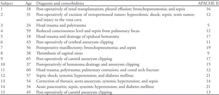
Only two patients failed to complete the study protocol. One had a relevant MBP and HR increase in the 30º inclination and was immediately returned back to 0º and improved without any other interventions. However, no variables could be recorded, so this subject was withdrawn from the study. he other patient had signiicant clonus at 50º, preventing the assessment of thoracoabdominal cirtometry and respiratory muscle strength (PImax and PEmax) at this inclination level. he patient was repositioned back into bed, and the parameters normalized. he other 13 patients completed the full protocol without reporting any discomfort or showing instability. he admission diagnoses and comorbidities are shown in table 1.
Considering neurological level and alertness (GCS and blinking relex), the patients underwent no statistically signiicant changes during the procedures, as shown in table 2.
Regarding the respiratory variables (Table 2), RR decreased at 30º and underwent a mild increase at 50º; however, these diferences were not statistically signiicant.
Chest cirtometry increased in the 30º inclination and then was decreased at 50º; however, neither change was statistically signiicant. Abdominal cirtometry showed gradually increasing abdominal circumference, with no statistical signiicance.
increase in inspiratory strength (p = 0.0218). When the inclinations were compared, a statistically signiicant increase was identiied only for the comparison between 50º and 0º (p = 0.025). PEmax’s behavior was similar to PImax’s; however, the observed increase was not statistically signiicant. VC underwent a statistically signiicant increase during the maneuver (p = 0.024) regarding the comparison of the values at 50º versus
0º (p = 0.003). he analysis of VE showed an initial
drop and then an increase; however, the changes were not statistically signiicant for the diferent angle
inclinations. Regarding TV, a statistically signiicant and gradual increase was observed during the maneuver (p = 0.012). Comparing the diferent angle inclinations, signiicant diferences were found for 30º versus 0º (p = 0.040) and 50º versus 0º (p =0.017).
Regarding the hemodynamic variables (table 2), MBP trended to increase (p = 0.051); the comparison of 50º versus 0º was statistically signiicant (p=0.016). he analysis of HR showed a gradual increase (p=0.001) for the comparisons between 30º and 0º (p=0.025), between 50º and 0º (p=0.001) and between 50º and 30º (p=0.002).
Table 2 - Neurological, respiratory and hemodynamic variables in the 0º, 30º and 50º positions
Variables 0° 30° 50° p value for the
sample
Glasgow coma scale 11 11 11 1
Blinking relex (presence) 14 14 14 1
Respiratory rate (ipm) 30.8 ± 9.2 26.6 ± 7.1 27.1 ± 6.5 0.203 Chest cirtometry (cm) 1.2 ± 0.5 1.3 ± 0.6 1.1 ± 0.5 0.631 Abdomen cirtometry (cm) 1.6 ± 1.0 1.8 ± 1 2.0 ± 1 0.110 Maximal inspiratory pressure (cm H2O) -59.7 ± 18.0 -62.2 ± 20.9 -67.1 ± 23.6* 0.021
Maximal expiratory pressure (cm H2O) 44.7 ± 30.2 47.2 ± 32.5 49.6 ± 32.5 0.244 Vital capacity (mL) 1612.1 ± 753.4 1802.1 ± 992.3 1932.8 ± 883.6* 0.024
Minute volume (mL) 11167.1 ± 3360.4 10886.4 ± 3301.6 11288.5 ± 2583.3 0.883 Tidal volume (mL) 381.4 ± 124.3 420.1 ± 137.0* 443.3 ± 144.8* 0.012
Mean blood pressure (mm Hg) 98.7 ± 17.5 102.6 ± 18.3 102.7 ± 18.3* 0.051
Heart rate (bpm) 93.1 ± 14.9 99.0 ± 16.6* 104.5 ± 18.2*/** 0.001
* p< 0.05 versus 0°; ** p< 0.05 versus 30°.
Table 1 - Demographic information
Subject Age Diagnosis and comorbidities APACHE II
1 18 Post-operatively of renal transplantation; pleural efusion; bronchopneumonia; and sepsis 16 2 31 Post-operatively of excision of retroperitoneal tumor; hypovolemic shock; sepsis; testis tumor;
and injury to the vena cava
12
3 24 Head trauma and polytrauma 5
4 59 Reduced consciousness level and sepsis from pulmonary focus 12
5 18 Head trauma and drainage of epidural hematoma 17
6 43 Post-operatively of cerebral aneurysm clipping 11 7 56 Postoperative maxillectomy; bronchopneumonia; and sepsis 19
8 38 hrombosis of sagittal sinus 9
9 65 Post-operatively of carotid aneurysm clipping 17
DISCUSSION
No changes were observed at the neurological level.
he observed changes in f, VE, PEmax, chest cirtometry
and abdomen cirtometry were not statistically signiicant. Regarding PImax and VC, a statistically signiicant increase was seen in the comparison between the 50º and 0º inclinations. However, TV increased for the comparisons between 30º and 0º and between 50º and 0º. Mean blood pressure increased only for the comparison of 50º versus 0º. Heart rate increased for all angle inclinations.
his study showed that during the procedure, there were no changes in neurological variables as assessed with the GCS and blinking relex; however, the study by Velar and Júnior reported signiicant improvement in consciousness level and alertness in patients undergoing 70º orthostatism for approximately 20 seconds. According to these authors, this result occurred because the alert systems were less stimulated
than during orthostatism.(8)
Our HR indings diverged from those in the literature. In this study, no diferences were found for this variable,
in agreement with previous studies;(10-12) however,
all of the previous studies were conducted in healthy subjects. However, Gisolf studied orthostatism with the support of nine mathematical models simulating the cardiorespiratory system and found reduced HR after ive minutes in an orthostatic position.(13) In contrast, in
the study by Chang et al., in 15 intensive care patients, HR increased with 70º inclination maintained for
ive minutes.(9) Bourdin et al. assessed the ICU’s most
frequently acquired physiotherapy measurements, as well as orthostatism’s efects, in 20 critically ill patients and reported a statistically signiicant HR increase
during passive orthostatism.(14)
Regarding cirtometry, no changes in chest or abdominal circumference were detected. We could not ind any articles in the literature reporting on this variable during passive orthostatism. In a study in which respiratory mechanics were assessed in 28 mechanically ventilated patients, it was found that the more verticalized the patient was, higher pulmonary compliance and expansibility were. his inding was ascribed to a smaller dependent zone in this position, assuming that free zones are more compliant and
thus favoring respiratory mechanics.(15) Another study
reported on thoracic and abdominal expansibility, both in supine and active orthostatic positions, in healthy subjects. horacic expansion was greater
during orthostatism, and abdominal expansion was greater in the supine position. his change was likely due to downward movement of the viscera, allowing for
improved thoracic expansion.(10)
his study showed statistically signiicant diferences only for PImax and not for PEmax. In a study by Fiz et al. assessing PImax and PEmax in supine, seated and standing positions in 10 obese patients and 10 normal body mass index subjects, it was observed that the values were higher for the standing position in comparison to other positions; therefore, they were in agreement with
our PImax increase.(16) In another study by Roquejani
et al., the inluence of body position was assessed on the respiratory muscle strength of healthy subjects. Orthostatic position was not assessed; however, more verticalized positions showed no diferences for PImax
and PEmax in comparison with other positions.(17)
he analysis of VC showed statistically signiicant increases when the 50º and 0º inclinations were compared, in agreement with other authors’ indings. Already in 1927, Wilson suggested that VC decreased due to a pulmonary congestion in the supine position and the diaphragm elevation decreased due to the pressure of abdominal viscera, consequently reducing
the volume of the chest.(18) Blair and Hickman described,
in their study of 11 healthy subjects, that residual volume (RV), residual functional capacity (RFC) and total pulmonary capacity (TPC) were signiicantly reduced when changing from an orthostatic to a supine
position.(19) In the same article, the authors stated that
these changes apparently resulted from progressive diaphragm elevation, presumed to occur due to the pressure of the abdominal viscera and decreased rest
diaphragm tonus in the supine position.(19) During the
irst studies of human neural drive to the diaphragm, it was identiied that muscle activity was increased by an average of four- to ive-fold when the subjects changed
from supine to orthostatic positions.(20) his inding
means that the neural drive to the diaphragm likely increases considerably to compensate for diferent loads on the diaphragm in diferent positions, perhaps due to increased diaphragm proprioceptive relexes stimulating the subjects to breath more deeply, increasing the VC.(10,21)
In our study, VE showed an initial decrease,
followed by an increase, with orthostatism; however, this diference was not signiicant. In agreement with
this study, Butler et al.(10) found no diference for V
E
units were studied separately, aiming to assess the neural activity of the diaphragm and the intercostal muscles in the supine position; no change was found for the frequency of discharge in either muscle of the
motor units.(10) However, in other studies in which V
E
was compared between diferent positions, increased
values were found during orthostatism.(9,11-13)
Regarding TV, this study showed a statistically
signiicant increase with time between 30º and 0º and between 50º and 0º. In a case reported by Dean and Ross, a mechanically ventilated patient started training on an orthostatic board during the ifth day after ICU admission. he patient was initially positioned at 15º for ive minutes and then at 45º for 10 minutes. he study identiied improved ventilation parameters and increased pulmonary volumes using radiographic analysis; however, the lack of quantitative data prevented the determination
of the efects of the position on ventilation.(22) Other
authors have described similar results.(9,11-13) However,
the mechanics behind the ventilation change are not fully understood.
According to Yoshizaki,(11) the aferent signs that
originate from the lower limbs and that are related to maintaining posture are activated during orthostatism. his information is projected to the respiratory center in the brain, resulting in increased ventilation. In his study, contraction of the gastrocnemius muscle, detected by electroneuromyography, supported this possibility.(11)
he efort necessary to support the position on the orthostatic board can also increase energy expenditure,
thereby increasing ventilation.(23) However, some studies
have shown that this increased O2 consumption occurs
when the subject is not irmly ixed to the board, resulting in increased muscle activity.(11)
Another possible mechanism for the TV increase is that changing from a supine to an orthostatic position may increase RFC and diaphragm mobility, lowering the abdominal contents. his RFC change modiies the point at which each tidal volume occurs on the pressure-volume curve, resulting in increased respiratory system compliance and implying higher inspired volumes in an orthostatic position.(9,12)
Orthostatic positioning may promote increased ventilation due to vestibular stimulation. Vestibular nerve stimulation in anesthetized cats was reported to increase phrenic nerve and respiratory muscle activity, indicating that there is some vestibular stimulation in
breathing.(24) However, in human models, vestibular
stimulation failed to show ventilation efects.(25)
In addition, several types of chest wall receptors have
been reported to be responsible for ventilation increases. In the supine position, C pulmonary ibers are activated as a result of the increased volume of pulmonary blood, leading to tachypnea and reduced alveolar ventilation. However, intercostal muscles are activated during inclination and might induce hyperventilation
(increased RR and TV).(26) he patient’s alertness during
orthostatism might also support the theory of increased
ventilation;(9) however, in our study, the alertness level
did not change with orthostatism.
Finally, the inclination procedure increases the patient’s anxiety, thereby stimulating sympathetic activity. his process may have efects on breathing. Sympathetic nerve stimulation is known to cause
bronchodilation, increase RR and reduce TV.(27) In
this study, the respiratory response to orthostatism was characterized by increased TV and unchanged RR during the intervention, suggesting that other factors might inluence ventilation, as sympathetic stimulation alone would have reduced TV, as previously reported in healthy young subjects. Similarly, the increased MBP should have been associated with reduced RR and TV in response to increased stimulation of arterial
baroreceptors;(28) therefore, it was not in agreement with
this study’s indings. here is evidence that increased aferent signs from baroreceptors inhibit breathing
via an integrated central mechanism.(28) However, in
disagreement with this study, the trial by Buttler found no TV or neural drive changes in the respiratory muscles during active orthostatism.(10)
In agreement with the literature, HR and MBP showed statistically signiicant increases with inclination.(29,30)
there is peripheral vasoconstriction when the body is
changed from a supine to orthostatic position.(30)
Gravitational stress is also responsible for increasing the secretion of hormones such as noradrenalin, adrenalin and aldosterone, contributing to increases in
HR and MBP.(29) Orthostatism can also cause a signiicant
reduction of cardiac vagal activity in comparison with the supine position. In contrast, sympathetic activity can signiicantly increase during elevation. hese indings support the provisional hypothesis regarding the inluence of body posture on autonomic nervous system regulation. It could be said that orthostatism induces increased sympathetic tonus and decreased parasympathetic tonus.(30)
his study has limitations that include its small sample size, which was due to diiculty in screening subjects who it the inclusion criteria. his limitation impaired the observation of more subtle changes in the assessed parameters. he exclusion of two subjects from some variables’ analyses as a result of instability might also have contributed to the lack of signiicant changes in respiratory parameters, which failed to show relevant changes. Additionally, the patients were not assessed after the intervention; therefore, it is not possible to say for how long the changes were maintained.
CONCLUSION
It is concluded that orthostatism does not change consciousness level or alertness; however, it provides improved TV, VC, and PImax, and it increases HR and MBP in critically ill patients who are restricted to bed but who have clinical conditions that allow the maneuver.
RESUMO
Objetivo: Analisar o nível de consciência, efeitos pulmona-res e hemodinâmicos em pacientes intensivos durante a posição ortostática.
Métodos: Estudo realizado de abril de 2008 a julho de 2009 na unidade de terapia intensiva adulto do HC-UNICAMP. Foram incluídos quinze pacientes que estiveram mecanicamente ventila-dos por mais de sete dias; traqueostomizaventila-dos; em nebulização inter-mitente; pressão inspiratória máxima inferior a -25cmH2O; índice de Tobin inferior a 105; drive ventilatório preservado, ausência de sedativos; pressão parcial de oxigênio arterial maior que 70mmHg; saturação de oxigênio maior que 90% e estabilidade hemodinâmi-ca. Os parâmetros avaliados, nas inclinações de 0°, 30° e 50°, foram o nível de consciência; relexo de blinking; cirtometria tóraco-abdo-minal; capacidade vital; volume corrente; volume minuto ; força da musculatura respiratória e sinais vitais.
Resultados: Não houvealteração do nível neurológico. A fre-qüência respiratória (f) e VE reduziram-se em 30° com posterior aumento em 50°, no entanto, essas alterações não foram estatistica-mente signiicativas. A cirtometria abdominal e a pressão expiratória máxima apresentaram aumento, novamente sem signiicância esta-tística. Em relação à pressão inspiratória máxima e a capacidade vital observou-se aumento estatisticamente signiicante na comparação entre as angulações 50º e 0°. Já o volume corrente aumentou ao longo do tempo, na comparação entre as angulações 30º e 0°, e entre 50º e 0°. A pressão arterial média sofreu incremento somente na compara-ção entre 50° e 0°. A freqüência cardíaca elevou-se ao longo do tempo e quando comparada entre 30°e 0°, 50° e 0°, e 50° e 30°.
Conclusão: O ortostatismo passivo proporcionou melhora do volume corrente, capacidade vital , pressão inspiratória máxima, e aumento da frequência cardíaca e pressão arterial média em pacien-tes críticos.
Descritores: Serviço hospitalar de isioterapia; Reabilitação; Unidades de terapia intensiva
REFERENCES
1. Adam S, Forrest S. ABC of intensive care: other supportive care. BMJ. 1999;319(7203):175-8. Review.
2. Perme C, Chandrashekar R. Early mobility and walking program for patients in intensive care units: creating a standard of care. Am J Crit Care. 2009;18(3):212-21. 3. Jerre G, Silva TJ, Beraldo MA, Gastaldi A, Kondo C, Leme
F, et al. Fisioterapia no paciente sob ventilação mecânica. J Bras Pneumol. 2007;33(Supl 2): 142-50.
4. Needham DM. Mobilizing patients in the intensive care unit: improving neuromuscular weakness and physical function. JAMA. 2008;300(14):1685-90.
5. Morris PE, Herridge MS. Early intensive care unit mobility: future directions. Crit Care Clin. 2007;23(1): 97-110.
6. Chang AT, Boots R, Hodges PW, Paratz J. Standing with assistance of a tilt table in intensive care: a survey of Australian physiotherapy practice. Aust J Physiother. 2004;50(1):51-4.
7. Luque A, Martins CGG, Silva MSS, Lanza FC, Gazzotti MR. Prancha ortostática nas unidades de terapia intensiva da cidade de São Paulo. Mundo Saúde.
8. Vellar CM, Forti Júnior G. Ortostatismo passivo em pacientes comatosos na UTI – um estudo preliminar. Rev Neurociênc. 2008;16(1):16-9.
9. Chang AT, Boots RJ, Hodges PW, homas PJ, Paratz JD. Standing with the assistance of a tilt table improves minute ventilation in chronic critically ill patients. Arch Phys Med Rehabil. 2004;85(12):1972-6.
frequencies of single motor units in human diaphragm and parasternal muscles in lying and standing. J Appl Physiol. 2001;90(1):147-54.
11. Yoshizaki H, Yoshida A, Hayashi F, Fukuda Y. Efect of posture change on control of ventilation. Jpn J Physiol. 1998;48(4):267-73.
12. Chang AT, Boots RJ, Brown MG, Paratz JD, Hodges PW. Ventilatory changes following head-up tilt and standing in healthy subjects. Eur J Appl Physiol. 2005;95(5-6):409-17.
13. Gisolf J, Wilders R, Immink RV, van Lieshout JJ, Karemaker JM. Tidal volume, cardiac output and functional residual capacity determine end-tidal CO2 transient during standing up in humans. J Physiol. 2003;554(Pt 2):579-90. 14. Bourdin G, Barbier J, Burle JF, Durante G, Passant S,
Vincent B, et al. he feasibility of early physical activity in intensive care unit patients: a prospective observational one-center study. Respir Care. 2010;55(4):400-7.
15. Porto EF, Castro AAM, Leite JRO, Miranda SV, Lancauth A, Kumpel C. Análise comparativa da complacência do sistema respiratório em três diferentes posições no leito (lateral, sentada e dorsal) em pacientes submetidos à ventilação mecânica invasiva prolongada. Rev Bras Ter Intensiva. 2008;20(3):213-9.
16. Fiz JA, Aguilar X, Carreres A, Barbany M, Formiguera X, Izquierdo J, Morera J. Postural variation of the maxi mum inspiratory and expiratory pressures in obese patients. Int J Obes. 1991;15(10):655-9.
17. Roquejani AC, Araújo S, Oliveira RARA, Dragosavac D, Falcão ALE, Terzi RGG, Kousour C. Inluência da posição corporal na medida da pressão inspiratória máxima (PImáx) e da pressão expiratória máxima (PEmáx) em voluntários adultos sadios. Rev Bras Ter Intensiva. 2004;16(4):215-8. 18. Wilson WH. he Inluence of posture on the volume of
the reserve air. J Physiol. 1927;64(1):54-64.
19. Blair E, Hickam JB. he efect of change in body position on lung volume and intrapulmonary gas mixing in normal
subjects. J Clin Invest. 1955;34(3):383-9.
20. Druz WS, Sharp JT. Activity of respiratory muscles in upright and recumbent humans. J Appl Physiol. 1981;51(6):1552-61.
21. Druz WS, Sharp JT. Electrical and mechanical activity of the diaphragm accompanying body position in severe chronic obstructive pulmonary disease. Am Rev Respir Dis. 1982;125(3):275-80.
22. Dean E, Ross J. Oxygen transport: he basis for contemporary cardiopulmonary physical therapy and its optimization with body positioning and mobilization. Phys her Pract. 1992;1:34-44.
23. Miyamoto Y, Tamura T, Himura T, Nakamura T, Higuchi J, Mikami T. he dynamic response of the cardiopulmonary parameters to passive head-up tilt. Jpn J Physiol. 1982;32(2):245-58.
24. Rossiter CD, Hayden NL, Stocker SD, Yates BJ. Changes in outlow to respiratory pump muscles produced by natural vestibular stimulation. J Neurophysiol. 1996;76(5):3274-84.
25. Lee CM, Wood RH, Welsch MA. Inluence of head-down and lateral decubitus neck lexion on heart rate variability. J Appl Physiol. 2001;90(1):127-32.
26. Duron B. Postural and ventilatory functions of intercostals muscles. Acta Neurobiol Exp (Wars). 1973;33(1):355-80. 27. Bechbache RR, Chow HH, Duin J, Orsini EC. he
efects of hypercapnia, hypoxia, exercise and anxiety on the pattern of breathing in man. J Physiol. 1979;293:285-300. 28. Nishino T, Honda Y. Changes in pattern of breathing
following baroreceptor stimulation in cats. Jpn J Physiol. 1982;32(2):183-95.
29. László Z, Rössler A, Hinghofer-Szalkay HG. Cardiovascular and hormonal changes with diferent angles of head-up tilt in men. Physiol Res. 2001;50(1):71-82.