Positive end-expiratory pressure increases strain in
patients with ALI/ARDS
Pressão expiratória inal positiva aumenta o estiramento em
pacientes com LPA/SDRA
INTRODUCTION
Mechanical ventilation may induce or worsen lung injury by causing overdistension and by the cyclic recruitment and derecruitment (R/D) of unstable alveoli.(1-5) Experimental data have shown that using positive
end-expiratory pressure (PEEP) and a low tidal volume may protect against ventilator-induced lung injury (VILI).(6,7) The protective role of a low tidal
volume has been confirmed in large clinical trials of patients with acute lung injury and acute respiratory distress syndrome (ALI/ARDS).(8-11)
However, while recent trials have demonstrated that high levels of PEEP decrease refractory hypoxemia, they have not demonstrated any mortality benefit.(3,12-14)
Recently, various authors have proposed that stress and strain on the lung
Guillermo Bugedo1, Alejandro
Bruhn1, Tomás Regueira1, Carlos
Romero1, Jaime Retamal1, Glenn
Hernández1
1. Pontiicia Universidad Católica de Chile. Santiago, Chile.
ABSTRACT
Objective: he objective of this study was to assess the efects of positive end-expiratory pressure on recruitment, cyclic recruitment and derecruitment and strain in patients with acute lung injury and acute respiratory distress syndrome using lung computed tomography.
Methods: his is an open, controlled, non-randomized interventional study of ten patients with acute lung injury and acute respiratory distress syndrome. Using computed tomography, single, basal slices of the lung were obtained during inspiratory and expiratory pauses at a tidal volume of 6 ml/kg and a positive end-expiratory pressure of 5, 10, 15 and 20 cmH2O. he densities of the lung parenchyma were measured in Hounsield units. he values for positive end-expiratory pressure-induced recruitment, cyclic recruitment and derecruitment and strain were then calculated.
Results: Increasing levels of positive
end-expiratory pressure were correlated with increased recruitment and global strain (p < 0.01), which was signiicantly correlated with plateau pressure (r2 =
0.97, p < 0.01). In addition, increasing levels of positive end-expiratory pressure systematically increased strain along the sternovertebral axis.
Conclusion: While strain is an adverse efect of positive end-expiratory pressure, the decision use positive end-expiratory pressure with any patient should be balanced against the potential beneits of recruitment. Due to the small number of patients in this study, the present data should be treated as hypothesis generating and is not intended to limit the clinical application of a high level of positive end-expiratory pressure in patients with severe hypoxemia.
Keywords: Positive end-expiratory pressure; Respiration, artiicial; Respiratory distress syndrome, adult; Tomography, x-ray computed
his study was conducted in the Departamento de Medicina Intensiva, Pontiicia Universidad Católica de Chile - Santiago, Chile.
Funding: Departamento de Medicina Intensiva, Pontiicia Universidad Católica de Chile, and Fondo Nacional de Desarrollo Cientíico y Tecnológico (FONDECyT) 11060350.
Conlicts of interest: None.
Submitted on November 29, 2011 Accepted on February 22, 2012
Corresponding author:
Guillermo Bugedo
PO Box 114-D, Santiago, Chile. Postal Code: 6510260
parenchyma is the ultimate cause of VILI.(4,15,16) Stress is
measured by transpulmonary pressures, which are largely heterogeneous along diferent lung regions. Strain refers to alveolar cell deformation that is induced by changes in transpulmonary pressures. Several approaches have been proposed to assess strain at a patient’s bedside. he group of Gattinoni deined strain as the relative diference between the gas volume at end inspiration and the resting volume of the lung.(15,17) Although this deinition does not account
for alveolar surface area, which can vary depending on the degree of recruitment, it can be easily used in conjunction with computed tomography as a surrogate for clinical strain.
By recruiting unstable alveoli, high levels of PEEP improve oxygenation and may protect against cyclic R/D. However, high levels of PEEP can also increase strain across the lung parenchyma.(3,18,19) he objective of the
present study was to assess the efects of PEEP on strain, recruitment and cyclic R/D in patients with ALI/ARDS.
METHODS
his is an open, controlled, non-randomized, interventional CT-scan study of ten patients with ALI/ARDS who were mechanically ventilated. he institutional ethics committee approved the study protocol, and written informed consent was obtained from each patient’s next of kin. While under continuous infusions of midazolam and morphine, patients were transferred to the CT-scan facility room and connected to a Siemens 900-C ventilator (Siemens-Elema AB, Sweden). Pancuronium 0.1 mg/kg was administered before the procedure to avoid increased ventilatory eforts. Prior to initiating the protocol, gentle tracheal suctioning was followed by a short recruiting maneuver, which consisted of increasing the PEEP above 20 cmH2O to obtain plateau pressures between 40 and 45 cmH2O for a period of one minute. Continuous electrocardiogram monitoring was performed, and invasive blood pressure and oxygen saturation parameters were continuously measured with a portable monitor (LightSolo, Datex-Ohmeda, Helsinki, Finland).
CT- scan protocol
A conventional CT scan (GE Light Speed QX/I, GE Medical Systems, Wisconsin, USA) was performed from the neck to the lung bases and took 8-mm thick slices during an inspiratory pause. he matrix was 512 x 512, which gives a voxel size of approximately 2 –3 mm3. A single
CT slice approximately 2–3 cm above the diaphragmatic dome, arbitrarily selected by the corresponding author
(GB) to avoid the appearance of the diaphragm even at the lowest pressures, was used for the entire protocol.
Patients were on volume-controlled ventilation throughout the study. Ideal body weight was calculated according to the ARDSnet study.(10) Baseline parameters
included a tidal volume of 6 ml/kg, a PEEP of 10 cmH2O, a respiratory rate of 25 and a FiO2 set to obtain oxygen saturation greater than 92%. Single CT sections were taken both at expiratory and inspiratory pauses at a PEEP of 10, 5, 15 and 20 cmH2O following the stabilization of mean airway pressures. his task was generally accomplished after 45 to 60 seconds, so each PEEP level was usually completed within two minutes. Mean airway pressure, plateau pressure (Ppl) and expiratory tidal volume (Vt) were measured by the ventilator at each PEEP level. Static compliance (Cst) was calculated at a PEEP of 10 cmH2O, given that Cst = Vt / Ppl – PEEP.
Image processing
he CT images were downloaded to an optical disk and processed using the Maluna® (Mannheim, Germany) software.(20) We used methods similar to those
of other CT studies, describing the lung parenchyma according to its density in Hounsield units (HU).(21,22)
First, the lung contour on each image was traced, and then, the total CT volume (TVCT) and average density (HCT) were measured.
With these data, we obtained lung weight (WCT) and gas volume (GVCT) values for each lung slice:
GVCT = TVCT x (HCT / −1000)
WCT = TVCT − GVCT.
Hyperinlated tissue was deined as -1000 to -901 HU, normally aerated tissue as -900 to -501 HU, poorly aerated tissue (PAT) as -500 to -101 HU and non-aerated tissue (NAT) as -100 to +100 HU.(21) For
data analysis, we used the weight (expressed in grams) of each compartment.
Recruitment induced by the increase in PEEP was deined as the decrease in NAT at expiration compared
with that at a PEEP of 5 cmH2O. his value was
expressed as a percentage of the lung slice weight:
Recruitment = 100 x (NAT exp PEEP 5 - NAT exp PEEP x) / total weight exp PEEP 5
Strain was calculated according to Valenza et al.:(23)
Strain = (GVCT end insp PEEP x - GVCT end exp PEEP x) / GVCT end-exp PEEP 5
R/D = 100 x [(NAT end exp / total weight exp) – (NAT end insp / total weight insp)]
Recruitment, strain and cyclic R/D were calculated for each PEEP level both for the entire slice (referred to as global) and regionally by splitting the slice into ten compartments along the sternovertebral axis.
Statistical analysis
Clinical characteristics and ventilatory data are presented as the mean ± SD. Comparisons in the amount of gas and tissue at diferent levels of PEEP were evaluated by ANOVA for repeated measures or by paired t-test. Linear regression was used to correlate strain with airway pressures and recruitment with R/D. he level of statistical signiicance was set at p < 0.05.
RESULTS
Ten patients (4 M, 6 F, 51 ± 16 y.o.) with ALI/ARDS (Pa/FiO2 80–285) were included in the study. Clinical characteristics and baseline parameters are shown in table 1. he distribution of lung volumes and plateau and mean airway pressures at diferent levels of PEEP are shown in igure 1.
Recruitment and cyclic R/D
In all patients, recruitment increased signiicantly with increasing levels of PEEP (Table 2). However, cyclic R/D was highly variable with increasing levels
of PEEP, though overall a decreasing trend with higher PEEP levels was observed (p = 0.056; Figure 2 and Table 2). Regional R/D was predominant in the middle levels and decreased when the PEEP level increased to 20 cmH2O (Figure 3).
We found no correlation between PEEP-induced
Table 1 - Characteristics of the patients
Gender (M/F)
Age (Years)
Diagnosis Type of
ARDSa
APACHE II Pa/FiO2 Day on
MV
Oxygenation index
Static compliance (ml/cmH2O)
Outcome
M 52 Pneumonia P 22 128 2 11.7 39.6 D
F 24 Pancreatitis EP 15 84 1 27.5 18.9 S
F 72 Pneumonia P 24 232 1 7.8 27.1 D
F 64 Postoperative pneumonia P 15 285 5 6.3 21.7 S
F 37 Peritonitis EP 18 183 3 8.6 16.9 S
F 60 Pneumonitis P 18 80 5 27.5 31.1 D
M 57 Pneumonia P 14 193 8 6.4 46.5 S
F 30 Peritonitis EP 12 173 2 8.7 25.6 S
M 67 Postoperative pneumonia P 11 275 1 5.8 38.1 S
M 51 Pneumonia P 17 128 12 16.5 26.9 S
4M/6F 51 ± 16 16 ± 4 181 ± 80 4.0 ± 3.6 13.3 ± 4 29.2 ± 9.6
a Type of ALI/ARDS according Gattinoni L, Pelosi P, Suter PM, Pedoto A, Vercesi P, Lissoni A. Acute respiratory distress syndrome caused by
pulmonary and extrapulmonary disease. Diferent syndromes? Am J Respir Crit Care Med. 1998;158(1):3-11.
P- pulmonary; EP - extrapulmonary; ARDS - acute respiratory distress syndrome; APACHE II - acute lung injury/acute respiratory distress syndrome; Pa/FiO2 - oxygen partial pressure/oxygen inspiratory fraction; MV- mechanical ventilation; S - survival; D - death. Values are given as the mean ± SD.
recruitment and changes in cyclic R/D (delta R/D) when increasing PEEP from 5 to 20 cmH2O (Figure 4).
Strain
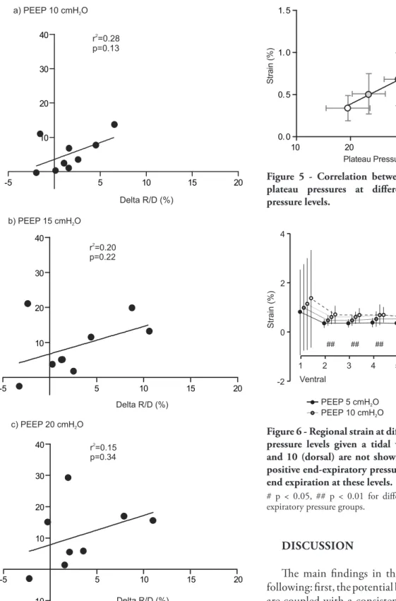
Global strain increased with higher levels of PEEP (p < 0.01). Global strain at various PEEP
levels was highly correlated with plateau pressure (r2 = 0.97, p < 0.01) (Figure 5). Regional strain
at a PEEP of 5 cmH2O was distributed evenly
along the sternovertebral axis. Increasing PEEP levels systematically increased strain along the sternovertebral axis (Figure 6).
Table 2 - Weight (g) of each lung compartment during expiration and inspiration at diferent positive end-expiratory pressure levels
PEEP Cycle Hyperinlated
(-1000 to -901)
Normal aeration (-900 to -501)
Poorly aerated (-500 to -101)
Non-aerated (-100 to +100)
Total weight R/Da
(expiration-inspiration)
Recruitmentb
(versus PEEP 5)
5 Expiration 0.10 ± 0,24 14.40 ± 7.04 10.75 ± 4.34 28.27 ± 14.32 53.52 ± 16.77 6.04 ± 5.83% Baseline
Inspiration 0.16 ± 0.36 14.87 ± 7.48 12.63 ± 3.97 23.67 ± 11.26 51.33 ± 16.34
10 Expiration 0.09 ± 0.23 14.56 ± 6.98 12.63 ± 4.16 24.15 ± 13.05* 51.44 ± 15.78 4.16 ± 4.84% 4.13 ± 3.46%
Inspiration 0.16 ± 0.24 14.06 ± 7.74 14.13 ± 3.97 20.65 ± 10.50 48.98 ± 15.18
15 Expiration 0.15 ± 0.27 14.26 ± 7.88 13.46 ± 4.04 22.03 ± 12.45* 49.90 ± 15.69* 3.16 ± 6.04% 6.70 ± 6.29%*
Inspiration 0.35 ± 0.49 13.25 ± 8.57 14.22 ± 3.84 19.05 ± 10.66 46.87 ± 15.82
20 Expiration 0.27 ± 0.44 14.50 ± 9.25 15.13 ± 3.13 20.43 ± 11.75* 50.33 ± 15.88 2.27 ± 2.75% 8.92 ± 9.18%*
Inspiration 0.53 ± 0.74* 13.32 ± 8.97 15.23 ± 2.92 18.16 ± 11.18 47.24 ± 16.34
a Recruitment and derecruitment (R/D) is the fractional decrease in non-aerated tissue between inspiration and expiration at each positive
end-expiratory pressure level.
b Recruitment is the decrease in non-aerated tissue at expiration compared to a positive end-expiratory pressure of 5 cmH2O.
* p < 0.05 versus a positive end-expiratory pressure of 5. All data are given as the mean ± SD. Also shown are the mean ± SD values for opening and closing, and expiratory recruitment a,b.
PEEP - positive end-expiratory pressure.
Figure 2 - Global cyclic recruitment and derecruitment at diferent positive end-expiratory pressure levels. here were no signiicant diferences between the groups.
Figure 3 - Regional distribution of cyclic recruitment and derecruitment at diferent positive end-expiratory pressure levels given a tidal volume of 6 ml/kg.
DISCUSSION
he main indings in the present study include the following: irst, the potential beneits of PEEP recruitment are coupled with a consistent increase in strain; second, global strain, a physical measure of alveolar stretching, was closely related to plateau pressure. However, due to the small number of patients in the study, the present data should be treated as hypothesis generating and not intended to limit the clinical application of a high level of PEEP in patients with severe hypoxemia.
Figure 4 - Correlation between the efects of positive end-expiratory pressure on recruitment and cyclic recruitment and derecruitment (R/D) at positive end-expiratory pressure levels of 10, 15 and 20 cmH2O. Recruitment and delta R/C were calculated relative to a positive end-expiratory pressure of 5 cmH2O.
Figure 5 - Correlation between strain and corresponding plateau pressures at diferent positive end-expiratory pressure levels.
Figure 6 - Regional strain at diferent positive end-expiratory pressure levels given a tidal volume of 6 ml/kg. Levels 9 and 10 (dorsal) are not shown because most patients, at a positive end-expiratory pressure of 5 cmH2O, had no gas at end expiration at these levels.
Current literature on ventilator-induced lung injury suggests that lung injury is primarily caused by overdistension and cyclic R/D.(4,5) he role of PEEP
in preventing VILI is controversial, as only tidal-volume reduction has demonstrated a clear impact on the survival of patients with ALI/ARDS.(10,13,14) A
recent meta-analysis suggested that higher levels of PEEP, beyond those that are used to decrease refractory hypoxemia, might have a survival beneit in patients with ARDS.(24)
In patients with ALI/ARDS, Caironi et al. demonstrated that high levels of PEEP decreased cyclic R/D in patients with highly recruitable lungs but increased strain independent of lung recruitment.(25) In
their study, cyclic R/D was not directly measured but estimated from hypothetical curves that were derived from previous studies.(26) By measuring non-aerated
tissue both at expiration and inspiration, we were able to directly measure recruitment, cyclic R/D, and strain. hus, we demonstrated that, in all patients, an increase in the levels of PEEP decreases the amount of non-aerated tissue at end expiration (recruitment) but also increases strain. he efect of PEEP on cyclic R/D was very heterogeneous, although a trend toward less cyclic R/D with higher levels of PEEP levels was observed. he most likely explanation for this inding is type-II error due to our small sample size of non-selected ALI/ ARDS patients.
Another important clinical inding derived from this protocol includes that global strain, a physical measure of alveolar stretching, was closely related to plateau pressure. We recognized that transpulmonary pressure, and not plateau pressure, is the distending force of the lung.(27) However, as the measurement of
transpulmonary pressures is complicated and is not routinely performed in most ICUs worldwide, plateau pressure may still have clinical value for assessing strain at the bedside. Although we cannot suggest a threshold value for plateau pressure, it appears logical, based on experimental and clinical studies, that it should be a highly controlled variable when ventilating patients with ALI/ARDS. Our data are consistent with the data from Hager et al., who utilized data from the ARDSnet trial, and demonstrated that mortality increased linearly with plateau pressure.(28)
Several human studies have reported the simultaneous onset of alveolar recruitment and overdistension in patients with ALI/ARDS at PEEP levels ranging from 10 to 20 cmH2O.(18,29,30) he linear
correlation we observed between strain and plateau
pressure (Figure 5) might be due to another variable because recruitment and overdistension were not observed, and both phenomena occurred in the same proportion.(31) However, given the small number of
patients and values for plateau pressure, we are unable to generalize with regard to these data. Moreover, even in healthy lungs, the strain-pressure relationship is not linear.(32)
Whether strain is more important than cyclic R/D in inducing further alveolar damage is a topic of debate.(25) he demonstration that bullae are prevalent
in dependent regions of the lung suggests that cyclic R/D also has a major impact on VILI.(33) Recent trials
comparing higher versus lower levels of PEEP in patients with ARDS demonstrated higher plateau pressures but a similar rate of mortality in patients with higher levels of PEEP.(12-14,24) Once again, however, diferent rates
of lung recruitment among patients may explain the confounding results of these trials.(20,25,34)
Limitations of our study
First, our study included a small number of patients with ALI/ARDS who were mechanically ventilated for various periods of time, so these results should be treated as hypothesis generating and not intended to limit the clinical application of a high level of PEEP in patients with severe hypoxemia. Larger trials in patients with diferent types of ALI/ARDS,(34) based on
the measurement of inlammatory mediators, may help identify the real impact of PEEP on strain and lung function.
Second, although we did not perform a whole lung CT for every level of PEEP, it is widely accepted that a single, basal CT section adequately represents the whole lung.(21,35) Several authors have used this
approach to avoid excessive radiation exposure.(36) As
faster spiral-CT machines become available, whole-lung-CT clinical trials using different levels of airway pressure could be performed at acceptable radiation levels.(37)
hird, we compared static expiratory and end-inspiratory CT results to infer changes in density distributions that occurred during mechanical ventilation. However, this approach may have some limitations. Experimental data suggest that recruitment and derecruitment are time dependent and therefore, static images taken after prolonged inspiratory and expiratory holds may not adequately relect the lung during uninterrupted mechanical ventilation.(38,39)
has the potential to dynamically assess changes in density distribution during continuous mechanical ventilation.(40)
Finally, as previously stated, strain does not refer to gas volumes but to changes in the linear dimension of alveolar cells. In a lung that is fully open, measures of strain and gas volume may have a near-perfect correlation. In contrast, in a heterogeneous lung—as in human ARDS—this may not be totally equivalent. It would be also interesting to evaluate the impact of a stronger recruitment maneuver prior to setting the PEEP level, which may favor recruitment and decrease strain.(41)
CONCLUSIONS
Strain is systematically increased by increasing levels of PEEP and also correlates with plateau pressure. Thus, strain is clearly an adverse effect of PEEP, which for any patient should be balanced against the potential benefits of recruitment and prevention of cyclic R/D. Due to the small number of patients in this study, the present data should be treated as hypothesis generating and are not intended to limit the clinical application of a high level of PEEP in patients with severe hypoxemia.
Acknowledgments
he authors thank Pietro Caironi, Massimo Cressoni and Luciano Gattinoni for their invaluable help with the analysis of the CT images and manuscript writing.
RESUMO
Objetivo: O objetivo deste estudo foi avaliar os efeitos da pressão expiratória inal positiva no estiramento, recrutamento e recrutamento e desrecrutamento cíclico avaliados por tomogra-ia computadorizada pulmonar em pacientes com lesão pulmo-nar aguda/síndrome do desconforto respiratório agudo.
Métodos: Trata-se de um estudo aberto, controlado, não randomizado, de intervenção, em pacientes com lesão pulmonar aguda/síndrome do desconforto respiratório agudo. Foram rea-lizados cortes simples de tomograia computadorizada durante pausas inspiratórias e expiratórias com um volume corrente de 6 ml/kg e níveis de pressão expiratória inal positiva de 5, 10, 15 e 20 cmH2O. Medimos as densidades do parênquima pul-monar em unidades Hounsield e calculamos o recrutamento, recrutamento e desrecrutamento cíclico induzidos pela pressão expiratória inal positiva, assim como o estiramento.
Resultados: O aumento dos níveis de pressão expiratória inal positiva aumenta de forma consistente o recrutamento e o estiramento globais (p<0,01), o que se correlacionou de forma signiicante com a pressão de platô (r2=0,97; p<0,01). O
au-mento dos níveis de pressão expiratória inal positiva auau-mentou sistematicamente a distensão alveolar em todo o eixo esterno-vertebral.
Conclusão: A distensão alveolar é um efeito adverso da pressão expiratória inal positiva que deve ser ponderado em qualquer paciente em relação ao seus potenciais benefícios no recrutamento. Em razão do número reduzido de pacientes, estes dados devem ser considerados como geradores de hipótese e não limitar a aplicação de valores elevados de pressão expiratória i-nal positiva em pacientes com hipoxemia grave.
Descritores: Respiração com pressão positiva; Respiração artiicial; Síndrome do desconforto respiratório do adulto; Tomograia computadorizada por raios x
REFERENCES
1. Pinhu L, Whitehead T, Evans T, Griiths M. Ventilator-associated lung injury. Lancet. 2003;361(9354):332-40. Review.
2. Fan E, Needham DM, Stewart TE. Ventilatory management of acute lung injury and acute respiratory distress syndrome. JAMA. 2005;294(22):2889-96.
3. Slutsky AS, Imai Y. Ventilator-induced lung injury, cytokines, PEEP, and mortality: implications for practice and for clinical trials. Intensive Care Med. 2003;29(8):1218-21.
4. Albaiceta GM, Blanch L. Beyond volutrauma in ARDS: the critical role of lung tissue deformation. Crit Care. 2011;15(2):304.
5. Nardelli LM, Garcia CS, Pássaro CP, Rocco PR. Entendendo os mecanismos determinantes da lesão
pulmonar induzida pela ventilação mecânica. Rev Bras Ter Intensiva. 2007;19(4):469-74.
6. Dreyfuss D, Saumon G. Ventilator-induced lung injury: lessons from experimental studies. Am J Respir Crit Care Med. 1998;157(1):294-323.
7. Muscedere JG, Mullen JB, Gan K, Slutsky AS. Tidal ventilation at low airway pressures can augment lung injury. Am J Respir Crit Care Med. 1994;149(5):1327-34. 8. Amato MB, Barbas CS, Medeiros DM, Magaldi RB,
Schettino GP, Lorenzi-Filho G, et al. Efect of a protective-ventilation strategy on mortality in the acute respiratory distress syndrome. N Engl J Med. 1998;338(6):347-54. 9. Villar J, Kacmarek RM, Pérez-Méndez L, Aguirre-Jaime A.
10. Ventilation with lower tidal volumes as compared with traditional tidal volumes for acute lung injury and the acute respiratory distress syndrome. he Acute Respiratory Distress Syndrome Network. N Engl J Med. 2000;342(18):1301-8.
11. Amato MB, Carvalho CR, Vieira S, Isola A, Rotman V, Moock M, et al. Ventilação mecânica na lesão pulmonar aguda/síndrome do desconforto respiratório agudo. Rev Bras Ter Intensiva. 2007;19(3):374-83.
12. Brower RG, Lanken PN, MacIntyre N, Matthay MA, Morris A, Ancukiewicz M, Schoenfeld D, hompson BT; National Heart, Lung, and Blood Institute ARDS Clinical Trials Network. Higher versus lower positive end-expiratory pressures in patients with the acute respiratory distress syndrome. N Engl J Med. 2004;351(4):327-36. 13. Meade MO, Cook DJ, Guyatt GH, Slutsky AS, Arabi YM,
Cooper DJ, Davies AR, Hand LE, Zhou Q, habane L, Austin P, Lapinsky S, Baxter A, Russell J, Skrobik Y, Ronco JJ, Stewart TE; Lung Open Ventilation Study Investigators. Ventilation strategy using low tidal volumes, recruitment maneuvers, and high positive end-expiratory pressure for acute lung injury and acute respiratory distress syndrome: a randomized controlled trial. JAMA. 2008;299(6):637-45. 14. Mercat A, Richard JC, Vielle B, Jaber S, Osman D, Diehl
JL, Lefrant JY, Prat G, Richecoeur J, Nieszkowska A, Gervais C, Baudot J, Bouadma L, Brochard L; Expiratory Pressure (Express) Study Group. Positive end-expiratory pressure setting in adults with acute lung injury and acute respiratory distress syndrome: a randomized controlled trial. JAMA. 2008;299(6):646-55.
15. Chiumello D, Carlesso E, Cadringher P, Caironi P, Valenza F, Polli F, et al. Lung stress and strain during mechanical ventilation for acute respiratory distress syndrome. Am J Respir Crit Care Med. 2008;178(4):346-55.
16. Protti A, Cressoni M, Santini A, Langer T, Mietto C, Febres D, et al. Lung stress and strain during mechanical ventilation: any safe threshold? Am J Respir Crit Care Med. 2011;183(10):1354-62. Erratum in Am J Respir Crit Care Med. 2012;185(1):115.
17. Valenza F, Guglielmi M, Maioletti M, Tedesco C, Maccagni P, Fossali T, et al. Prone position delays the progression of ventilator-induced lung injury in rats: does lung strain distribution play a role? Crit Care Med. 2005;33(2):361-7.
18. Rouby JJ. Lung overinlation. he hidden face of alveolar recruitment. Anesthesiology. 2003; 99(1):2-4.
19. Herrera MT, Toledo C, Valladares F, Muros M, Díaz-Flores L, Díaz-Flores C, Villar J. Positive end-expiratory pressure modulates local and systemic inlammatory responses in a sepsis-induced lung injury model. Intensive Care Med. 2003;29(8):1345-53.
20. Gattinoni L, Caironi P, Cressoni M, Chiumello D, Ranieri VM, Quintel M, et al. Lung recruitment in patients with the acute respiratory distress syndrome. N Engl J Med.
2006;354(17):1775-86.
21. Gattinoni L, Caironi P, Pelosi P, Goodman LR. What has computed tomography taught us about the acute respiratory distress syndrome? Am J Respir Crit Care Med. 2001;164(9):1701-11.
22. Bugedo G, Bruhn A, Hernández G, Rojas G, Varela C, Tapia JC, Castillo L. Lung computed tomography during a lung recruitment maneuver in patients with acute lung injury. Intensive Care Med. 2003;29(2):218-25.
23. Valenza F, Guglielmi M, Irace M, Porro GA, Sibilla S, Gattinoni L. Positive end-expiratory pressure delays the progression of lung injury during ventilator strategies involving high airway pressure and lung overdistention. Crit Care Med. 2003;31(7):1993-8.
24. Briel M, Meade M, Mercat A, Brower RG, Talmor D, Walter SD, et al. Higher vs lower positive end-expiratory pressure in patients with acute lung injury and acute respiratory distress syndrome: systematic review and meta-analysis. JAMA. 2010;303(9):865-73.
25. Caironi P, Cressoni M, Chiumello D, Ranieri M, Quintel M, Russo SG, et al. Lung opening and closing during ventilation of acute respiratory distress syndrome. Am J Respir Crit Care Med. 2010;181(6):578-86.
26. Crotti S, Mascheroni D, Caironi P, Pelosi P, Ronzoni G, Mondino M, et al. Recruitment and derecruitment during acute respiratory failure: a clinical study. Am J Respir Crit Care Med. 2001;164(1):131-40.
27. Talmor D, Sarge T, Malhotra A, O’Donnell CR, Ritz R, Lisbon A, et al. Mechanical ventilation guided by esophageal pressure in acute lung injury. New Engl J Med. 2008;359(20):2095-104.
28. Hager DN, Krishnan JA, Hayden DL, Brower RG; ARDS Clinical Trials Network. Tidal volume reduction in patients with acute lung injury when plateau pressures are not high. Am J Respir Crit Care Med. 2005;172(10):1241-5. 29. Puybasset L, Gusman P, Muller JC, Cluzel P, Coriat P,
Rouby JJ. Regional distribution of gas and tissue in acute respiratory distress syndrome. III. Consequences for the efects of positive end-expiratory pressure. CT Scan ARDS Study Group. Adult Respiratory Distress Syndrome. Intensive Care Med. 2000;26(9):1215-27.
30. Dambrosio M, Roupie E, Mollet JJ, Anglade MC, Vasile N, Lemaire F, Brochard L. Efects of positive end-expiratory pressure and diferent tidal volumes on alveolar recruitment and hyperinlation. Anesthesiology. 1997;87(3):495-503. 31. Carvalho AR, Spieth PM, Pelosi P, Vidal Melo MF, Koch T,
Jandre FC, et al. Ability of dynamic airway pressure curve proile and elastance for positive end-expiratory pressure titration. Intensive Care Med. 2008;34(12):2291-9. 32. Perchiazzi G, Rylander C, Vena A, Derosa S, Polieri
D, Fiore T, et al. Lung regional stress and strain as a function of posture and ventilatory mode. J Appl Physiol. 2011;110(5):1374-83.
Fumagalli R, Tagliabue M. Lung structure and function in diferent stages of severe adult respiratory distress syndrome. JAMA. 1994;271(22):1772-9.
34. Garcia CS, Pelosi P, Rocco PR. Síndrome do desconforto respiratório agudo pulmonar e extrapulmonar: existem diferencas? Rev Bras Ter Intensiva. 2008;20(2):178-83. 35. Neumann P, Hedenstierna G. Ventilation-perfusion
distributions in diferent porcine lung injury models. Acta Anaesthesiol Scand. 2001;45(1):78-86.
36. Albaiceta GM, Taboada F, Parra D, Luyando LH, Calvo J, Menendez R, Otero J. Tomographic study of the inlection points of the pressure-volume curve in acute lung injury. Am J Respir Crit Care Med. 2004;170(10):1066-72. 37. Picano E. Informed consent and communication of risk
from radiological and nuclear medicine examinations: how to escape from a communication inferno. BMJ. 2004;329(7470):849-51.
38. Markstaller K, Eberle B, Kauczor HU, Scholz A, Bink A, helen M, et al. Temporal dynamics of lung aeration determined by dynamic CT in a porcine model of ARDS. Br J Anaesth. 2001;87(3):459-68.
39. Neumann P, Berglund JE, Mondéjar EF, Magnusson A, Hedenstierna G. Efect of diferent pressure levels on the dynamics of lung collapse and recruitment in oleic-acid-induced lung injury. Am J Respir Crit Care Med.1998;158(5 Pt 1):1636-43.
40. Bruhn A, Bugedo D, Riquelme F, Varas J, Retamal J, Besa C, et al. Tidal volume is a major determinant of cyclic recruitment-derecruitment in acute respiratory distress syndrome. Minerva Anestesiol. 2011;77(4):418-26. 41. Borges JB, Okamoto VN, Matos GF, Caramez MP, Arantes