Sepsis-related acute respiratory distress syndrome
in children with cancer: the respiratory dynamics
of a devastating condition
INTRODUCTION
Adult cancer patients with acute respiratory distress syndrome (ARDS) have a signiicantly higher risk of death compared with those without cancer. Additionally, these patients are more critically ill and are likely to have pneumonia and sepsis as a result of ARDS.(1) Adults who develop sepsis-related ARDS present PaO2/FiO2 ratios (partial pressure of oxygen in arterial blood/fraction of inspired oxygen) that are signiicantly lower than those with non-sepsis-related ARDS; they also have higher mortality at 28 and 60 days, experience
Rodrigo Genaro Arduini1, Orlei Ribeiro de Araujo1, Dafne Cardoso Bourguignon da Silva1, Andreza Almeida Senerchia2, Antonio Sergio Petrilli3
1. Intensive Care Unit, Grupo de Apoio ao Adolescente e à Criança com Câncer, Instituto de Oncologia Pediátrica, Universidade Federal de São Paulo - São Paulo (SP), Brazil.
2. Department of Clinical Research, Grupo de Apoio ao Adolescente e à Criança com Câncer, Instituto de Oncologia Pediátrica, Universidade Federal de São Paulo - São Paulo (SP), Brazil. 3. Department of Pediatric Oncology, Grupo de Apoio ao Adolescente e à Criança com Câncer, Instituto de Oncologia Pediátrica, Universidade Federal de São Paulo - São Paulo (SP), Brazil.
Objective: To evaluate the clinical
course and respiratory parameters of mechanically ventilated children with cancer sufering from sepsis-related acute respiratory distress syndrome.
Methods: his 2-year prospective,
longitudinal, observational cohort study enrolled 29 children and adolescents. Clinical data, measurements of blood gases and ventilation parameters were collected at four diferent time points. Fluctuations between measurements as well as diferences in estimated means were analyzed by linear mixed models in which death within 28 days from the onset of acute respiratory distress syndrome was the primary endpoint.
Results: here were 17 deaths within
28 days of acute respiratory distress syndrome onset and another 7 between 29 - 60 days. Only 5 patients survived for more than 60 days. Nine (31%) patients died as a direct consequence of refractory hypoxemia, and the others died of multiple organ failure and catecholamine-refractory shock. In 66%
Conflicts of interest: None.
Submitted on July 3, 2016 Accepted on October 27, 2016
Corresponding author: Orlei Ribeiro de Araujo
Grupo de Apoio ao Adolescente e à Criança com Câncer Instituto de Oncologia Pediátrica da Universidade Federal de São Paulo
Rua Botucatu, 743
Zip code: 04023-062 - São Paulo (SP), Brazil E-mail: [email protected]
Responsible editor: Jefferson Pedro Piva
Síndrome do desconforto respiratório agudo relacionada à sepse
em crianças com câncer: dinâmica respiratória de uma condição
devastadora
ABSTRACT
Keywords: Respiration, artiicial;
Respiratory distress syndrome, adult; Sepsis; Neoplasms; Child
of the measurements, the tidal volume required to obtain oxygen saturation equal to or above 90% was greater than 7mL/kg. he estimated means of dynamic compliance were low and were similar for survivors and non-survivors but with a negative slope between the irst and inal measurements, accompanied by a negative slope of the tidal volume for non-survivors. Non-survivors were signiicantly more hypoxemic, with PaO2/FiO2 ratios showing lower estimated means and a negative slope along the four measurements. Peak, expiratory and mean airway pressures showed positive slopes in the non-survivors, who also had more metabolic acidosis.
Conclusions: In most of our
children with cancer, sepsis and acute respiratory distress syndrome progressed with deteriorating ventilation indexes and escalating organic dysfunction, making this triad nearly fatal in children.
fewer intensive care unit (ICU)-free and ventilator-free days, and exhibit lower successful extubation rates.(2) Sepsis and respiratory failure account for approximately 2/3 of hemato-oncology patients admitted to the pediatric intensive care unit (PICU),(3) but little is known about the clinical course of ARDS in this group. Children with cancer who develop ARDS are extremely ill, and the mortality is unacceptably high (64.7% in one study).(4)
Despite eforts in basic and clinical research, ARDS mortality remains relatively unchanged. Various strategies have been attempted to revert hypoxemia, including recruitment maneuvers, ventilation modes, inhaled vasodilators and extracorporeal membrane oxygenation. Although these interventions improved oxygenation,
none was able to improve mortality.(5) here are no
efective therapies, and clinical tests show limited success: only the prone position and the use of low tidal volumes (TV) demonstrated consistent evidence of mortality reduction.(6) he prone position is usually applied without major diiculties, but the use of low TV depends on pulmonary conditions and is therefore not possible for all children with cancer, ARDS and sepsis.
Given the severity of the disease, its high mortality rate and the low number of studies available, it is relevant to improve the knowledge on this topic. he aim of this study was to evaluate the clinical course and respiratory parameters of mechanically ventilated children with cancer sufering from sepsis-related acute respiratory distress syndrome.
METHODS
After approval by the Ethics Committee (Universidade Federal de São Paulo - UNIFESP - Nº 0031/11) and with a waiver of informed consent, this prospective, longitudinal, observational cohort enrolled 29 children with malignant diseases and sepsis-related ARDS who required mechanical ventilation for more than 24 hours and were admitted to the PICU from February 2011 to January 2013. Sepsis was deined as systemic inlammatory response syndrome caused by suspected or proven infection,(7) sepsis-related ARDS was deined as ARDS developing in patients with sepsis,(2) and ARDS was deined according to the European American Consensus Conference criteria. he data were retrospectively analyzed to conirm the diagnosis according to the Berlin deinitions.(8) No interventions or blood sample collections were performed in these patients in addition to the usual protocol for standard care. he ventilation protocol used in the ICU follows the guidelines of the III Brazilian consensus on mechanical ventilation.(9)
Pediatric Logistic Organ Dysfunction (PELOD) scores, white blood cell counts, values of ventilator settings and measurements (peak pressure [PP], positive end-expiratory pressure [PEEP], TV per kg and mean airway pressure [MAP]) and arterial blood gases were collected at four points: (1) at the time of endotracheal intubation; (2) at the moment of the ARDS diagnosis, (3) at the lowest PaO2/FiO2 ratio throughout the whole period of mechanic ventilation; and (4) at the last blood sample analysis before the outcome. Fixed temporal variations were not established due to the risk of death at any time of clinical course. Sex, age, weight, platelet count, hemoglobin, and coagulation tests were collected at the time of ARDS diagnosis.
he oxygenation index (OI) was calculated according to Ortiz et al.: OI = FiO2 x MAP/PaO2.(10) Dynamic compliance (Cdyn) was calculated as TV/(PIP - PEEP).
Normal Cdyn values are 1.1 to 2.0mL/cmH2O/kg in
healthy infants.(11)
Data were analyzed with Statistical Package for Social Science (SPSS) v. 20.0 (IBM Corp., Armonk, NY, USA) and Minitab 17 (Minitab Inc., State College, PA, USA). Considering that blood gas data and ventilation parameters were obtained in repeated measurements, and these usually result in correlated errors, linear mixed models were used to evaluate luctuations between measurements and diferences in estimated means; the outcome “death within 28 days” (more likely related to ARDS) was the ixed efect. In mixed models, the intercept is the predicted value of the dependent variable when all of the independent variables are zero; thus, in our models, intercepts represent the estimated mean value of a measurement at the baseline, or irst measurement. Mixed models can also handle diferent temporal variations between repeated measures.(12) ROC curves were used to obtain cutof values of Pa02/FiO2 ratios and OI as predictors of survival or death within 28 days, both in the sample and in Monte Carlo simulations. A threshold for statistical signiicance was set at p < 0.05.
RESULTS
consequence of refractory hypoxemia. he others died of multiple organ failure and catecholamine-refractory shock, and no deaths could be directly attributed to cancer or hematologic disease in the study period. Two non-survivors had failure of two organs or systems in addition to the lungs, and the others had 3 or more failures of organs or systems. All patients received vasoactive drugs and sedation. hree patients received recruitment maneuvers, and the prone position was used in two (all non-survivors). High frequency oscillatory ventilation was not used. Non-survivors remained in the ICU for a mean of 20.4 days (SD 6.49), and the median time of invasive mechanical ventilation (IMV) was 11.5 days (interquartile range 25th - 75th: 9 - 14.7).
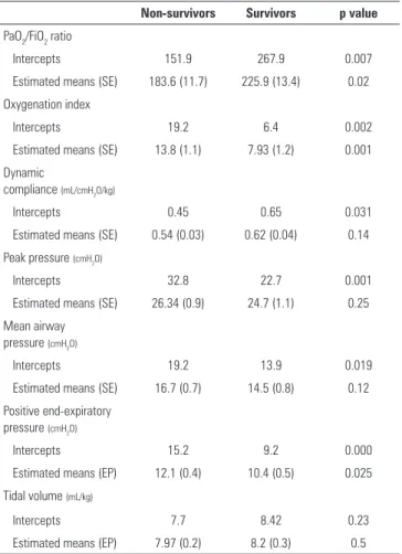
Table 2 shows data for the estimated means and intercepts observed in mixed models for ventilator settings and measurements. he PaO2/FiO2 ratio showed a signiicant diference in the intercepts and estimated means. here was also a diference in the slope, with a steep drop (-112 in the estimated mean diference) between the initial measurement observed and that corresponding to the worst PaO2/FiO2 ratio. In a logistic regression model with the worst values of PaO2/FiO2 as predictors of death within 28 days, the observed odds ratios were 0.9793 (p = 0.022); i.e., each unit increase in the ratio corresponded to an increase of 2% in the chance of survival. Values equal to or greater than 100 showed diagnostic sensitivity of 75% for survival within 28 days as well as a speciicity of 70%, with an area under the ROC curve (AUC) of 0.8; p = 0.005. In a Monte Carlo simulation with 10,000 patients in each group, constructed from a random distribution based on the values of the sample, the sensitivity was 56% with a speciicity of 85% and AUC = 0.77 (p < 0.0001).
he oxygenation index showed a signiicant diference in the intercepts and estimated means. he slope was positive, with a signiicant mean diference of +12.7 between the irst and last measurement (p = 0.009) for non-survivors. One value equal to or greater than 14.5 showed diagnostic sensitivity of 69% to death within 28 days as well as speciicity of 83%, with an area under the ROC curve of 0.83; p = 0.003. In a Monte Carlo simulation with 10,000 patients in each group, sensitivity was 73% with a speciicity of 53% and AUC = 0.73 (p < 0.0001).
here were signiicant diferences in the intercepts for peak pressures and MAP, but not in the estimated means. he slope was positive for PP, with a signiicant mean diference of +11.7cmH20 between the initial and inal measurements (p = 0.007) for the non-survivors. he
mean diference in MAP (+6cmH20 between the initial
and inal measurement) was not signiicant (p = 0.059).
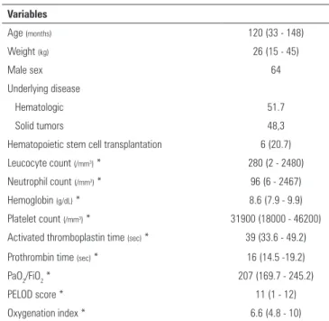
Table 1 - Demographic and clinical data
Variables
Age (months) 120 (33 - 148)
Weight (kg) 26 (15 - 45)
Male sex 64
Underlying disease
Hematologic 51.7
Solid tumors 48,3
Hematopoietic stem cell transplantation 6 (20.7)
Leucocyte count (/mm3) * 280 (2 - 2480)
Neutrophil count (/mm3) * 96 (6 - 2467)
Hemoglobin (g/dL) * 8.6 (7.9 - 9.9)
Platelet count (/mm3) * 31900 (18000 - 46200)
Activated thromboplastin time (sec) * 39 (33.6 - 49.2)
Prothrombin time (sec) * 16 (14.5 -19.2)
PaO2/FiO2 * 207 (169.7 - 245.2)
PELOD score * 11 (1 - 12)
Oxygenation index * 6.6 (4.8 - 10)
PaO2/FiO2 - partial pressure of oxygen in arterial blood/fraction of inspired oxygen; PELOD - pediatric logistic organ dysfunction. Values are listed as medians and interquartile ranges (25-75), (%) and N (%).* at acute respiratory distress syndrome diagnosis.
Tidal volumes per kg were similar both in the intercepts and estimated means for non-survivors and survivors, but there was a negative slope between the initial measurement and that corresponding to the worst PaO2/FiO2 ratio (9 and 7.37, p = 0.003). Among the measurements, 55% were performed in pressure controlled ventilation, while 45% were volume controlled. Figure 1 illustrates the observation that in 66% of the measurements, the TV required to obtain oxygen saturation equal to or above 90% was greater than 7mL/kg.
here was a positive slope (mean diference of +3.13cmH20, p = 0.002) between the irst and inal PEEP measurement for the non-survivors. Figures 2 to 4 show plots of ventilator parameters and oxygenation indices. here was no signiicant luctuation in inspiratory times.
Estimated means of dynamic compliance were low and were similar for survivors and non-survivors, but there was a diference in the intercepts and a negative slope between the irst and inal measurements in the non-survivors (-0,26mL/cmH2O/kg, p = 0.02).
Table 2 - Estimated means and intercepts for ventilator settings and measurements in mixed models
Non-survivors Survivors p value
PaO2/FiO2 ratio
Intercepts 151.9 267.9 0.007
Estimated means (SE) 183.6 (11.7) 225.9 (13.4) 0.02
Oxygenation index
Intercepts 19.2 6.4 0.002
Estimated means (SE) 13.8 (1.1) 7.93 (1.2) 0.001
Dynamic
compliance (mL/cmH2O/kg)
Intercepts 0.45 0.65 0.031
Estimated means (SE) 0.54 (0.03) 0.62 (0.04) 0.14
Peak pressure (cmH2O)
Intercepts 32.8 22.7 0.001
Estimated means (SE) 26.34 (0.9) 24.7 (1.1) 0.25
Mean airway pressure (cmH2O)
Intercepts 19.2 13.9 0.019
Estimated means (SE) 16.7 (0.7) 14.5 (0.8) 0.12
Positive end-expiratory pressure (cmH2O)
Intercepts 15.2 9.2 0.000
Estimated means (EP) 12.1 (0.4) 10.4 (0.5) 0.025
Tidal volume (mL/kg)
Intercepts 7.7 8.42 0.23
Estimated means (EP) 7.97 (0.2) 8.2 (0.3) 0.5
PaO2/FiO2 - partial pressure of oxygen in arterial blood/fraction of inspired oxygen. SE - standard error.
were 25.5 and 21.6 for survivors and non-survivors, respectively, with estimated means of 25.9 (SE 0.9) and 22 (SE 0.8, p = 0.002). For the base excess, the intercepts were 1.71 and -2.6, with estimated means of 1.6 (SE 1.1) and -2.71 (SE 0.9, p = 0.002).
he partial pressure of carbon dioxide presented similar estimated means in survivors (42.5mmHg, SE 1.4) and non-survivors (42.6, SE 1.6), with a positive slope between the initial measurement and that corresponding to the worst PaO2/FiO2 ratio (mean diference +13.9mmHg, p < 0.0001). Hypercapnia was observed in 22% of the measurements in 14 patients. he PaO2 also showed similar means between survivors (89mmHg, SE 3.7) and non-survivors (88, SE 3.2), with a strong negative slope between the irst measurement and the one corresponding to the worst PaO2/FiO2 ratio (-31.4mmHg, p < 0.0001). he oxygen saturation levels were also similar in their intercepts (95.4% and 94.5), with a negative slope between the initial and the worst measurement (-4.27, p < 0.0001).
Figure 1 - Plot of current tidal volumes per kg and concomitant oxygen saturation values.
Figure 2 - Best fit lines of the mean airway pressure and tidal volume values in non-survivors (continuous line and triangles) and survivors (dashed line and circles). The X-axis represents the four time points of observation. MAP - mean airway pressure; TV - tidal volumes.
Figure 3 - Best fit lines of peak inspiratory pressure and positive end-expiratory pressure values in non-survivors (continuous line and triangles) and survivors (dashed line and circles). PP - peak inspiratory pressure; PEEP - positive end-expiratory pressure.
Figure 4 - Best fit lines of partial pressure of oxygen in arterial blood/fraction of inspired oxygen as well as oxygenation index values in non-survivors (continuous line and triangles) and survivors (dashed line and circles). PaO2/FiO2 - partial pressure of oxygen in arterial blood/fraction of inspired oxygen; OI - oxygenation index.
(SE 1025, p = 0.17), with a positive slope for neutrophils between the irst measurement and that corresponding to the worst PaO2/FiO2 ratio (mean diference: +2843, p = 0.042). here were no signiicant diferences in the intercepts and slope for total leukocytes count.
he PELOD score showed a diference in the intercepts (16.7 in non-survivors within 28 days and 7 in survivors (p = 0.005)). he estimated means were also diferent for non-survivors (13.2, SE 1.3) and survivors (7.5, SE 1.6, p = 0.037), with a positive slope for non-survivors and a mean diference of +6.94 between the irst and last measurements (p = 0.01).
DISCUSSION
Most of our patients progressed with escalating losses in organ function along with the deterioration of both oxygenation and lung compliance, relected in the increasing demand for higher pressures and worsening of ventilation indexes. Despite the fact that only a minority of patients died as a direct consequence of respiratory failure, the authors cannot minimize the role that it played in the dying process; data show that the deterioration of gas exchange was able to discriminate patients who would die within 28 days, with some sensitivity and speciicity. Although the study design does not enable conclusions about causality, it seems fair to say, based on the strength of association, that respiratory failure was a key part of this process. Ben-Abraham et al. studied 17 children with ARDS and hematological malignancies admitted to the PICU and placed under IMV. Signiicant diferences were observed between survivors and non-survivors after the
third day of hospitalization when comparing PP, PEEP and ventilation index values.(4)
Despite the great depletion in granulocytes, lymphocytes and monocytes, patients receiving chemotherapy are able to maintain elevated levels of inlammatory cytokines in sepsis, particularly interleukins 6 and 8;(13) this suggests that production and excretion by macrophages and dendritic cells are preserved. By receiving almost the totality of cardiac output, lungs are exposed to a great number of inlammatory mediators secreted by these cells in peripheral organs, in addition to the local production by alveolar macrophages and activated endothelial cells.(14) Most of our patients were neutropenic at the time of ARDS diagnosis, with a median of 96 neutrophils, which seems to demonstrate that pulmonary inlammatory events can be initiated without the participation of these cells. It is worth noting that in subsequent days, increased neutrophil counts coincided with the worst PaO2/FiO2 ratio values observed.
he hallmark of ARDS injury is alveolar inlammation, with inlux of protein-rich luid and surfactant inactivation. Compliance reduction is a consequence of alveolar collapse and subsequent exclusion of poorly aerated areas from the gas exchange. In this situation, small TV can cause a dramatic rise in airway pressure. We observed a pronounced negative slope in TV in our non-surviving patients, relecting the progressively worse compliance. Parenchymal injury is difuse, but not uniform, and normal areas can be present among cysts and consolidations. Elevated TV and peak pressures can promote overdistension of these normal areas, with subsequent inlammatory injury, this time induced by mechanical ventilation and similar to ARDS.(15)
In addition to aggravating lung injury, mechanical ventilation can also result in hemodynamic imbalance that can lead to the development of multiple organ
failure.(16) By means of more subtle mechanisms,
cytokines to the interstitium. hese mechanisms of injury involving inlammatory pathways, in which cytokines play a key role, have been termed biotrauma.(17) Not only mediators are translocated but also lipopolysaccharides and bacteria.(18)
Immunosuppression has been recognized as a
key pathophysiological mechanism in sepsis.(19) In
children with cancer, this mechanism is an addition to immunosuppression of the disease itself and to the depletion of immune system cells, contributing to a somber prognosis.
A possible confounding variable in this study is the fact that all patients received multiple transfusions due to anemia and thrombocytopenia. Transfusion-related acute lung injury, whose pathogenesis is related to the infusion of donor antibodies that recognize leukocyte antigens in the transfused host, or to the infusion of lipids and other biological response modiiers that accumulate during storage or processing of blood components, could act synergistically with other risk factors for acute lung injury; it could also overlap with ARDS. Even if there were a temporal relationship between transfusion and a new episode of hypoxemia, it would probably be attributed to a worsening of ARDS.(20) An interesting line of research could evaluate whether a less liberal transfusion policy has an impact on this group of patients.
A less aggressive ventilation strategy based on low TV (5 - 7mL/kg), with plateau pressures lower than or equal to 30cmH2O, has been efective in reducing mortality in adults with ARDS.(21) It also leads to a reduction in the inlammatory response not only in the lung but also in plasma, conirming that the systemic dissemination of the events originated in the lungs.(15) Unfortunately, this protective strategy is based on maintaining oxygen saturation in the lower limit of normal (approximately 90%); as our data show, to maintain these saturation levels the majority of our patients required TV greater than 7mL/kg due to low lung compliance. We believe that mechanical ventilation contributed to the worsening of lung injury and high mortality. Due to the severity of the condition, we believe that aggressive measures should be attempted in order to lower mortality to a certain degree. A strategy combining permissive hypoxemia and supranormal cardiac output (by optimizing the preload and vasoactive drugs) could meet the tissue oxygen
consumption demand without the burden of increasing PaO2.(22) Recruitment maneuvers have limited application in our patients due to thrombocytopenia with the risk of pulmonary bleeding, and due to severe hemodynamic instability. Risk of bleeding is a theoretical concern, because these maneuvers have caused ultrastructural damage with detachment of the alveolar epithelium in animal models.(23) However, we have no evidence from evaluations of this complication in human studies, particularly in cancer
patients. Hypotension is a common complication.(24)
he best method to perform these maneuvers has not been deined, and this is also a limitation.(25) he fact
that mean values of PCO2 were normal in our sample
illustrates the tendency to over-correct the hypercapnia, despite the recommendation to permit it. he scarce use of the prone position in our patients can be attributed to severe hemodynamic instability and a high dependency on airway and vascular access.(26)
his study was limited because it was single-center and observational, and involved a small sample. However, we believe it is important to show the darker side of a probably frequent clinical condition in the pediatric oncology ICU that is rarely studied. Eforts should be made to better understand ARDS in the context of sepsis in children with cancer.
CONCLUSION
Most of our children with cancer, sepsis and acute respiratory distress syndrome progressed with deterioration in ventilation indexes accompanied by catastrophic organic dysfunction, making this triad nearly fatal in children who required mechanical ventilation. Protective ventilation strategies could be hindered by the diiculty of maintaining acceptable oxygenation with tidal volumes lower than 7mL/kg.
Authors’ contributions
Objetivo: Avaliar a evolução clínica e os parâmetros respira-tórios de crianças com câncer submetidas à ventilação mecânica que apresentavam síndrome do desconforto respiratório agudo relacionada à sepse.
Métodos: Este estudo longitudinal, prospectivo e
observa-cional de coorte com duração de 2 anos incluiu 29 crianças e adolescentes. Dados clínicos, avaliações de gasometria sanguínea e parâmetros ventilatórios foram coletados em quatro momen-tos diferentes. As lutuações entre as avaliações e as diferenças entre as médias estimadas foram analisadas por meio de mode-los lineares mistos, tendo como parâmetro primário (endpoint) a ocorrência de óbito dentro de 28 dias após o início da síndrome do desconforto respiratório agudo.
Resultados: Ocorreram 17 óbitos dentro de 28 dias após o
início da síndrome do desconforto respiratório agudo, e outros 7 entre 29 e 60 dias. Apenas cinco pacientes sobreviveram por mais de 60 dias. Nove (31%) pacientes faleceram como conse-quência direta de hipoxemia refratária, e os demais em razão de falência de múltiplos órgãos e choque refratário a catecolaminas.
Em 66% das avaliações, o volume corrente demandado para ob-ter saturação de oxigênio igual ou acima de 90% foi superior a 7mL/kg. As médias estimadas de complacência dinâmica fo-ram baixas e similares para sobreviventes e não sobreviventes, porém com inclinação negativa da reta entre a primeira e última avaliações, acompanhada por uma inclinação negativa da reta para volume corrente nos não sobreviventes. Os não sobrevi-ventes tiveram signiicantemente mais hipoxemia, com relações PaO2/FiO2 que demonstravam médias mais baixas e inclinação negativa da reta nas quatro avaliações. As pressões pico, expira-tória e média das vias aéreas demonstraram inclinações positivas na reta para os não sobreviventes, que também apresentaram mais acidose metabólica.
Conclusões: Na maioria de nossas crianças com câncer, a
sepse e a síndrome do desconforto respiratório agudo evoluíram com deterioração dos índices ventilatórios e progressiva disfun-ção de órgãos, o que tornou esta tríade praticamente fatal em crianças.
RESUMO
Descritores: Respiração artiicial; Síndrome do desconforto
respiratório do adulto; Sepse; Neoplasias; Criança
REFERENCES
1. Soubani AO, Shehada E, Chen W, Smith D. The outcome of cancer patients with acute respiratory distress syndrome. J Crit Care. 2014;29(1):183.e7-183.e12.
2. Sheu CC, Gong MN, Zhai R, Chen F, Bajwa EK, Clardy PF, et al. Clinical characteristics and outcomes of sepsis-related vs non-sepsis-related ARDS. Chest. 2010;138(3):559-67.
3. Demaret P, Pettersen G, Hubert P, Teira P, Emeriaud G. The critically-ill pediatric hemato-oncology patient: epidemiology, management, and strategy of transfer to the pediatric intensive care unit. Ann Intensive Care. 2012;2(1):14.
4. Ben-Abraham R, Weinbroum AA, Augerten A, Toren A, Harel R, Vardi A, et al. Acute respiratory distress syndrome in children with malignancy-can we predict outcome? J Crit Care. 2001;16(2):54-8.
5. Collins SR, Blank RS. Approaches to refractory hypoxemia in acute respiratory distress syndrome: current understanding, evidence, and debate. Respir Care. 2011;56(10):1573-82.
6. Tonelli AR, Zein J, Adams J, Ioannidis JP. Effects of interventions on survival in acute respiratory distress syndrome: an umbrella review of 159 published randomized trials and 29 meta-analyses. Intensive Care Med. 2014;40(6):769-87.
7. Goldstein B, Giroir B, Randolph A; International Consensus Conference on Pediatric Sepsis. International pediatric sepsis consensus conference: definitions for sepsis and organ dysfunction in pediatrics. Pediatr Crit Care Med. 2005;6(1):2-8. Review.
8. Fanelli V, Vlachou A, Ghannadian S, Simonetti U, Slutsky AS, Zhang H. Acute respiratory distress syndrome: new definition, current and future therapeutic options. J Thorac Dis. 2013;5(3):326-34.
9. Fioretto JR, Freddi NA, Costa KN, Nobrega RF. I Consenso brasileiro de ventilação mecânica em pediatria e neonatologia. Ventilação mecânica na lesão pulmonar aguda (LPA)/Síndrome do desconforto respiratório agudo (SDRA) [Internet]. São Paulo: Associação de Medicina Intensiva Brasileira; 2012 [cited 2015 Aug 28]. Available at: http://www.sbp.com. br/src/uploads/2015/02/I-CONSENSO-BRASILEIRO-DE-VENTILACAO-MECANICA-EM-PEDIATRIA-E-NEONATOLOGIA.pdf
10. Ortiz RM, Cilley RE, Bartlett RH. Extracorporeal membrane oxygenation in pediatric respiratory failure. Pediatr Clin North Am. 1987;34(1):39-46. 11. Morrow B, Futter M, Argent A. A recruitment manoeuvre performed after
endotracheal suction does not increase dynamic compliance in ventilated paediatric patients: a randomised controlled trial. Aust J Physiother. 2007;53(3):163-9.
12. Garson GD. Fundamentals of hierarchical linear and multilevel modeling. Garson GD. In: Hierarchical linear modeling: guide and applications [Internet]. 2013 [cited 2015 Aug 28]. Available at: http://www.sagepub. com/sites/default/files/upm-binaries/47528_ch_1.pdf.
13. Karakurt DG, Demirsoy U, Corapcioglu F, Oncel S, Karadogan M, Arisoy ES. Do proinflammatory cytokine levels predict serious complication risk of infection in pediatric cancer patients? Pediatr Hematol Oncol. 2014;31(5):415-24.
14. Zimmerman GA, Albertine KH, Carveth HJ, Gill EA, Grissom CK, Hoidal JR, et al. Endothelial activation in ARDS. Chest. 1999;116(1 Suppl):18S-24S. 15. Ranieri VM, Suter PM, Tortorella C, De Tullio R, Dayer JM, Brienza A, et
al. Effect of mechanical ventilation on inflammatory mediators in patients with acute respiratory distress syndrome: a randomized controlled trial. JAMA. 1999;282(1):54-61.
16. Del Sorbo L, Slutsky AS. Acute respiratory distress syndrome and multiple organ failure. Curr Opin Crit Care. 2011;17(1):1-6.
17. Halbertsma FJ, Vaneker M, Scheffer GJ, van der Hoeven JG. Cytokines and biotrauma in ventilator-induced lung injury: a critical review of the literature. Neth J Med. 2005;63(10):382-92.
18. Slutsky AS, Ranieri VM. Ventilator-induced lung injury. N Engl J Med. 2013;369(22):2126-36. Erratum in: N Engl J Med. 2014;370(17):1668-9. 19. Hotchkiss RS, Monneret G, Payen D. Immunosuppression in sepsis: a novel
understanding of the disorder and a new therapeutic approach. Lancet Infect Dis. 2013;13(3):260-8.
20. Benson AB, Moss M, Silliman CC. Transfusion-related acute lung injury (TRALI): a clinical review with emphasis on the critically ill. Br J Haematol. 2009;147(4):431-43.
22. Martin DS, Grocott MP. Oxygen therapy in critical illness: precise control of arterial oxygenation and permissive hypoxemia. Crit Care Med. 2013;41(2):423-32.
23. Rzezinski AF, Oliveira GP, Santiago VR, Santos RS, Ornellas DS, Morales MM, et al. Prolonged recruitment manoeuvre improves lung function with less ultrastructural damage in experimental mild acute lung injury. Respir Physiol Neurobiol. 2009;169(3):271-81.
24. Fan E, Checkley W, Stewart TE, Muscedere J, Lesur O, Granton JT, et al. Complications from recruitment maneuvers in patients with acute lung injury: secondary analysis from the lung open ventilation study. Respir Care. 2012;57(11):1842-9.
25. Rotta AT, Piva JP, Andreolio C, Carvalho WB, Garcia PC. Progress and perspectives in pediatric acute respiratory distress syndrome. Rev Bras Ter Intensiva. 2015;27(3):266-73.