Corresponding Author: 2011 Mar-Apr;19(2):309-16
www.eerp.usp.br/rlae
Corresponding Author:
Adriana Sousa Carvalho de Aguiar Rua Alexandre Baraúna, 1115 Bairro: Rodolfo Teóilo
CEP: 60430-160 Fortaleza, CE, Brasil E-mail: [email protected]
Association of the Red Reflex in Newborns with Neonatal Variables
1Adriana Sousa Carvalho de Aguiar
2Lorena Barbosa Ximenes
3Ingrid Martins Leite Lúcio
4Lorita Marlena Freitag Pagliuca
5Maria Vera Lúcia Moreira Leitão Cardoso
6The aim of this study was to investigate the results of the red reflex test and to associate
these results with neonatal variables. This descriptive study was conducted with 190
newborns in a public maternity hospital. A total of 187 infants presented no alteration
and three presented suspect results. Different shades of reflex color were observed: 50
(26.3%) presented red; 34 (17.9%) orange-red, 92 (48.4%) orange, 11 (5.8%) light yellow
and three (1.6%) milky white spots. Statistically significant associations between the color
gradient instrument and neonatal variables were found: weight (p=0.03), gestational age
(p=0.019) and oxygen therapy (p=0.024). Nurses trained to practice and evaluate this test
may become professionals in the potential for the prevention of childhood blindness.
Descriptors: Eye Health; Blindness; Pediatric Nursing; Neonatal Screening.
1 Paper extracted from to Undergraduate Nursing Program Thesis “Prevenção da cegueira infantil atravé do teste do reflexo vermelho”,
presented to Departamento de Enfermagem, Universidade Federal do Ceará, CE, Brasil. This research was supported by Conselho Nacional de Desenvolvimento Científico e Tecnológico (CNPq), process # 620117/2008-7.
2 RN, Master’s Student in Nursing, Departamento de Enfermagem, Universidade Federal do Ceará, CE, Brasil. Scholarship holder of
Fundação Cearense de Apoio ao Desenvolvimento Científico (FUNCAP). E-mail: [email protected].
3 RN, Ph.D. in Nursing, Associate Professor, Departamento de Enfermagem, Universidade Federal do Ceará, CE, Brazil. E-mail:
4 RN, Ph.D. in Nursing, Full Professor, Faculdade Integrada da Grande Fortaleza, CE, Brazil. E-mail: [email protected].
5 RN, Ph.D. in Nursing, Full Professor, Departamento de Enfermagem, Universidade Federal do Ceará, CE, Brazil. E-mail: [email protected]. 6 RN, Ph.D. in Nursing, Associate Professor, Departamento de Enfermagem, Universidade Federal do Ceará, CE, Brazil. E-mail:
Associação do relexo vermelho em recém-nascidos com variáveis neonatais
Objetivou-se investigar o resultado do teste do relexo vermelho, conhecido como teste do olhinho, e associar as impressões do relexo com variáveis neonatais. Este é um estudo
descritivo, quantitativo, realizado com 190 recém-nascidos de uma maternidade pública,
dos quais 187 apresentaram resultado não alterado e três suspeitos. Observaram-se
diferentes nuances de coloração do relexo: em 50 (26,3%) a coloração do relexo
apresentou-se vermelha; 34 (17,9%) laranja-avermelhado; 92 (48,4%) alaranjado;
11 (5,8%) amarelo claro e três (1,6%) com manchas esbranquiçadas.
Encontraram-se associações estatisticamente signiicantes entre o instrumento gradiente de cores
e variáveis neonatais: peso (p=0,03), idade gestacional (p=0,019) e oxigenoterapia
(p=0,024). Enfermeiros capacitados para prática e avaliação desse teste podem se
tornar proissionais em potencial para a prevenção da cegueira infantil.
Descritores: Saúde Ocular; Cegueira; Enfermagem Pediátrica; Triagem Neonatal.
Asociación del relejo rojo con variables neonatales en recién nacidos
Se objetivó investigar el resultado de la prueba del relejo rojo, conocido como prueba del ojito, y asociar las impresiones del relejo con variables neonatales. Se trata de un
estudio descriptivo, cuantitativo, realizado con 190 recién nacidos de una maternidad
pública; de los cuales 187 presentaron resultados no alterados y tres sospechosos. Se
observaron diferentes matices de coloración del relejo: en 50 (26,3%) se presentó
rojo; 34 (17,9%) naranja rojizo; 92 (48,4%) anaranjado; 11 (5,8%) amarillo claro y
tres (1,6%) con manchas blanquecinas. Se encontraron asociaciones estadísticamente
signiicativas entre el instrumento gradiente de colores y las variables neonatales:
peso (p=0,03), edad de gestación (p=0,019) y oxigenoterapia (p=0,024). Enfermeros
capacitados para la práctica y evaluación de esa prueba pueden tornarse profesionales
en potencial para la prevención de la ceguera infantil.
Descriptores: Salud Ocular; Ceguera; Enfermería Pediátrica; Tamizaje Neonatal.
Introduction
Studies indicate that approximately 75% of cases
of blindness are preventable, which would imply an
improvement in quality of life, and a considerable
reduction in the economic and social costs of specialist
treatment and rehabilitation programs(1).
The red relex test (RRT) constitutes a great
ally when considering measures designed to prevent
childhood blindness. The use of the red relex as
a screening test, inserted in several scenarios of
nursing care for the newborn (NB) i.e. in the hospital
environment or in primary health care, contributes to the
early identiication of vision problems, making effective
interventions possible(2).
This test constitutes part of the physical
examination, is low cost, of simple application and
eficient, preferably indicated in the irst days of the life
of the infant(3-4). It searches for leukocoria as the main
clinical sign, a condition in which the pupil presents a
white color, commonly seen in congenital cataracts, in
retinoblastomas and in advanced stage retinopathyof
The importance of this test is to evaluate the quality
of the transparent media of the eye, with the details
of the internal structures being evaluated through
fundoscopy by the ophthalmologist. When the focus
of the ophthalmoscope light is aligned directly along
the visual axis of the pupillary space, a homogeneous
orange - reddish glow is relected. This indicates that the
internal structures of the eye (cornea, crystalline lens
and vitreous) are transparent, allowing the retina to be
affected by light in a normal way. When there is any
alteration, it is not possible to observe the relex or its
quality is bad(3,6-7).
The coloration of the relex observed may be inluenced by several factors including, the incidence
of light, the pigmentation and the stage of retinal
development. Likewise, other variables may be related
such as, the use of oxygen, the gestational age of the
newborn and its position at the time of the test, which
effects the alignment of the visual axis(8). Therefore it is
necessary to know all these variables, since, in addition
to being considered risk factors for the development of
vision problems, they can be related to the result found
in the red relex test.
In one study, where the aim was to train nurses
of a maternity hospital to perform the red relex test,
through the application of an educational method, 240
examinations were conducted. It was observed that the
sector that favored the largest number of evaluations of
this test was the low-risk neonatal unit, with 101 NBs
evaluated (42%), followed by the rooming-in unit with
60 (25%) and the normal delivery center with 51 (21%).
These were considered propitious environments due to
the ease of leaving the infants in darkness, due to them
being sectors where the NB is in a satisfactory general
condition and due to them making prior contact possible
with the mothers in order to offer guidance. In addition,
the routine of these units allowed the investigation
of the neonatal history in the medical records. Of the
different sectors, the one with the lowest number of
evaluations was the high-risk neonatal unit (1.7%), due
to the unstable health status of the newborn(9).
It is noteworthy that literature regarding the ocular
health of the NB with emphasis on detection of vision
problems is scarce and the contents related to this
thematic are barely addressed in the disciplines of the
undergraduate courses in Nursing(2).
However, the use of the red relex test as a
compulsory routine is growing. Through initiatives
of the Ministry of Health and Health Secretariats of
the States, this test has established itself gradually
as a strategy for the promotion of ocular health that
requires commitment and multidisciplinary participation
to reduce avoidable blindness. To this end, the need is
highlighted for the preparation and training of human
resources, which arise from isolated initiatives in
some Brazilian municipalities, through research and
education for physicians (pediatricians, neonatologists,
ophthalmologists) and nurses(5,10).
This study aimed to investigate the outcome of
the red relex test in newborns and to associate the appearance of the visualized relex with neonatal
variables.
Methods
This is a descriptive study with a quantitative
approach, developed in a large Public Maternity Hospital,
a reference center for the city of Fortaleza and the State
of Ceará. Data collection was conducted from November
2006 to February 2007.
To calculate the sample size, the variable “ocular
alteration or suspected of ocular disease” was elected,
calculated with an prevalence of approximately 10%(11). The formula for inite populations was employed:
n = t
2
5% x P x Q x N
e2 (N-1) +t2
5% x P x Q
Key: t is the value of the Student’s t distribution
(t5%=1.96); P is the prevalence of children with ocular
alteration (P= 10%); Q=100% - P, i.e. Q=90%); e is
the sampling error (e=4%); N is the population size.
Considering that the collection occurred over a four month
period and that there are an average of 600 births per
month, based on data provided by the institution where
the research took place, then N=2400. The sample size
was 190 (n=190).
The inclusion criteria were NBs hospitalized in the
Neonatal Unit of low and medium risk, the
“Kangaroo-mother” ward and the Rooming-in unit, since these NBs
did not present unstable clinical conditions and handling
restrictions, as for example, infants in incubators,
intubated or on mechanical ventilation.
A structured form was used as a means of collecting
data, composed of variables related to neonatal history
(weight, gestational age, therapeutics use, conditions
at birth) and a monocular ophthalmoscope for the
performance of the red relex test. The completion of
the data form occurred by consulting the records in the
Rev. Latino-Am. Enfermagem 2011 Mar-Apr;19(2):309-16.
To assist in the evaluation of the examination and
description of the ocular relex found in the infants an instrument called the red relex test color gradient was
used, composed of shades of color distributed in gradients
of red, orange and yellow, which vary in intensity (light
to dark) and appearance (homogeneous, the presence
or absence of mottling or milky white spots). Every
shade of color presented in the instrument is assigned a
code for identiication, so that variations in the red color of the ocular relex are grouped into R1 to R18, orange
into L1 to L21 and yellow into A1 to A16.
The color gradient was intended to facilitate the
registration and description of the characteristics of the
relex visualized in the test, making them the closest to the indings observed by the examiner using an
ophthalmoscope.
This instrument was developed from the authors’
experiences with the red relex test, which demonstrated
the need to create a resource that would be used to
assist in the identiication and comparison with the relex
found in the examination, as there are color variations
of the relex within the range of normality, which are not
necessarily red, but that can be considered unchanged.
Through the records of the appearances found as result
of this test the red relex test color gradient instrument
was created, which has been perfected(2,9,12).
The method for applying the red relex test used
in this study was based on a structured model created
by an author who used it to train nurses in the ield of
Neonatology, as part of his doctoral thesis(9).
In this study the following terms were adopted:
normal, altered, and suspect to classify the relex
visualized in the test, highlighting the observation
“presence or absence of leukocoria”. The relexes
considered suspect or altered were referred for
specialized evaluation by an ophthalmologist partner of
the research group.
The data were tabulated in EXCEL, described
and analyzed using the software SPSS version 11.0
and based on descriptive statistics, using absolute
and relative frequency, and presented in the form of
tables. The indings encountered in the red relex test and identiied in the color gradient instrument were
associated with neonatal history variables (weight,
gestational age, conjunctivitis, use of oxygen therapy, of
phototherapy and blood transfusion) adopting the χ2 test
and the maximum verisimilitude method. For all tests
the signiicance level was set at 95%.
The study followed the ethical considerations
laid out in resolution 196/86 of the National Health
Council(13) and was approved by the Ethics Committee
of the Institution in which it was performed, through the
oficial notice No. 130/06 and protocol No. 28/06. The
mothers or guardians of the NBs were informed of the
study aims and signed the Terms of Free Prior Informed
Consent authorizing the vision evaluation of their child.
Results
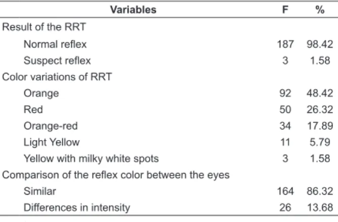
Of the total newborns evaluated using the red
relex test, in 187 (98.42%) a normal, homogeneous, relex was observed, with leukocoria absent, presenting
the following color variations: 92 (48.42%) orange, 50
(26.32%) red; 34 (17.89%) orange-red and 11 (5.79%)
light yellow.
Only three (1.58%) of the newborns presented a
suspect relex. For these, besides the relevance of their
life histories and health conditions, a non-standard
relex coloration was noticed, prominently yellow with
lighter spots (milky white) in the center. These infants,
considered at risk, were included in the retinopathy
of prematurity screening and treatment program of
the institution. Table 1, below, presents the result and
description of the relex, regarding the color appearance,
found in the test.
Table 1 - Distribution of newborns (NBs) concerning the
result and the description of the color variations of the
red relex test (RRT), Ceará, Brazil, 2007
Variables F %
Result of the RRT
Normal reflex 187 98.42
Suspect reflex 3 1.58
Color variations of RRT
Orange 92 48.42
Red 50 26.32
Orange-red 34 17.89
Light Yellow 11 5.79
Yellow with milky white spots 3 1.58 Comparison of the reflex color between the eyes
Similar 164 86.32
Differences in intensity 26 13.68
n=190
Comparing the coloration intensity of the ocular
relex between the pupils there were slight differences
sometimes in the right eye, sometimes in the left eye.
The infants examined presented various shades of
ocular relex color, which were described and recorded
with the aid of the RRT color gradient instrument.
These results were then grouped into four categories,
denominated Grads, which included the variations of
the relexes visualized in the test and identiied in the
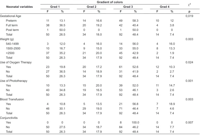
Thus, grouped in the category Grad 1 were the
shades of red of the ocular relex observed and were identiied as R1, R2, R4 and R6 in the RRT color gradient
instrument. In the Grad 2 category, the color orange-red
identiied by L1 and L17 and in the Grad 3 category the variations of the color orange of the relex encountered
in the test, represented by L2, L4, L6, L9, L12 and L18
in the gradient. These three categories represented
Table 2 - Association between the red relex test color gradient instrument and the neonatal variables, Ceará, 2007
the variations in color of RRT normality. Grouped in the
Grad 4 category were the shades of yellow (A1, A3) of
the relex observed, which in some cases, depending
on the history of the neonate, may be present without
alterations. However, this category also included the
relex color of yellow with milky white spots at the center
(A9), found in the test and considered suspect.
Neonatal variables
Gradient of colors
χ2
Grad 1 Grad 2 Grad 3 Grad 4
F % F % F % F % p
Gestational Age 0,019
Preterm 11 13.1 14 16.6 49 58.3 10 12
Full term 38 36.5 20 19.2 42 40.4 4 3.8
Post term 1 50.0 0 0 1 50.0 0 0
Total 50 26.5 34 18.0 92 48.4 14 7.4
Weight (g) 0.003
540-1499 3 12.0 4 16.0 14 56.0 4 16.0
1500-2500 10 16.7 9 15.0 33 55.0 8 13.3
>2500 37 35.2 21 20.0 45 42.9 2 1.9
Total 50 26.3 34 17.9 92 48.4 14 7.4
Use of Oxygen Therapy 0.024
Yes 23 19.8 20 17.2 61 52.6 12 10.3
No 27 36.5 14 18.9 31 41.9 2 2.7
Total 50 26.3 34 17.9 92 48.4 14 7.4
Use of Phototherapy 0.001
Yes 10 13.3 15 20.0 39 52.0 11 14.7
No 40 34.8 19 16.5 53 46.1 3 2.6
Total 50 26.3 34 17.9 92 48.4 14 7.4
Blood Transfusion 0.003
Yes 4 10.8 5 13.5 21 56.8 7 18.9
No 46 30.1 29 19.0 71 46.4 7 4.6
Total 50 26.3 34 17.9 92 48.4 14 7.4
Conjunctivitis
Yes 0 0 0 0 8 100.0 0 0 0.007
No 50 27.5 34 18.7 84 46.2 14 7.7
Total 50 26.3 34 17.9 92 48.4 14 7.4
n=190
As shown in Table 2, it was veriied that there was a statistically signiicant association between the RRT color
gradient and the neonatal variables (gestational age
p=0.019) (birth weight p=0.003) (use of oxygen therapy
p=0.024); (phototherapy p=0.001) (blood transfusion
p=0.003) and (conjunctivitis p=0.007). These variables
are considered risk factors for the development of vision
problems and therefore may have a relationship with the
outcome of the red relex test.
It should also be noted in Table 2, that the sample
of this study was composed primarily of full term NBs
(104 - 55.03%) with adequate birth weight (105 -
55.26%), and, regarding the appearance of the ocular
relex observed, the shades of color referring to the
categories Grad 1 and Grad 3 prevailed.
Among those who presented the orange coloration
of the ocular relex (Grad 3) (92 - 48.4%), a similar
result was observed between the preterm infants (49 -
58.3%) and the full term infants (42 to 40.4%). A relex
of a clearer appearance, with little intensity, brown, with
less shine, was more often observed in the former, unlike
the full term infants.
The results also showed that a higher prevalence of
a pale yellow ocular relex, sometimes with milky white
spots over the center (Grad 4), among the premature
NBs (10 - 12%) and in those weighing less than 2500g
(12 - 14%).
Among the NBs who underwent oxygen therapy,
there was a predominance of the orange relex (Grad 3) with variations of intensity in 61 (52.6%) of them.
From relating the colors of the gradient to the use of
oxygen therapy, a signiicant relationship (p=0.024) was found, although this signiicance was not evident when a
Rev. Latino-Am. Enfermagem 2011 Mar-Apr;19(2):309-16.
Discussion
The red relex test, although considered a simple
examination, requires attention during its realization, as
the examiner needs to verify the relex characteristics
carefully and also to observe, in the evaluation,
variables ranging from the birth conditions of the infant,
to the material environmental and human resources.
Other important aspects when performing this test are
the acquisition by the examiner of knowledge on the
ocular anatomy and the investigation of factors related
to the maternal and neonatal history which could have
relationships with alterations in vision and with the result
of the red relex.
A study conducted in 2002 which contemplated
the ophthalmological evaluation of 667 newborns,
conducted in a Rooming-in unit, using the red relex test
(direct ophthalmoscopy), showed that the main ocular
alteration found through the test was opacity of the
cornea(14).
The literature says that the view of the fundus
should be clear, i.e. there should be no opacity in the
ocular media, such as dark shadows or black spots that
block the red relex(6). Classiication of terms such as “normal or present” were adopted when the relex in
both eyes was similar in color, intensity, clarity and no
opacities or white spots existed in any area, either in one
or both relexes observed. The result of “abnormal or
absent” was characterized in an opposite way to this(15). Authors also report that the different relex between the
eyes can mean large differences in the refractive power
of the eyes, i.e. a degree that can lead to the need for
glasses(8).
The indings of the present study denote a color appearance variation in the relex among the infants with a normal ocular relex, being sometimes pale,
sometimes darker or lighter, sometimes with areas like
cobwebs or with the presence of mottling.
Thus, it is clear that the term referred to in literature
as “red relex”, although universal, did not always
correspond closely to what was found when performing
the test where, in reality, color variations from orange to
red were often veriied, relative to the characteristics of
the fundus and retina.
In general, the fundus of premature infants,
depending on gestational age, appears pale due to the
blood vessels that provide pigmentation to the retina not
being fully developed. Hence the fact that a yellow or clear
relex is commonly observed with an ophthalmoscope
in preterm infants(6). Other authors also emphasize that
in children with low eye pigmentation (Caucasian), the
relex has a red-orange coloration. In children with a
more intense eye pigmentation, as in black race infants,
the relex has a darker color (red-maroon)(16).
In premature infants, the remnant of the tunica
vasculosa lentis is often visible in front or behind the
lens, or even in both positions. The remnant is usually
absorbed over time by the children, however, rarely it
stays and appears as a complete or partial “cobweb”(17).
A study which examined 47 newborns who
presented factors associated with prematurity, low birth
weight, hypoxia and ventilatory support, found only one
of these NBs had an altered ocular relex, with eight classiied as suspect. In seeking the diagnostic reports
of the ophthalmologist, it was found that one of the
infants presented total retinal detachment, with a totally
opaque relex observed, and eight developed retinopathy
of prematurity(12).
Retinopathy of prematurity (ROP), a leading cause
of preventable blindness in children, is a vasoproliferative
disorder of the retina that affects premature NBs of low
birth weight. It has as risk factors, luctuations in oxygen
levels, septicemia and blood transfusion. At an advanced
stage there may be retinal detachment in the NB and
detection of the presence of leukocoria, thus becoming
a noticeable sign in the red relex test(18-19).
In another study, in which the sample consisted of
180 newborns hospitalized in a public maternity hospital,
24 (13%) presented a relex classiied as suspect.
Contact was sought with those responsible for providing
follow-up of the care process of these children, referring
them to specialized evaluation by an ophthalmologist
collaborator of the study. The visual indings concerning the realization of the red relex test in this study were conirmed and better detailed with the completion of the
fundoscopy and retinal mapping. Other doubtful aspects
for the authors regarding the perception of the color of
the relex, when viewed using the ophthalmoscope, were
also discussed and considered as variations of normality,
i.e. variations in normal retinal development, which is
presented in the process of vascularization(2).
It is noteworthy that the preterm infants, due to
risk factors that surround them, are found to be more
vulnerable to develop alterations of vision when compared
to the full term infants. Hence the probable inluence of
gestational age and birth weight in the results regarding
the color of the relex observed in this study.
Regarding the color gradient and use of oxygen
therapy, a statistically signiicant association was found,
the duration of oxygen use was sought. This inding
may be based on the fact that the sample consisted
predominantly of full term infants who required few
days of oxygen. However, in another study(20) where the sample was exclusively of premature NBs, a signiicant
association (p=0.028) was found between the result of
the RRT and the duration of oxygen therapy.
The literature argues that oxygen, due to being
a vasoconstrictor, increases ischemia stimulating the
formation of tortuous neovessels. These cause scarring
of the retina that can lead to displacement and blocking
of the vision. Therefore the less weight the premature
infant has, the more likely it is that they will develop
ocular problems, such as retinopathy, because they
will be exposed to oxygen for longer and subjected to
constant luctuations in their blood pressure(21).
Another study that developed a systematic vision
examination with a sample of 114 premature newborns,
using the red relex test, identiied 13 (11%) with a suspect relex. By relating this to neonatal history variables, a statistically signiicant association was
obtained between the outcome of the RRT and the Apgar
score in the irst minute of life (p=0.041)(10).
Although the literature on the subject is growing,
there has been some dificulty to discuss some of the
variables presented in more detail, such as the use of
phototherapy, blood transfusion and conjunctivitis.
However, studies have highlighted, as factors that may
make the development of retinal alterations possible, in
addition to prematurity and low birth weight, hypoxia or
hyperoxia, blood transfusions, infections, and exposure
to intense light(18).
In view of the results presented, it is necessary to
draw attention to the importance of precautions such
as, for example, monitoring, during administration, of
the oxygen concentration to prevent its toxic effects on
the visual system and the use of eye protection during
phototherapy to prevent the incidence of light causing
the degeneration of the retina(18,22).
Conclusion
This study is relevant not only for the promotion
of ocular health, but also for the overall health of the
child. Finally, the role of vision in normal physical and
cognitive development is an indisputable fact, due to the
interferences caused by its privation in the quotidian life
of the human being. Given this, it is emphasized that to
prevent alterations of vision in the infantile population,
strategies to promote ocular health should be targeted
from the pregnancy, prenatal and neonatal periods.
Due to this being a little explored topic, particularly
in nursing studies, it was necessary to use speciic
literature of other areas to deepen the discussion on the
subject.
Based on the data obtained it can be concluded that
of the total of newborns evaluated using the red relex test, the majority presented a normal relex, noting how
the coloration and intensity varied between orange, red
and light yellow. Only three newborns revealed a suspect
relex, characterized by yellow with lighter (whitish)
spots in the center.
Statistically signiicant associations were found between the shades of color identiied in the gradient for the realization of the red relex test, and some variables
of neonatal history, such as: weight (p=0.03), gestational
age (p=0.019), use of oxygen therapy (p=0.024) and
phototherapy (p=0.001). Furthermore, as evidenced
among premature infants, there was a predominance of
the relex color of orange with low intensity and of light
yellow, due to incomplete vascularization of the retina
at birth. A similar result occurred in neonates of low
weight.
The indings showed that there is not a standard color of the relex visualized. Although the term used in
the literature to refer to the test may wrongly suggest
that the relex is observed only in red, in reality this may
present variations depending on several factors, among
them the neonatal history variables that will affect the
characteristics of the fundus and the retina.
References
1. Manica NB, Correia ZMS, Macon IM, Telichebesky
N, Loch LF. O que os pediatras conhecem sobre
afecções oculares na criança? Arq Bras Oftalmol.
2003;66(4):489-92.
2. Aguiar ASC, Cardoso MVLML, Lúcio IML. Teste do
reflexo vermelho: forma de prevenção à cegueira na
infância. Rev Bras Enferm. 2007;60(5):541-5.
3. Reis PP. Reflexo vermelho. Textos científicos Sociedade
Mineira de Pediatria. [internet]. 2005. [acesso: 15 março
2008]. Disponível em: http://www.smp.org.br
4. Graziano RM. Prevenção da cegueira no
recém-nascido pré-termo. [internet]. 2006. [acesso: 15 março
2009]. Disponível em: http://www.paulomargotto.com.
br/index_sub.php?tipo=27
5. Sociedade Cearense de Pediatria. Saúde da criança
- teste do olhinho precisa ser lei. Boletim Informativo
Rev. Latino-Am. Enfermagem 2011 Mar-Apr;19(2):309-16.
[acesso em: 03 março 2010]. Disponível em: http://
www.socep.org.br/novo/pdf/infojanfev2009.pdf
6. Jarvis C. Exame físico e avaliação de saúde. 3ed. Rio
de Janeiro: Guanabara Koogan; 2002.
7. Weber J. Semiologia: guia prático para a enfermagem.
5ed. Rio de Janeiro: Guanabara Koogan; 2007.
8. Bonotto LB. A importância do Exame de Reflexo
Vermelho. [Internet]. 31 ago 2006 [acesso: 20 março
2010]. Disponível em: http://www.oftalmopediatria.
com/texto.php.
9. Lúcio IML. Método Educativo para a prática do teste do
reflexo vermelho no cuidado ao recém-nascido. [tese de
doutorado]. Fortaleza (CE): Departamento de Enfermagem
da Universidade Federal do Ceará; 2008. 137 p.
10. Cardoso MVL, Lúcio IML, Aguiar ASC. Aplicação do
teste do reflexo vermelho no cuidado neonatal. Rev
Rene. 2009;10(1):81-7.
11. Endriss D, Ventura LM, Diniz JR, Celino AC, Toscano
J. Doenças oculares em neonatos. Arq Bras Oftalmol.
2002;65(5):551-5.
12. Costa KAB, Cardoso MVLML, Lúcio IML. Avaliação
visual em recém-nascidos no ambiente hospitalar. Rev
Paul Enferm. 2005;24(2):23-9.
13. Ministério da Saúde (BR). Diretrizes e normas
regulamentadoras de pesquisa envolvendo seres
humanos: resolução 196/96. Brasília (DF): Conselho
Nacional de Saúde; Ministério da Saúde; 1996. 12 p.
14. Wasilewski D, Zago RJ, Bardal AMC, Heusi,
TM, Carvalho FP, Maciel LF, et al. Importância da
avaliação oftalmológica em recém-natos. J Pediatr.
2002;78(3):209-12.
15. American Academy of Pediatrics. Red reflex
examination in neonates, infants, and children.
Pediatrics. 2008;122(6):1401-4.
16. Tamura MY, Teixeira LF. Leucocoria e Teste do Reflexo
Vermelho. Einstein. 2009; 7(3):376-82.
17. Voughan D, Asbury T, Riordan – Eva P. Oftalmologia
geral. 15ed. São Paulo: Atheneu;2003.
18. Margotto PR. Assistência ao recém-nascido de risco.
2ed. Brasília: Hospital Anchieta;2006.
19. Tamez RN, Silva MJP. Enfermagem na UTI neonatal.
4ed. Rio de Janeiro: Guanabara Koogan;2009.
20. Lúcio IML, Cardoso MVLML, Almeida PC. Investigação
do reflexo vermelho em recém-nascidos e sua relação
com fatores da história neonatal. Rev Esc Enferm USP.
2007;41(2):222-8.
21. Ferreira RC. Retinopatia da prematuridade como
diagnosticar. [Internet] [acesso: 10 março 2010].
Disponível em:
http://deficienciavisual.com.sapo.pt/sd-retinopatiaprematuro.html
22. Ferreira ALC, Nascimento RM, Veríssimo RCSS.
Irradiance of phototherapy equipment in maternity
wards in Maceió. Rev. Latino-Am. Enfermagem.
2009;17(5):695-700.
Received: Apr. 20th 2010