05 artigo 570
ORIgINAL ARtICLE
1 – Orthopedist at the Knee Surgery Center, National Institute of Traumatology and Orthopedics (INTO), Rio de Janeiro, RJ, Brazil.
2 – Orthopedist and Trainee in the Knee Surgery Center, National Institute of Traumatology and Orthopedics (INTO), Rio de Janeiro, RJ, Brazil. 3 – Orthopedist and Head of the Knee Surgery Center, National Institute of Traumatology and Orthopedics (INTO), Rio de Janeiro, RJ, Brazil. Work performed at the Knee Surgery Center, National Institute of Traumatology and Orthopedics (INTO), Rio de Janeiro, RJ, Brazil. Correspondence: Praia do Flamengo 66, Bloco B, Sala 1313, 22210-030 Rio de Janeiro, RJ. E-mail: [email protected] Work received for publication: August 3, 2011; accepted for publication: September 16, 2011.
ASSESSmENT OF TIBIAL SLOPE ANgLE AND PATELLAR HEIgHT
AFTER mEDIAL-OPENINg TIBIAL OSTEOTOmY
Alan de Paula Mozella1, Marcos Areias Vieira Costa2, Hugo Alexandre de Araujo Barros Cobra3
ABstRACt
Objective: To measure the variation in posterior tibial slope angle and patellar height in patients who underwent proximal tibial valgus-producing osteotomy using the medial-opening wedge technique. Methods: Anteroposterior panoramic radio-graphs of the lower limbs and lateral radioradio-graphs of the knee obtained before and after tibial valgus-producing osteotomy on 46 patients with unicompartmental arthrosis of the knee were analyzed. Results: In 23 patients, an external fixator was used to gradually apply a medial-opening wedge; and in the other 23, a blocked plate with a stop bar was applied as a fixation method. Patients with tricompartmental knee disease and those who underwent osteotomy to treat fracture seque-lae were excluded from this study. After surgery, the mean
INtRODUCtION
High tibial osteotomy as a treatment for medial unicompartmental osteoarthrosis was first reported by Jackson and Waugh in 1958, who performed os-teotomy distally to the anterior tuberosity of the tibia (TAT). Subsequently, Coventry popularized this the-rapy by using osteotomy proximally to the anterior tuberosity of the tibia, in accordance with the techni-que recommended by Gariepy in 1964(1-4).
Patients with unicompartmental gonarthrosis and varus mechanical alignment of the limb were tradi-tionally treated with lateral-closing osteotomy, and several studies showed that 60 to 90% of the results were good, with 10-year follow-up(5,6).
Over the last two decades, osteotomy using a me-dial-opening wedge technique, either with gradual or
with immediate opening, has become the technique of choice because of advantages such as a lower fi-bular nerve injury rate, non-violation of the proximal tibiofibular joint, greater precision in correcting the mechanical axis, maintenance of the bone stock and less alteration to the morphological characteristics of the proximal tibia. Several series of patients who underwent medial-opening osteotomy have shown cli-nical results comparable to those presented by lateral--closing osteotomy(7,8).
Because of the bone defect created during the pro-cedure, fixation is an important factor in the final result from this technique. This stabilization may be achieved by means of rigid internal devices like lo-cked plates, with or without a tab, or by means of external fixation.
increase in the tibial slope was 1.7 degrees (p < 0.01) in the group in which the blocked plate with a stop bar was used; and 2.7 degrees (p < 0.05) in the group in which the external fixator was used. There was no statistical difference between the groups regarding the increase in the posterior tibial slope. Conclusion: The patellar height did not present any change in the cases in which the plate was used, when measured using the Insall-Salvati method, but it presented a decrease in 11 ca-ses (47.8%) when the Caton-Deschamps method was applied. The same tendency was observed regarding change in the patellar height in the cases in which the external fixator was used, such that a decrease was observed in eight cases (34.7%) only when measured using the Caton-Deschamps method.
Keywords - Osteotomy; Tibia; Knee; Patella
The authors declare that there was no conflict of interest in conducting this work
Another indication for high tibial osteotomy is for correction of load imbalance in situations of ligament instability, in patients with varus buckling, thereby changing the axial alignment, reducing the varus de-formity and avoiding overloading on the ligament reconstruction(9).
Although tibial osteotomy is initially used for cor-recting deformities in the coronal plane, alterations to the posterior slope of the tibial plateau(10-12) and to the
patellar height(13,14) often occur concomitantly, thus
giving rise to biomechanical alterations in the knee and changes in contact pressure on the cartilage, with consequences that are still uncertain.
MAtERIAL AND MEtHODs
The aim of this study was to evaluate changes to the posterior slope angle of the proximal tibia and changes to patellar height of patients who underwent high tibial osteotomy by means of the open-wedge te-chnique, performed at the Knee Surgery Clinic of the National Institute of Traumatology and Orthopedics, between January 2006 and December 2008.
This study included patients who underwent os-teotomy to treat osteoarthrosis or unicompartmental overloading of the knee, who presented varus devia-tion of the mechanical axis. Patients with tricompart-mental disease of the knee, those undergoing oste-otomy to treat sequelae of fractures and those with previous surgery on the extensor mechanism were excluded from this study.
For retrospective analysis, the 46 patients were divided into two groups:
• Group I: formed by 23 patients who underwent
high tibial osteotomy using the immediate-opening wedge technique, with autologous bone grafting and fixation using a Puddu® plate (Arthrex,
Na-ples, Florida, USA), in accordance with the stan-dard technique.
• Group II: formed by 23 patients who underwent
high tibial osteotomy using the gradual-opening medial wedge technique, by means of fixation us-ing an Ortofix® external fixator, in accordance with
the standard technique.
This study was submitted for evaluation and was approved by the Research Ethics Committee of the National Institute of Traumatology and Orthopedics, and was conducted at the Knee Surgery Center of this institution.
RADIOgRAPHIC EVALUAtION
The radiographic evaluation consisted of AP pa-noramic radiographs with bipedal weight-bearing and pre and postoperative radiographs of the knee in true lateral view, at 30 degrees of flexion.
The AP radiographs were used to measure the ti-biofemoral angle, while the lateral radiographs were used to measure the posterior slope of the tibial pla-teau and the patellar height. The measurements were made on postoperative radiographs that showed con-solidation of the osteotomy.
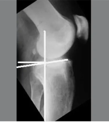
The tibial slope was measured using a method ba-sed on the posterior cortical line of the proximal tibia. A reference line at the level of the tibiofemoral joint was traced out perpendicularly to the posterior corti-cal line of the tibia. Another line was traced out using the most prominent points on the anterior and poste-rior margins of the tibial plateau. The angle formed by these two lines was taken to be the posterior slope angle of the proximal tibia. The angle measurements were made by two different examiners. If these were not concordant, the simple arithmetic mean was taken as the measurement (Figure 1).
Measurements on the pre and postoperative pa-tellar height were made using the Insall-Salvati and Caton-Deschamps methods.
stAtIstICAL ANALysIs
Statistical analysis was performed with the aim of assessing the degree of significance of the parameters measured. The Shapiro-Wilk W test was performed to evaluate the normality and the Levene test to evaluate the homoscedasticity of the variances. The posterior slope angles of the tibial plateau and the pre and pos-toperative patellar height indices in both groups were considered to present normal distribution, and Student’s t test was used to compare the means. The Statistica 8.0 software was used to perform the statistical calculations.
REsULts
Group I was formed by 23 patients (20 males and three females); their mean age was 41.2 years, with a range from 19 to 50 years. Surgery was performed on the right side in seven cases, and on the left side in 16 patients.
The mean preoperative angular deviation was five degrees of varus (ranging from 1 to 10°). The mean correction achieved in this group was 12.3 degrees. The mean postoperative tibiofemoral angle in this group was 7.3° of valgus.
The preoperative posterior slope of the proximal tibia ranged from two to 16 degrees, with a mean of 8.2° (sd = 3.76). There was a mean increase of 1.7° in the posterior slope angle (p < 0.01), and thus the post-operative mean posterior slope was 9.9°(sd = 3.32). In two cases, there were reductions in the plateau slope, of up to four degrees. The greatest increase in tibial slope was six degrees (Figure 2 A and B).
Although it was observed that greater corrections of angular deformity presented a tendency towards greater increases in the tibial slope angle, this correla-tion was not shown to be statistically positive.
The patellar height in group I did not present any variation from before to after the operation when sured using the Insall-Salvati method. When mea-sured using the Caton-Deschamps method, 11 cases (47.8%) evolved with a low patella after the surgery (Figure 3 A and B).
Group II was formed by 23 patients (19 males and four females); their mean age was 41.7 years, with a range from 25 to 53 years. The surgery was performed on the right side in five cases and on the left side in 18 patients.
The mean preoperative angular deviation was 5.9 degrees of varus (ranging from 2 to 12°). The mean correction obtained in this group was 12.9 degrees.
The mean postoperative tibiofemoral angle in this group was 7.0° of valgus.
The posterior slope angle of the proximal tibia ranged from two to 18 degrees, with a mean of 9.1° (sd = 3.05). There was a mean increase of 2.7 degrees in the posterior slope angle (p < 0.05) and thus, the mean postoperative posterior slope was 11.8 degrees (sd = 4.14). In two cases, there was a reduction in the slope of the tibial plateau. The greatest variation in the slope angle was an increase of six degrees, and reductions of the slope angle of the order of three degrees were also observed (Figure 4 A-C).
Comparison of the variation in anteroposterior tibial slope from before to after the operation, between group I and group II, did not show any statistically significant differences (p > 0.05).
As noted in group I, there was no positive correlation in group II between the degree of cor-rection of the angular deformity and increases in tibial slope (Figure 5).
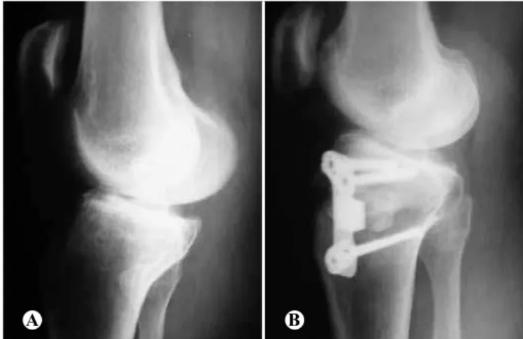
Figure 2 – (A) AP radiograph; (B) Lateral view of a patient in group I who underwent
osteotomy and fixation using a locked plate with tab (Puddu® plate).
A B
Figure 3 – (A) Preoperative radiograph; (B) Postoperative radiograph showing reduction of
the patellar height in a patient who underwent osteotomy and fixation using a Puddu® plate.
In group II, there was no change in patellar height between pre and postoperative measurements when evaluated using the Insall-Salvati method. However, when measured using the Caton-Deschamps method, eight cases (34.7%) were seen to have evolved with a low patella after the surgery.
DIsCUssION
Several recent studies have indicated that the normal anteroposterior slope of the joint surfa-ce of the proximal tibia ranged from seven to ten degrees(10,15-17). In our series, these data did not
pre-sent any discrepancy.
High tibial osteotomy, either with a lateral-closing or a medial-opening wedge, causes modification of the proximal tibial slope parameters, as shown by renowned authors(6,18). Most studies have identified
increases in the tibial slope angle after osteotomy with a medial-opening wedge; our study is therefore con-cordant with the literature.
Hernigou et al(19) and Koshino et al(20) developed
mathematical models for correcting genu varum, but they did not demonstrate any method for maintaining the posterior slope angle in the sagittal plane after high tibial osteotomy had been performed.
Noyes et al(21) used three-dimensional geometrical
analysis on the proximal tibia and advocated that, in order to maintain the posterior slope of the joint surface of the tibial plateau, the anterior space of the opening wedge should be half of the posterior space of the wedge.
Several authors have corroborated the study by Noyes et al(21), thus demonstrating that while
lateral--closing wedge osteotomy is being performed, there is often a decrease in the degree of posterior slope, by an average of five degrees from before to after the operation. Other studies have shown mean increases of between three and four degrees in the slope angle, during medial-opening osteotomy(16,22).
Our study is concordant with the work by Marti
et al(23) and Griffin et al(11), showing increases in the
posterior slope angle of the tibial plateau of approxi-mately two degrees, with a 95% confidence interval, following medial-opening osteotomy.
The consequences of changes in the sagittal plane following high tibial osteotomy remain a topic under debate in the literature. Some authors, such as Herni-gou et al(19), have argued that changes to the slope of
the proximal tibia might accelerate the process of joint degeneration, through alterations to knee kinematics and biomechanics.
Biomechanical studies developed in a different manner by Griffin et al(15) and Brinkman et al(22)
showed that there was a linear relationship between tibial translation and anteroposterior slope of the
Figure 4 – (A) Patient who underwent gradual-opening high tibial osteotomy tibial using
an Ortofix® fixator; (B) Postoperative AP radiograph; (C) Postoperative lateral radiograph.
Figure 5 – Correlation between degree of angular correction and tibial slope in groups I and II.
A B
C
correction
0 5 10 15 20 20
-20
0 5 10 15 20 25 correction y = 0.2143x... 10
0 -10
Correlation between
correction and slope...
tibia. Thus, an increase in slope would cause greater tibial translation.
An increase in posterior slope of the proximal ti-bia would also, as shown in biomechanical studies, lead to increased tibiofemoral contact pressure on the anterior portion of the tibial plateau and decreased pressure on the posterior femoral condyle(22).
Changes to patellar height can be seen following high tibial osteotomy, as highlighted by different authors(17,24). There are several different explanations
for this occurrence: shortening of the patellar tendon due to the healing process subsequent to surgical ma-nipulation; bone neoformation at the insertion of the patellar tendon; relative shortening after tibial trans-lation due to change to the slope of the tibial plateau following osteotomy, as postulated by Kaper et al(25);
alteration of the height of the joint line; or transfer of the tibial tuberosity.
Measurement of patellar height using the Insall--Salvati index correlates more strongly with the length of the patellar tendon, while measurement using the Caton-Deschamps method represents the relative height of the patella, in consideration with the interline(26).
In performing medial-wedge high tibial osteotomy,
there is an increase in the distance between the an-terior tibial tuberosity and the joint line, and conse-quently elevation of the interline in relation to the patella. Thus, indices like Blackburn-Peel and Caton--Deschamps present changes, as shown by authors like Noyes et al(21) and Wright et al(27), and our study
was concordant with these data.
CONCLUsIONs
Valgus-producing tibial osteotomy using the me-dial-opening wedge technique caused a statistically significant change to the posterior slope angle of the proximal tibial joint surface. There was no statistical difference in this increase between groups fixed using a plate or using an external fixator.
No positive correlation was observed between the degree of correction of the varus deformity and the variation in the tibial slope angle or alteration of pa-tellar height.
The patellar height did not present modifications when measured using the Insall-Salvati method. However, when measured using the Caton-Deschamps method, a reduction in patellar height was observed, most notably in the group fixed with a plate.
REFERENCEs
1. Coventry MB. Osteotomy about the knee for degenerative and rheumatoid arthritis. J Bone Joint Surg Am. 1973;55(1):23-48.
2. Coventry MB. Osteotomy of the upper portion of the tibia for degenerative ar-thritis of the knee. A preliminary report. J Bone Joint Surg Am. 1965;47:984-90. 3. Coventry MB. Upper tibial osteotomy. Clin Orthop Relat Res. 1984; (182):46-52. 4. Coventry MB, Ilstrup DM, Wallrichs SL. Proximal tibial osteotomy. A critical
long-term study of eighty-seven cases. J Bone Joint Surg Am. 1993;75(2):196-201. 5. Akizuki S, Shibakawa A, Takizawa T, Yamazaki I, Horiuchi H. The long-term outcome of high tibial osteotomy: a ten- to 20-year follow-up. J Bone Joint Surg Br. 2008;90(5):592-6.
6. El-Azab H, Halawa A, Anetzberger H, Imhoff AB, Hinterwimmer S. The effect of closed- and open-wedge high tibial osteotomy on tibial slope: a retrospec-tive radiological review of 120 cases. J Bone Joint Surg Br. 2008; 90(9):1193 7. Franco V, Cerullo G, Cipolla M, Gianni E, Puddu G. Open wedge high tibial
osteotomy. Techn Knee Surg. 2002;1(1):43-53.
8. Sprenger TR, Doerzbacher JF. Tibial osteotomy for the treatment of varus gonarthrosis. Survival and failure analysis to twenty-two years. J Bone Joint Surg Am. 2003;85(3):469-74.
9. Dejour H, Neyret P, Boileau P, Donell ST. Anterior cruciate reconstruction com-bined with valgus tibial osteotomy. Clin Orthop Relat Res. 1994;(299):220-8. 10. Hohmann E, Bryant A. Closing or opening wedge high tibial osteotomy: watch
out for the slope. Op Techn Orthop. 2007;17:38-45.
11. Giffin JR, Vogrin TM, Zantop T, Woo SL, Harner CD. Effects of increasing tibial slope on the biomechanics of the knee. Am J Sports Med. 2004;32(2):376-82. 12. Rodner CM, Adams DJ, Diaz-Doran V, Tate JP, Santangelo SA, Mazzocca AD, et al.
Medial opening wedge tibial osteotomy and the sagittal plane: the effect of increasing tibial slope on tibiofemoral contact pressure. Am J Sports Med. 2006;34(9):1431-41. 13. Scuderi GR, Windsor RE, Insall JN. Observations on patellar height after
proximal tibial osteotomy. J Bone Joint Surg Am. 1989;71:245-8.
14. Singerman R, Davy DT, Goldberg VM. Effects of patella alta and patella infera on patellofemoral contact forces. J Biomech. 1994;27(8):1059-65.
15. Giffin JR, Stabile KJ, Zantop T, Vogrin TM, Woo SL, Harner CD. Importance
of tibial slope for stability of the posterior cruciate ligament deficient knee. Am J Sports Med. 2007;35(9):1443-9.
16. Yanasse RH, Cavallari CE, Chaud FL, Hernandez AJ, Mizobuchi RR, Laraya MH. Measurement of tibial slope angle after medial opening wedge high tibial osteotomy: case series. Sao Paulo Med J. 2009;127(1):34-9.
17. Brouwer RW, Bierma-Zeinstra SM, van Koeveringe AJ, Verhaar JA. Patellar height and the inclination of the tibial plateau after high tibial osteotomy. The open versus the closed-wedge technique. J Bone Joint Surg Br. 2005;87(9):1227-32. 18. Sterett WI, Miller BS, Joseph TA, Rich VJ, Bain EM. Posterior tibial slope after
medial opening wedge high tibial osteotomy of the varus degenerative knee. J Knee Surg. 2009;22(1):13-6.
19. Hernigou P, Medevielle D, Debeyre J, Goutallier D. Proximal tibial osteotomy for osteoarthritis with varus deformity. A ten to thirteen-year follow-up study. J Bone Joint Surg Am. 1987;69(3):332-54.
20. Koshino T, Murase T, Saito T. Medial opening-wedge high tibial osteotomy with use of porous hydroxyapatite to treat medial compartment osteoarthritis of the knee. J Bone Joint Surg Am. 2003;85(1):78-85.
21. Noyes FR, Goebel SX, West J. Opening wedge tibial osteotomy: the 3-triangle method to correct axial alignment and tibial slope. Am J Sports Med. 2005;33(3):378-87. 22. Brinkman JM, Lobenhoffer P, Agneskirchner JD, Staubli AE, Wymenga AB, van
Heerwaarden RJ. Osteotomies around the knee: patient selection, stability of fixation and bone healing in high tibial osteotomies. J Bone Joint Surg Br. 2008;90(12):1548-57. 23. Marti CB, Gautier E, Wachtl SW, Jakob RP. Accuracy of frontal and sagittal plane correction in open-wedge high tibial osteotomy. Arthroscopy. 2004;20(4):366-72. 24. Closkey RF, Windsor RE. Alterations in the patella after a high tibial or distal
femoral osteotomy. Clin Orthop Relat Res. 2001;(389):51-6.
25. Kaper BP, Bourne RB, Rorabeck CH, Macdonald SJ. Patellar infera after high tibial osteotomy. J Arthroplasty. 2001;16(2):168-73.
26. Scuderi GR, Windsor RE, Insall JN. Observations on patellar height after proximal tibial osteotomy. J Bone Joint Surg Am. 1989;71(2):245-8. 27. Wright J, Heck D, Hawker G, Dittus R, Freund D, Joyce D,et al. Rates of tibial osteotomies