796
Rev Soc Bras Med Trop 49(6):796-798, November-December, 2016 doi: 10.1590/0037-8682-0172-2016
Case Report
Corresponding author: Dr. Felipe Caino.
e-mail: [email protected]
Received 23 May 2016
Accepted 1 August 2016
Abdominal mucormycosis in a child: a case report
Felipe Rezende Caino de Oliveira
[1],[2], Nimara Grace Cardoso Batista Couto
[1],
Juliana de Oliveira Bastos
[1], José Colleti Junior
[2]and Werther Brunow de Carvalho
[3][1]. Unidade de Terapia Intensiva Pediátrica, Hospital Martagão Gesteira, Salvador, Bahia, Brasil. [2]. Unidade de Terapia Intensiva Pediátrica, Hospital Santa Catarina, São Paulo, Brasil. [3]. Instituto da Crianca , Departamento de Pediatria, Universidade de São Paulo, São Paulo, Brasil.
Abstract
A 2-year-old Brazilian female child from the countryside in Bahia State presented with pain in the right fl ank of the abdomen, accompanied by a daily fever for about 2 weeks before admission. A large mass in the abdomen was resected by the surgical team. The biopsies revealed the mass was an intra-abdominal mucormycosis. However, the diagnosis was late, and despite treatment (amphotericin B) initiation, the patient eventually died.
Keywords: Mucormycosis. Pediatric abdominal pain. Amphotericin B.
INTRODUCTION
Mucormycosis is a rare fungal infection caused by fungi of the Zygomycetes class Mucorales and Entomophthorales order. It usually occurs in tropical area and presents as a subcutaneous infection. Mucormycosisis caused by opportunistic pathogens and rarely causes disease in immunocompetent patients, yielding mainly processes that cause neutropenia or neutrophil dysfunction. After aspergillosis and candidosis, it is the third most common invasive fungal infection, representing 8.3-13.0% of all fungal infections found in autopsies of hematologic patients(1) (2).
Clinical manifestations are variable, including rhinocerebral commitment, often 44-49% of the reported cases; localized or generalized primary cutaneous involvement (10-19%); lung (10-11%); disseminated (6-11%); and gastrointestinal (2-11%)(3)(4).
Gastrointestinal presentation of mucormycosis is uncommon and rarely diagnosed in living patients. In such cases, the diagnosis is late, and the mortality rate is high, at approximately 85%(3). Only 25% of cases of gastrointestinal mucormycosis are diagnosed antemortem, and the disease is reported mainly in premature infants, newborns, patients, and malnourished children with oncological diseases, diabetes mellitus, or a history of corticosteroid use(5) (6). It can be acquired by ingesting pathogens in foods such as fermented porridge, alcoholic beverages and drinks derived from corn, herbs contaminated with spores, and homeopathic remedies(7).
Mucormycosis rarely appears in children. In an analysis of all reports of pediatric mucormycosis cases published until 2004, 157 patients (64% male) with an average age of 5years were identifi ed(8). Twenty-eight (18%) patients had hematological diseases, and 9 (6%) had undergone transplantation. A series of an additional 30 pediatric cases from 2004 to 2008 was also reported in 2009(9).
We report an unusual gastrointestinal mucormycosis presentation in a previously healthy child, whose diagnosis was challenging to the entire team.
CASE REPORT
A 2-year-old Brazilian female child, weighing 12kg (z-score: 0), from the countryside (Ribeira do Pombal, Bahia State) presented with pain in the right fl ank of the abdomen, accompanied by daily fever for about 2 weeks before admission. She was attended by different doctors in her hometown; however, only symptomatic drugs for pain were prescribed, and the symptoms increased. The patient’s condition worsened, and dysuria appeared, associated with inappetence, in the last week before admission.
797
Caino de Oliveira FR et al. -Atypical presentation of mucormycosis in a previously healthy child
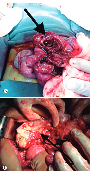
FIGURE 1. (A and B): Surgical fi nding of the abdominal mass (arrows). A
B
FIGURE 2. Pathological anatomy of the abdominal tumor revealing hyphae
with thick walls, chambers, and bud formation.
had an irregular shape, measured 9cm at its greatest diameter, and weighed 185g.
We initiated empirical treatment for tuberculosis due to the severity of clinical symptomsand the suspicion of visceral disease (insidious daily fever, loss of weight and appetite) as well as broad-spectrum antibiotic therapy for sepsis. In the meantime, the anatomopathological analysis results suggested mucormycosis (Figure 2): frequent hyphae with thick walls, irregular branching, chambers, shoots forming on refractile walls, and heavy staining by silver salts. In addition to vascular wall invasion in the abdominal region, the bone marrow immunophenotyping was suggestive of myelodysplasia with no other remarkable fi ndings. No other focus of mucormycosis was identifi ed in other organs.
Intravenous amphotericin B (1mg/kg/day) was started on hospital day 41. However, the patient already had multiple organ dysfunction at that time. Despite clinical support in the pediatric intensive care unit,her clinical condition deteriorated, and the patient died on day 54 after admission.
DISCUSSION
Mucormycosis is a rare and often life-threatening disease that is commonly seen in patients who have immunodefi ciency syndromes such as malignancies,undergone solid organs transplant, received long-term steroid therapy, and poorly controlled diabetes. However, no underlying condition is discovered in 19% of cases. Of these cases, 9% have gastrointestinal manifestations of the disease(10). In the reported case, the patient had no reported underlying comorbidities, which contributed to the delayed diagnosis. However, the bone marrow immunophenotyping revealed myelodysplasia, which could have been a sign of a previous underlying condition, such as a myelodysplastic syndrome, that would better explain the presence of mucormycosis in this child.
The most common presentation of abdominal mucormycosisis reported in adults, althoug husually in the abdominal wall instead of intraabdominally, which made the diagnosis in our patient more diffi cult.
798
Rev Soc Bras Med Trop 49(6):796-798, November-December, 2016
The therapeutic approach relies on an attempt to reverse or reduce the predisposing framework, facilitating surgical debridement and the immediate start of antifungal therapy.The most commonly used drug is liposomal or classical amphotericin B in high daily doses, which is 94% effective against Mucor spp,
with a minimum inhibitory concentration<1ug/mL.The suggested dose is 1.0mg/kg/day for conventional amphotericin B (sodium deoxycholate) and 5-7.5mg/kg/day for liposomal amphotericin B. The antifungal effi cacy of the combination of amphotericin B plus a triazole or caspofungin, for example, has yet to be demonstrated. The use of posaconazole, a second-generation triazole derivative, is considered a rescue option for patients refractory or intolerant to amphotericin B(12).
In conclusion, mucormycosis with anintra-abdominal presentation in pediatrics remains a disease that is poorly understood and with a high mortality, requiring a high degree of clinical suspicion for the diagnosis. Currently, the triad of clinical awareness, prompt initiation of treatment, and timely surgical intervention effectively controls the disease, avoids the main complications, and increases the chance of a better outcome.
Acknowledgments
We offer our deepest thanks to the institutions that provided technical support
for the development and implementation of this study.
Confl ict of Interest
The authors declare that there are no confl icts of interest.
REFERENCES
1. Prabhu RM, Patel R. Mucormycosis and entomophthoramycosis: a review of the clinical manifestations, diagnosis and treatment.
ClinMicrobiol Infect 2004; 10 (suppl 1):31-47.
2. Dromer F, McGinnis MR. Zygomycosis. In: Anaissie EJ, McGinnis
MR, Pfaller MA, editors. Clinical mycology. New York: Churchill
Linvigstone; 2003. p. 297-308.
3. Roden MM, Zaoutis TE, Buchanan WL, Knudsen TA, Sarkisova TA, Schaufele RL, et al. Epidemiology and outcome of zygomycosis: a review of 929 reported cases. Clin Infect Dis 2005; 41:634-653. 4. Bonifaz A, Macias B, Paredes-Farreira F, Arias P, Ponce RM,
Araiza J. Palatal zygomycosis: experience of 21 cases. Oral Dis 2008; 14:569-574.
5. Michalak DM, Cooney DR, Rhodes KH, Telander RL, Kleinberg F. Gastrointestinal mucormycoses in infants and children: a cause of gangrenous intestinal cellulitis and perforation. J Pediatr Surg 1980; 15:320-324.
6. Garg PK, Gupta N, Gautam V, Hadke NS. Gastric mucormycosis: unusual cause of gastric perforation in an immunocompetent patient. South Med J 2008; 101:449-450.
7. Ismail MH, Hodkinson HJ, Setzen G, Sofi anos C, Hale MJ. Gastricmucormycosis. Trop Gastroenterol 1990; 11:103-105. 8. Zaoutis TE, Roilides E, Chiou CC, Buchanan WL, Knudsen
TA, Sarkisova TA, et al. Zygomycosis in children: a systematic
review and analysis of reported cases. Pediatr Infect Dis J 2007; 26:723-727.
9. Roilides E, Zaoutis TE, Walsh TJ. Invasive zygomycosis in neonates
and children. Clin Microbiol Infect 2009; 15 (suppl 5): 50-54.
10. Marques SA, Camargo RMP, Abbade LPF, Marques MEA. Mucormicose: infecção oportunística grave em paciente imunossuprimido. Relato de caso. Diagn Tratamento 2010; 15:64-68. 11. Tarrand JJ, Lichterfeld M, Warraich I, et al. Diagnosis of invasive
septate mold infections. A correlation of microbiological culture
and histologic or cytologic examination.Am J ClinPathol 2003; 119: 854-856.
12. Spellberg B, Walsh TJ, Kontoyiannis DP, EdwardsJr J, Ibrahim AS.