• Nenhum resultado encontrado

PDF EN Jornal Brasileiro de Pneumologia 2 15 english

N/A
N/A
Protected

Academic year: 2018

Share "PDF EN Jornal Brasileiro de Pneumologia 2 15 english"

Copied!
4
0
0

Texto

(1)

180 Fortuna FP, Irion K, Wink C, Boemo JL

J Bras Pneumol. 2006;32(2):180-3

Mounier-Kuhn syndrome*

FABRÍCIO PICCOLI FORTUNA1, KLAUS IRION2, CESARE WINK3, JORGE LUIS BOEMO4

* Study carried out at the Hospital Geral de Caxias do Sul (HGCS, Caxias do Sul General Hospital), Caxias do Sul, Rio Grande do Sul, Brazil.

1. Pulmonologist. Chief Resident in the Pulmonology Department of the Hospital Geral de Caxias do Sul (HGCS, Caxias do Sul General Hospital), Caxias do Sul, Rio Grande do Sul, Brazil

2. Radiologist. PhD in Pulmonology from the Universidade Federal do Rio Grande do Sul (UFRGS, Federal University of Rio Grande do Sul), Porto Alegre, Rio Grande do Sul, Brazil

3. Resident in the Department of Pulmonology of the Hospital Geral de Caxias do Sul (HGCS, Caxias do Sul General Hospital), Caxias do Sul, Rio Grande do Sul, Brazil

4. Cardiologist. Captain and Physician-in-Chief of the Department of Clinical Medicine, Garrison Hospital at Bagé, Bagé, Rio Grande do Sul, Brazil

Correspondence to: Rua Moreira César 2.821, 7o andar, Ed. Gaudí - CEP: 95034-000, Caxias do Sul, RS, Brasil. Tel.: 55 54 3221-4195. E-mail: fabriciofortuna@terra.com.br

Submitted: 9 February, 2005. Accepted, after review: 16 June 2005.

ABSTRACT

Mounier-Kuhn syndrome, or tracheobronchomegaly, is a rare clinical entity characterized by abnormal dilation of the trachea and main bronchi. The diagnosis can usually be made by measuring the tracheal diameter. We report the case of a 40-year-old black man with refractory lower respiratory tract infection. Tracheobronchomegaly was confirmed through computed tomography.

Keywords: Tracheobronchomegaly/diagnosis; Tomography, X-ray computed; Tracheobronchomegaly/rehabilitation;

Case reports [publication type]

An inadequate clinical response to treatment in a case of community-acquired pneumonia should alert the clinician to a variety of factors that might be involved in the treatment failure. Such factors include the choice of an inappropriate antibiotic, the presence of uncommon pathogens and infectious complications at other sites, as well as alternative diagnoses of noncommunicable diseases or immune disorders. Occasionally, in these patients, some anatomical factor is found to be responsible. Among all the possible anatomical alterations capable of causing an increased risk of pneumonia, as well as creating difficulty in eradicating the established process, is congenital tracheobronchomegaly, or Mounier-Kuhn syndrome.

INTRODUCTION

The objective of the present study is to present a case of community-acquired pneumonia in which Mounier-Kuhn syndrome was identified as the cause of the treatment failure.

CASE REPORT

A 40-year-old black male patient was referred to the pulmonology clinic due to fatigue upon exertion and persistent dry cough three weeks after having been discharged from the hospital, where he had been admitted for treatment of pneumonia. The patient had presented, 40 days prior, a sudden onset of fever, a productive cough with a limited amount of hemoptysis, and

(2)

J Bras Pneumol. 2006;32(2):180-3

Mounier-Kuhn syndrome 181

dyspnea. He was diagnosed with pneumonia and was hospitalized, receiving intravenous cefazolin for seven days. He presented favorable evolution. The patient also reported having experienced three previous episodes of pneumonia, the first at the age of 22. The chest X-ray performed at the time of the evaluation revealed discrete areas of consolidation, of residual aspect, in the middle lobe, as well as a significant increase in the diameter of the trachea and of the main bronchi. The patient underwent empirical treatment with a bronchodilator for two weeks, with no improvement, and then returned for a reevaluation. The cough had worsened, becoming productive. The chest X-ray taken at that time revealed new areas of consolidation in both lower lobes and in the middle lobe. There was no alteration in the vital signs, and the patient presented good general status. A t e n - d a y c o u r s e o f o r a l e r y t h r o m y c i n w a s prescribed. Over the first three days, the cough and fatigue lessened. By the end of the ten-day period, the cough had almost completely disappeared.

A few days after the treatment with erythromycin, however, the patient again presented productive cough, accompanied by fever, and the X-ray

revealed no improvement in the consolidation. The patient was again hospitalized and was submitted to sputum smear microscopy, which revealed innumerable polymorphonuclear cells and abundant gram-positive cocci. Sputum culture was unavailable. The patient started being treated with intravenous oxacillin, and a satisfactory clinical response was obtained in a few days, which was the response that had been expected after the administration of oral cephalexin. After having received oral cephalexin for 48 h, the patient again presented fever, which led us to return to the intravenous treatment and transfer him to a hospital possessing more advanced equipment in order to perform a more elaborate etiological investigation. The new sputum smear microscopy revealed polymicrobial flora, and the patient was started on ceftriaxone combined with amikacin. This treatment continued for four weeks, by which time the symptoms had been completely resolved. At that point, the patient was submitted to a computed tomography scan of the chest, which revealed that the areas of consolidation, although smaller in size, persisted in both lower lobes, and confirmed the abnormal widening of the trachea and main bronchi (Figures 1, 2 and 3). In the lower

(3)

182 Fortuna FP, Irion K, Wink C, Boemo JL

J Bras Pneumol. 2006;32(2):180-3

lobes, there was bilateral cylindrical bronchiectasis, which is consistent with a diagnosis of congenital t ra c h e o b ro n c h o m e g a l y, o r M o u n i e r - K u h n syndrome. Pulmonary function tests revealed an increase in total lung capacity (123% of predicted) and residual volume (160% of predicted), with no other alterations. Currently, the patient remains asymptomatic, and has daily respiratory therapy sessions. Annual immunization against influenza has been recommended.

DISCUSSION

Congenital tracheobronchomegaly, or Mounier-Kuhn syndrome, is a rare clinical entity, described for the first time in 1932,(1) and characterized by

marked tracheobronchial dilation and lower respiratory tract infections. It is predominantly found in male individuals in their 40s or 50s. Although the etiology of Mounier-Kuhn syndrome remains unknown, it is believed that it is related to a lack of smooth muscle and elastic connective tissue in the trachea and main bronchi, leading to herniation and even to the formation of diverticula between the cartilaginous rings. A finding of bronchiectasis, such as in the case in question, is uncommon.(2-3)

Mounier-Kuhn syndrome is occasionally found in asymptomatic individuals. When symptomatic, it is characterized by recurrent pneumonia, sometimes evolving to chronic productive cough, occasional hemoptysis, and progressive dyspnea as a result of the pulmonary involvement. More rarely, massive hemoptysis,(4) spontaneous pneumothorax,

and digital clubbing can be observed. We believe that, in the case in question, the syndrome was responsible for the inappropriate response to the appropriate antibiotic therapy, which led us to suspect of a structural abnormality of the airways.

Diagnosis can usually be made by measuring the tracheal diameter, using only data from chest X-rays, in which the trachea can seen in profile and thus the diameter determined. Computed tomography of the chest, however, makes this measurement more precise. The limits are 3 cm for the transverse diameter of the trachea, and 2.4 cm and 2.3 cm for the transverse diameters of the main right and left bronchi, respectively. Pulmonary function tests typically reveal an increase in total lung capacity, to the detriment of residual volume, occasionally with signs of obstructive ventilatory disorder. Currently, there are no specific treatments for this condition, except for antibiotic therapy during the crises and respiratory therapy in order to eliminate secretions, if necessary. The use of permanent prostheses is reserved only for selected advanced cases since there are no precise indications for the practice.(5-6)

Other presentations of widening of the lower airways can be mistaken for Mounier-Kuhn syndrome.(7) Among such presentations are those

Figure 2 - Computed tomography of the chest in axial planes revealing marked dilatation of the main right and left bronchi, which measured 28 mm and 25 mm, respectively. The spaces between the cartilaginous rings can be clearly seen

(4)

J Bras Pneumol. 2006;32(2):180-3

Mounier-Kuhn syndrome 183

caused by congenital disorders such as Ehlers-Danlos syndrome and Williams-Campbell syndrome (the latter, a rare form of cystic bronchiectasis resulting from cartilage deficiency from the fourth to the sixth bronchial order, with normal trachea and main bronchi). Disorders such as sarcoidosis, usual interstitial pneumonia and cystic fibrosis cause severe fibrosis of the upper lobes, leading to tracheal retraction. Inflammatory processes of the airways, such as allergic bronchopulmonary aspergillosis, caused by the colonization of the airway with Aspergillus sp., are also involved in the widening of the airways. In addition, it is known that smoking, chronic obstructive pulmonary disease, and repeated infections can worsen the profile.

A careful evaluation of the airway anatomy is very important in patients presenting recurrent pneumonia, productive chronic cough, or, as in the case in question, incomplete response to appropriate antibiotic therapy for pneumonia. M o u n i e r - K u h n s y n d r o m e , a l t h o u g h r a r e , constitutes a diagnostic possibility in these patients and must be considered.

REFERENCES

1. Mounier-Kuhn P. Dilatation de la trachée: constatations radiographiques et bronchoscopiques. Lyon Med. 1932;150:106-9.

2. W i e s n e r B , M a d e r I , L e o n h a r d i J , S t r a u s s H J . [Tracheobronchomegaly Mounier-Kuhn syndrome case report and review of the literature]. Pneumologie. 1997;51(3):291-5.German.

3. Arranz Caso JA, Fernandez Frances J, Jimenez Jurado D, Manzano Espinosa L. [Mounier-Kuhn syndrome. 2 representative cases of its clinical spectrum]. Rev Clin Esp. 1996;196(4):237-9. Spanish.

4. Haro M, Vizcaya M, Jimenez Lopez J, Nunes A, Loeches N, Mansilla F. [Tracheobronchomegaly: an exceptional predisposing factor for pulmonary aspergillomas and massive hemoptysis]. Arch Bronconeumol. 2000;36(2): 103-5. Spanish.

5. Genta PR, Costa MV, Stelmach R, Cukier A. A 26-yr-old male with recurrent respiratory infections. Eur Respir J. 2003;22(3):564-7.

6. Woodiring JH, Howard RS II, Relun SR. Congenital tracheobronchomegaly (Mounier-Kuhn syndrome): a report of 10 cases and review of the literature. J Thorac Imaging. 1991;6(2):1-10.

Imagem

Figure 1 - Anteroposterior chest X-ray revealing areas of consolidation in both lower lobes (predominantly on the right side) and widening of the trachea and bronchi
Figure 3 - Computed tomography of the chest with multiplane three-dimensional reconstruction revealing the abnormal dimensions of the trachea and main bronchi

Referências

Documentos relacionados

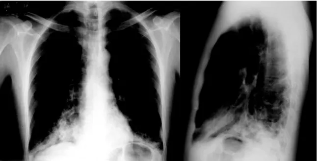
A computed tomography scan of the chest revealed atelectasis of the right upper lobe (caused by occlusion of the upper lobe bronchus) that extended up to the juxtacarinal portion of

The working diagnosis was superior vena cava syndrome, and the patient was submitted to computed tomography of the chest, which revealed a mass in the anterior mediastinum, with

A computed tomography scan of the chest revealed a 4-cm mass with heterogeneous content and pleural extension to the level of the lingula, as well as two micronodules in

Computed tomography of the chest revealed consolidation with interposed cavitation in the right upper lobe.. Fiberoptic bronchoscopy revealed purulent fluid within

Food biotechnology, Food safety, Food quality and safety, Process Engineering, Life science Technology, Nutrition & Food science, Food and nutrition, Food quality, safety

A computed tomography scan of the chest revealed a 4-cm mass with heterogeneous content and pleural extension to the level of the lingula, as well as two micronodules in

Ousasse apontar algumas hipóteses para a solução desse problema público a partir do exposto dos autores usados como base para fundamentação teórica, da análise dos dados

ECG, computed tomography scan, TTE) that identify and describe the size, type and setting of the lesion and general assessment. In our case, the patient had a cardiac tamponade due