AR
TIGO ORIGINAL / ORIGINAL AR
TICLE
INTRODUCTION
Inlammatory bowel disease (IBD) is a group of chronic conditions affecting the digestive tract in different locations(11). The mechanisms
under-lying the disease are not yet fully understood and its incidence has been increasing since the end of the past century, with important consequences for public health(26).
Crohn’s disease (CD) and ulcerative colitis (UC) are the most common presentations of IBD(12). Both
are characterized by chronic inlammation at intesti-nal and systemic level that seems to arise in response to an abnormal immunological reaction to the intestinal microbiota or other stimuli in susceptible individuals(3, 22). Throughout its course IBD is also
characterized by periods of activity and remission and features a wide variety of manifestations at intestinal and extra-intestinal sites(1, 8, 12). According
to some authors, 25%-30% of the patients diagnosed with IBD have at least one extra-intestinal
manifes-ORAL MUCOSA LESIONS AND manifes-ORAL
SYMPTOMS IN INFLAMMATORY BOWEL
DISEASE PATIENTS
Nuno
LARANJEIRA
1, Jorge
FONSECA
1,2, Tânia
MEIRA
2, João
FREITAS
2;
Sara
VALIDO
1and Jorge
LEITÃO
3ABSTRACT - Background - Inlammatory Bowel Disease is known for its extra intestinal manifestations, the oral cavity is no exception.
Objectives - The aim of this study was to evaluate the association between Inlammatory Bowel Disease and oral mucosa lesions and symptoms, and complementary to evaluate their possible relation with oral hygiene, smoking habits, drug therapy, duration and activity of the disease. Methods - Patients were selected from the Gastroenterology Clinic of a Portuguese tertiary referral hospital. This sample consisted of 113 patients previously diagnosed with ulcerative colitis or Crohn’s disease along with a control group of 58 healthy individuals that were accompanying the study group patients to their appointments. Clinical interviews and clinical examina-tions were performed for data collection. Results - The patients in the study group were more affected by oral symptoms (P=0.011), and showed a trend towards a higher incidence of oral mucosal lesions, even though statistical signiicance was not reached (8.8% versus 3.4% in the control group; P=0.159). Patients in active phase were the most affected. No differences were detected between Crohn’s disease and ulcerative colitis, or concerning smoking habits. The corticosteroid and immunosuppressant therapy seemed to increase the incidence of oral symptoms (P=0.052). The oral mucosa lesions increased and the oral symptoms decreased over the course of the disease, however without statistical signiicance. Conclusion - Oral mucosa’s lesions and oral symptoms were positively associated with Inlammatory Bowel Disease, mainly during disease activity periods and conceivably, associated with corticosteroid and immunosuppressant therapy.
HEADINGS - Inlammatory bowel diseases. Aphthous stomatitis. Mouth mucosa.
Declared conflict of interest of all authors: none Disclosure of funding: no funding received
1 Egas Moniz Interdisciplinary Research Center, CiiEM, Egas Moniz Health Science Institute, Almada; 2 Gastroenterology Service, Garcia de Orta Hospital, Almada; 3 Institute of Health Sciences, Portuguese Catholic University, Viseu. Portugal.
Correspondence: Nuno Laranjeira. Instituto Superior de Ciências da Saúde Egas Moniz, Campus Universitário, Quinta da Granja, Monte de Caparica, 2829 - 511, Caparica, Almada, Portugal. E-mail: [email protected]
tation, being the oral cavity one of the most affected areas(1, 8, 21). The oral lesions include affections in the
mucosa, periodontal tissues and tooth tissue, and have been said to play a role as an early indicator of IBD or at least as concomitant manifestations that could help with the diagnose(6, 10, 12, 19, 24).
In general, oral mucosal lesions described in the literature may arise either in patients with CD or UC and may be speciic or nonspeciic for these patholo-gies(6, 8, 24). Some authors cite diffuse swellings of the
oral and perioral tissues as the most prominent fea-tures(18) but there have been reports of a great variety
of pathological features in this patients(2, 15).
Further-more, it has been observed a higher prevalence of other oral symptoms as halitosis, nausea, xerostomia and regurgitation in patients with IBD(15).
METHODS
The study was conducted at General Gastroenterology and Inlammatory Disease Clinic of the Hospital Garcia de Orta (HGO), in Almada, Portugal. The protocol used for data collection was previously reviewed and approved by the Ethics Commission of HGO.
Patients who had been previously diagnosed with ulcer-ative colitis or Crohn’s disease and who agreed to participate in this study were selected, having healthy individuals which were accompanying the study group patients to their appoint-ments as the control group. Informed consent was obtained from all participants and the study design was in accordance with the Declaration of Helsinki.
Exclusion criteria were: patients under 18 years old, pregnant women, edentulous patients, patients with ixed orthodontic appliances, patients in need of prophylaxis for bacterial endocarditis, patients without a clear diagnosis of ulcerative colitis or Crohn’s disease and patients with other diseases that could interfere with the variables in study.
The assessment of disease activity was previously per-formed by the HGO gastroenterology team according to the Harvey-Bradshaw Index(14) for CD and according to the
Montreal Index of Activity(23) for UC. The diseases were
considered inactive for scores of 5 or less in the Harvey-Brad-shaw Index (CD) and for S0 ratings in the Montreal Index of Activity (UC).
The patients were divided in ive groups referring to the drug therapy: untreated; under salicylate therapy; under corticosteroids therapy; treated with immunosuppressant drugs - azathioprine and cyclosporine - and under biological therapy - inliximab and adalimumab. Patients treated with more than one drug were included in the group that most inluences the evolution of the disease. In increasing order: salicylates, corticosteroids, immunosuppressant drugs, bio-logical therapy.
Two groups were considered for the assessment of smoking habits: smokers and non-smokers. All individuals who reported smoking habits regardless of frequency were considered in the smokers group. Ex-smokers were classiied as non-smokers.
Disease duration was divided into three ranges: short term [up to 3 years]; average term [more than 3 to 9 years] and long term [longer than 9 years].
Clinical data
Patients admitted in the study answered a standardized questionnaire, in order to record the following information: age, gender, medical history, medication, smoking habits and oral hygiene habits.
Patients were also asked to indicate all the oral symp-toms they have experienced recently from a list containing: xerostomia, halitosis, dysphagia, regurgitation, dysgeusia and acid taste.
Oral examination
Data was collected by a team directed by the main
inves-tigator. Clinical observation was always performed by the same examiner, while another research team element pointed out data simultaneously, eliminating protocol differences and preserving the results agreement.
The search for oral lesions in the mucosa and surrounding soft tissues, started at the lips vermillion, then labial mucosa, followed by the buccal mucosa, gums, palate and tongue in this speciic order. All the oral lesions detected were then registered.
Statistical analysis
The data collected from the questionnaires and intraoral examination was inserted in Microsoft Excel™ and SPSS™ (Statistical Package for Social Sciences) for analysis and statistical processing.
To describe and summarize the data obtained in the sample, descriptive statistics measures were performed. Kolmogorov-Smirnov tests were used to test the normality of the sample; Spearman correlation tests, Fisher exact test and Kruskal-Wallis tests were then used to verify the cor-relation strength between these variables with a conidence level of 95%.
RESULTS
Sample characterization
Initially, 183 individuals were observed, from which 12 did not meet the established criteria. As a result, a inal number of 171 individuals were included in the study - 113 (66.1%) patients in the study group and 58 (33.9%) individuals in the control group.
The average age of the sample was 45.5±16.9 years, the minimum age was 19 years old and maximum 81 year old. Eighty-ive (49.7%) individuals were male and 86 (50.3%) were female. In the total sample there was 85.4% (n=146) of non-smoker subjects, and 14.6% (n=25) of smoker patients.
The IBD study group consisted of 57 (50.4%) males and 56 (49.6%) females, with a mean age of 44.5±17.2 years, with a minimum of 19 and maximum of 81 years old. 83.2% (n=94) of this group were non-smoker patients, against 16.8% (n=19) smokers.
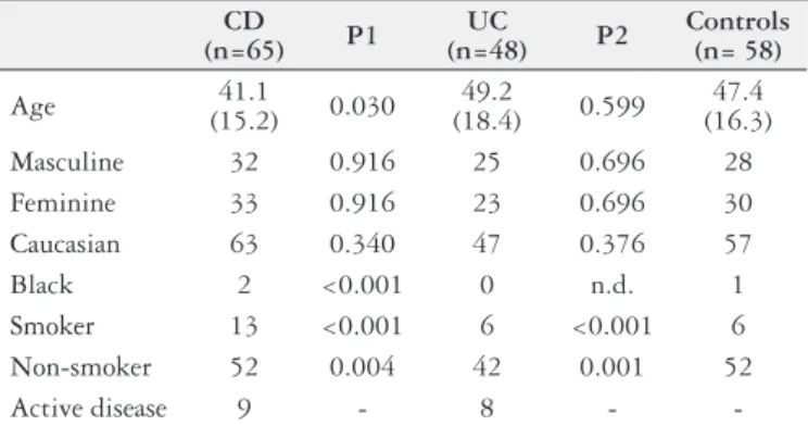
A total of 15% (n=17) of the 113 patients were under-going active disease, and 85% (n=96) were in remission. The most common diagnosis was Crohn’s disease in 57.5% (n=65), the remaining patients (n=48) were diagnosed with ulcerative colitis (Table 1).
The control group was composed of 28 (48.3%) male and 30 (51.7%) female individuals. The mean age was 47.4±16.3 years, with a minimum of 19 and maximum of 77 years old. Fifty-two (89.7%) were non-smoking, against 6 (10.3%) smokers (Table 1).
FIGURE 2. Distribution of lesions of the oral mucosa in the study and control groups.
TABLE 1. Demographic data of the groups (CD, UC and Control) for our sample
CD
(n=65) P1
UC
(n=48) P2
Controls (n= 58)
Age 41.1
(15.2) 0.030
49.2
(18.4) 0.599
47.4 (16.3)
Masculine 32 0.916 25 0.696 28
Feminine 33 0.916 23 0.696 30
Caucasian 63 0.340 47 0.376 57
Black 2 <0.001 0 n.d. 1
Smoker 13 <0.001 6 <0.001 6
Non-smoker 52 0.004 42 0.001 52
Active disease 9 - 8 -
-(χ) Standard deviation of respective datasets. (P1): signiicance of the differences between CD and Control; (P2): signiicance of the differences between control and UC.
2.50
2.00
1.50
1.00
0.50
0.50
S-OHI Teeth Brushing Dental floss Mouthwashes
Study Control
Clinical data
Case-control comparison
In the study group we detected the presence of aphthous ulcers (n=10), gingival swelling (n=1) and angular cheilitis (n=1). Only aphthous ulcers and gingival swelling were found on the control group individuals, (n=1) each. (Figure 2)
10.00%
9.00%
8.00%
7.00%
6.00%
5.00%
4.00%
3.00%
2.00%
1.00%
0.00%
Aphtous Ulcers Gingival Swelling Angular Queilitis
Control 1.70% 1.70% 0%
Study 8.80% 0.90% 0.90%
% of the sample
25.0%
20.0%
15.0%
10.0%
5.0%
0.0%
% of the sample
Halitosis Regurgitation Acid Taste Xerostomia Dysphagia Dysgeusia
Control 21.2% 20.4% 16.8% 12.4% 10.6% 9.7%
Study 13.8% 12.1% 8.6% 5.2% 1.7% 6.9%
FIGURE 1. Simpliied Oral Hygiene Index, number of daily brushings, interdental cleaning and use of mouthwashes in the study and control groups.
Lesions of the oral mucosa showed higher prevalence in the study group compared with the control group (8.8% versus 3.4%), even though this difference was not statistically signiicant (P=0.159).
The IBD group and the control group have also prevailed in several oral symptoms, such as halitosis, regurgitation, acid taste, xerostomia, dysphagia and dysgeusia. (Figure 3) The differences in the prevalence of oral symptoms in the study group when compared with the control group as presented in Figure 3, was statistically signiicant (P=0.011). Patients with IBD reported oral symptoms in 54.9% of cases compared with only 29.3% of the control group. Dysphagia (P=0.038) revealed to have a statistically signiicant association with IBD. Even when analyzed separately, none of the other symptoms showed statistical signiicance for this association.
FIGURE 3. Distribution of oral symptoms in the study and control groups.
Activity of IBD
The oral mucosa lesions revealed a higher prevalence in the active phase of the disease (35.3% versus 4.2%), (P<0.001). Aphthous ulcers proved to be the only type of affections with a statistically signiicant association to the active phase (P=0.001), with 35.3% of patients in the active stage diagnosed with this type of oral lesion versus 4.2% in remissive phase.
Patients in active phase reported oral symptoms in 70.6% of cases compared to only 52.1% of the patients in remis-sion. This represented a statistically signiicant difference (P=0.001). Dysphagia (P=0.018), regurgitation (P=0.044) and acid taste (P=0.038) revealed to have a statistically sig-niicant association with the active phase of IBD, even when analyzed separately.
Crohn’s disease vs ulcerative colitis
When analyzed separately neither of oral mucosa le-sions or symptoms had a statistically signiicant relation with CD or UC.
Pharmacotherapy
Untreated patients at the time of examination revealed no incidence of oral mucosa lesions (0%) and patients under corticosteroids treatment were the most affected by these lesions (22%). Nevertheless, considering all the different types of medication used by the patients, the pharmacolog-ical therapy of IBD did not show a statistpharmacolog-ically signiicant relation with the presence of lesions of the oral mucosa (P=0.711).
Individuals that were not under any type of medication also revealed less incidence of oral symptoms (38.5%). Patients taking salicylates were the least affected by these symptoms (50%), among medicated patients. On the op-posite, 66.7% of the patients under Immunossupressants and Corticosteroids reported oral symptoms. In spite of these results, there was no statistically signiicant relation between oral symptoms and the pharmacological therapy of IBD (P=0.052).
Smoking habits
Non-smokers were the most affected by lesions of the oral mucosa with 9.57% (n=9), compared to 5.26% (n=1) of the smokers group. This relation was not statistically signiicant (P=0.199).
When assessing the presence of oral symptoms between smoker patients and non-smoking patients no statistically signiicant relation was observed (P=0.595).
When analyzed separately neither of oral mucosa lesions or symptoms had a statistically significant relation with smoking habits.
Disease duration
The different types of oral mucosa lesions showed a slight increasing tendency in incidence over the course of IBD, however, not in a statistically signiicant way (P=0.670).
Oral symptoms decreased over the course of the dis-ease, still this decrease was also not statistically signiicant (P=0.720).
When analyzed separately neither of oral mucosa lesions or symptoms had a statistically signiicant relation with the disease duration.
DISCUSSION
This was the irst large study on oral health of Portuguese IBD patients, focusing a wide range of oral symptoms and mucosal lesions. In our study, patients within IBD group presented a higher prevalence of oral mucosal lesions when compared with the control group, supporting what was pre-viously reported in the literature(2, 5, 12, 15, 25), although in our
case without statistical signiicance. Patients with IBD often present more oral mucosal lesions than healthy controls, in accordance with the results of our study.
Few authors have studied the relation between IBD and its possible oral symptoms. The study by Katz and colleagues(15) was the irst to establish this relation in a
sample of 54 patients with IBD and 42 healthy subjects. As in our results, this study found that patients with IBD were more likely to experience oral symptoms when compared with a control group of healthy individuals. More recently, other authors have conirmed the same indings for patients with UC(7).
In the active phase of the disease, aphthous ulcers, proved to be the most common type of lesion in the oral
mucosa in our study (P=0.001), as already reported by
some authors(14, 18). In those studies, no further association
between any other type of oral mucosa lesions and the ac-tive phase of IBD was established. Although these data are in agreement with our results, other authors(5, 12) in similar
studies have not identiied a correlation between activity of IBD and any type of oral mucosa lesions. Other lesions previously cited as possible manifestations of IBD(4, 16)
were unusual in our sample and, therefore, did not reach statistical signiicance in the present study.
There was a greater presence of symptoms in active phase of IBD, 70.6% of cases in the active phase compared to only 52.1% of patients in remission, representing a statistically signiicant difference. A previous study, also pointed towards a higher incidence of oral manifestations in active period of the disease, however that study failed to demonstrate statistical signiicance(15).
In our sample 10.8% of CD patients presented oral mu-cosa lesions against 11.4% of UC patients. This difference was not statistically signiicant. Other authors had already reported no signiicant differences in prevalence of lesions between both diseases(13). However, there have been reports
in the literature considering that CD is more affected by oral lesions than UC(15, 17).
Although other studies(8, 9, 12, 19, 20, 24) associate
pyostoma-titis vegetans and orofacial granulomatosis with IBD, these data could not be conirmed in our population, since they showed no occurrence of these lesions. The recent phar-macotherapy of IBD had signiicant impact on the natural history and clinical presentation of patients. Eventually, the absence of some classical oral lesions in our study, with a large number of patients, may be result of the modern treatment of IBD.
Small differences can be found regarding oral symptoms between UC and CD, and those were not statistically signif-icant. Similar conclusions were already disclosed in Katz(15)
article, in which the author determined that even though both conditions showed a higher prevalence of oral symptoms compared with the control group, no signiicant differences existed between them.
higher incidence of oral symptoms. Conversely, immuno-suppressive therapy may be a surrogate marker of a more severe IBD and oral manifestations may be more frequent in those patients.
In our sample, non-smokers have been the most affected by oral mucosa lesions with 9.57% against 5.26% of smokers, although this difference was not statistically signiicant. 57.89% of smoking patients reported oral symptoms against 54.26% of non-smokers, which is not a statistically signiicant relation.
When considering lesions of the oral mucosa, we observed a trend towards increase in incidence over the course of IBD duration. For oral symptoms, there was a downward trend in incidence over the course of the disease, even though none of these relations proved to be statistically signiicant.
CONCLUSION
A positive association between inlammatory bowel dis-ease and oral mucosa lesions was established in this study, being IBD patients more affected, particularly in active phases of the disease. Aphthous ulcers were the most signif-icant expression of this group of manifestations.
The oral symptoms were also associated with IBD, with
dysphagia being the most reported symptom. Oral symp-toms proved to be even more prevalent in active periods of the disease.
The time since disease onset and pharmacological reg-imen may interfere with the incidence of oral lesions and symptoms, although in this study the differences detected were not statistically signiicant. Oral lesions seem to increase over time, while oral symptoms seem to decrease, what may be attributable to the fact that the ongoing drug therapy diminished these oral manifestations.
The association between IBD and oral mucosa lesions and symptoms represents a clinical challenge that should encourage dentists to plan a careful follow-up of these pa-tients, with the support of the gastroenterologist, in order to provide a better integrated treatment for these conditions.
Authors’ contributions
Laranjeira N, DMD, and Leitão J, PhD, MD, designed the study and participated in the interpretation of data, re-view, and approval of the manuscript. All authors had access to the data, contributed to the development of the content, reviewed each draft of the manuscript, and approved the inal content.
Laranjeira N, Fonseca J, Meira T, Freitas J, Valido S, Leitão J. Lesões das mucosas orais e sintomatologia oral em doentes com Doença Inlamatória Intestinal. Arq Gastroenterol. 2015,52(2):105-10.
RESUMO - Contexto - A doença inlamatória intestinal é conhecida por suas manifestações extraintestinais, a cavidade oral não é exceção. Objetivos - O
REFERENCES
1. Ardizzone S, Puttini PS, Cassinotti a, Porro GB. Extraintestinal manifestations of inlammatory bowel disease. Dig Liver Dis. 2008;40(Suppl 2):S253-9. 2. Asquith P, Thompson RA, Cooke WT. Oral manifestations of Crohn’s disease.
Gut. 1975;16(4):249-54.
3. Blumberg RS, Saubermann LJ, Strober W. Animal models of mucosal inlamma-tion and their relainlamma-tion to human inlammatory bowel disease. Curr Opin Immunol. 1999;11(6):648-56.
4. Boisnic S. Cheilitis. Rev Prat. 2002;52(4):370 4.
5. Brito F, de Barros FC, Zaltman C, Carvalho ATP, Carneiro AJDV, Fischer RG, et al. Prevalence of periodontitis and DMFT index in patients with Crohn’s disease and ulcerative colitis. J Clin Periodontol. 2008;35(6):555-60.
6. Daley TD, Armstrong JE. Oral manifestations of gastrointestinal diseases. Can J Gastroenterol. 2007;21(4):241-4.
7. Elahi M, Telkabadi M, Samadi V, Vakili H. Association of oral manifestations with ulcerative colitis. Gastroenterol Hepatol Bed Bench. 2012;5(3):155-60. 8. Fatahzadeh M. Inlammatory bowel disease. Oral Surg Oral Med Oral Pathol
Oral Radiol Endod. Elsevier Inc.; 2009;108(5):e1-10.
9. Flemmig TF, Shanahan F, Miyasaki KT. Prevalence and severity of periodon-tal disease in patients with inlammatory bowel disease. J Clin Periodontol. 1991;18(9):690-7.
10. Galbraith SS, Drolet B a, Kugathasan S, Paller AS, Esterly NB. Asymptomatic inlammatory bowel disease presenting with mucocutaneous indings. Pediatrics. 2005;116(3):e439-44.
11. Gassull M, Gomollón F, Hinojosa J, Obrador A. Enfermedad Inlamatoria Intestinal. 3rd ed. Madrid: Arán Ediciones; 2007.
12. Grössner-Schreiber B, Fetter T, Hedderich J, Kocher T, Schreiber S, Jepsen S. Prevalence of dental caries and periodontal disease in patients with inlammatory bowel disease: a case-control study. J Clin Periodontol. 2006;33(7):478-84. 13. Habashneh R a, Khader YS, Alhumouz MK, Jadallah K, Ajlouni Y. The
associ-ation between inlammatory bowel disease and periodontitis among Jordanians: a case-control study. J Periodontal Res. 2012;47(3):293-8.
14. Harvey RF, Bradshaw JM. A simple index of Crohn’s-disease activity. Lancet. 1980;1(8167):514.
15. Katz J, Shenkman a, Stavropoulos F, Melzer E. Oral signs and symptoms in relation to disease activity and site of involvement in patients with inlammatory bowel disease. Oral Dis. 2003;9(1):34-40.
16. Leão JC, Hodgson T, Scully C, Porter S. Review article: orofacial granulomatosis. Aliment Pharmacol Ther. 2004;20(10):1019-27.
17. Lourenço S V, Hussein TP, Bologna SB, Sipahi a M, Nico MMS. Oral manifes-tations of inlammatory bowel disease: a review based on the observation of six cases. J Eur Acad Dermatol Venereol. 2010;24(2):204-7.
18. Neville BW, Damm DD, Allen CM, Bouquot JE. Patologia Oral e Maxilofacial. 2nd ed. Koogan G, editor. Rio de Janeiro; 2004.
19. Ojha J, Cohen DM, Islam NM, Stewart CM, Katz J, Bhattacharyya I. Gingival involvement in Crohn disease. J Am Dent Assoc. 2007;138(12):1574-81. 20. Pittock S, Drumm B, Fleming P, McDermott M, Imrie C, Flint S, et al. The oral
cavity in Crohn’s disease. J Pediatr. 2001;138(5):767-71.
21. Rampton D. Inlammatory Bowel Disease Clinical Diagnosis and Manegement. London: Martin Dunitz; 2000.
22. Satsangi J, Morecroft J, Shah NB, Nimmo E. Genetics of inlammatory bowel disease: scientiic and clinical implications. Best Pract Res Clin Gastroenterol. 2003;17(1):3-18.
23. Satsangi J, Silverberg MS, Vermeire S, Colombel J-F. The Montreal classiication of inlammatory bowel disease: controversies, consensus, and implications. Gut. 2006;55(6):749-53.
24. Scheper HJ, Brand HS. Oral aspects of Crohn’s disease. Int Dent J. 2002;52(3):163-72. 25. Scully C, Cochran KM, Russell RI, Ferguson MM, Ghouri MA, Lee FD, et
al. Crohn’s disease of the mouth: an indicator of intestinal involvement. Gut. 1982;23(3):198-201.
26. Sleisenger MH. Pathophysiology of the gastrointestinal tract. In: Smith LH, Thie SO, editors. Pathophysiology: the biologic principles of disease. Philadelphia: The W. B. Sauders Co.; 1981. p. 1506-689.