Lara Maria HERRERA(a)
Raíssa Ananda Paim STRAPASSON(a) Luiz Eugênio Nigro MAZZILLI(a) Rodolfo Francisco Haltenhoff MELANI(a)
(a)University of São Paulo – USP, School of Dentistry, Departament of Social Odontology, São Paulo, SP, Brazil.
Differentiation between palatal rugae
patterns of twins by means of the Briñón
method and an improved technique
Abstract: Palatal rugae patterns are anatomic structures considered unique to each person. Monozygotic twins present similarities, however, Rugoscopy in particular, may contribute to their individualization for forensic purposes. The aims of this study were: to study the
palatal rugae classiications of Briñón; to propose improvements to facilitate use of this method, if pertinent; and to characterize palatal rugae in a sample of Brazilian monozygotic twins and singletons. Precise reproducibility of the two methods of Briñón, from 1982 and 2011, was prevented by poor intra-examiner agreement (70% and 13% respectively). Our proposed improvements to these methods,
although preliminary, were associated with better results. The most common palatal rugae patterns were types A, M, and Q. Palatal rugae
were conirmed to be unique to each individual, even in monozygotic twins. Furthermore, twins did not exhibit any special patterns that
might facilitate their differentiation from singletons.
Keywords: Forensic Sciences; Forensic Dentistry; Forensic Anthropology;
Palate; Twins.
Introduction
Palatal rugae are structures located on the palate and are formed in the third month of intrauterine life.1,2,3 In addition to aiding mastication, sense of taste, and facilitating proper placement of the tongue in the oral cavity, they may be used to establish human identity.1,4,5
The palatal ridges – their shape, position, number, and orientation – are unique to each individual.6,7 However, classiication of palatal rugae still represents a challenge because of the wide range of different methodologies available and, consequently, lack of standardization, as well as the subjective nature of interpreting these characteristics.8
With the purpose of improving recording and interpretation of palatal
rugae, Briñón9,10 proposed a classiication in 1982 (later updated in 2011)
similar to the ingerprint system. However, no studies testing or using either of these classiications have been published in the literature; thus,
their practical application remains unclear.
Several studies have sought to characterize ridge patterns in different populations,1,5,6,7,11,12 which could facilitate the identiication of groups in the event of a mass casualty incident.12 Research has also found that twins do Declaration of Interests: The authors
certify that they have no commercial or associative interest that represents a conflict of interest in connection with the manuscript.
Corresponding Author: Lara Maria Herrera [email protected]
DOI: 10.1590/1807-3107BOR-2017.vol31.0009
Submitted: Aug 24, 2015
not exhibit the same palatal rugae pattern.4,6,13,14 This
could be used to contribute to the their differentiation, as the similar physical characteristics of twins can
confuse identiication.
Within this context, the present study aimed to: study the palatal rugae classifications of Briñón;9,10 propose
improvements to these methods, seeking to facilitate their
use, if pertinent; and characterize palatal rugae patterns in a sample of Brazilian monozygotic twins and singletons.
Methodology
Study design and sample selection
This was a cross-sectional study. The sample, selected by a non-probabilistic convenience strategy, was composed of 10 pairs of adult monozygotic twins (4 men and 16 women, age 20 to 34 years) and 20 adult singletons (11 men and 9 women, age 22 to 35 years), all residing in the state of São Paulo, Brazil. The irst
part of this research project involved only twins, while the second and the third parts involved both twins and singletons.
None of the participants had distinguishing oral
clefts, lesions, inlammation, scar tissue, or grooves
in the palate. Individuals that were allergic to dental impression materials were not included.
Informed consent was obtained from each participant, and all procedures were performed in accordance with the research protocol approved by the
University of São Paulo School of Dentistry (FOUSP)
Research Ethics Committee, under administrative
procedure no. 1.014.974.
Rugoscopy record
An impression of the maxillary dental arch of each
individual was taken with irreversible hydrocolloid
material (alginate), and plaster casts were obtained.
Palatal ridges were delineated using a graphite pencil to facilitate their visualization and later analysis. Photographic records of the casts were obtained by
using two cameras (Nikon D80 and Nikon D3200)
positioned parallel to the surface of the palate.
The casts were classified by groups (twins and singletons) and numbered (from 1 to 20) by computer-based randomization (<www.random.org>). The numbers were
entered into a form containing the name, sex, and age
of the corresponding participant.
Analysis and classification of the palatal rugae
The rugoscopic structures of the twin subjects were
classiied in accordance with the methods of Briñón,9,10
as described in Figure 1. For each method, all casts were classiied six times, on different days, by a single observer (standard examiner). The sixth analysis was considered as the reference patterns (RP), and intra-examiner agreement coeficients were calculated.
Then, improvements to the methods were proposed and tested for the same sample of twins, by the same
examiner; intra-examiner agreement was calculated again. In addition, for a new sample of non-twin subjects, four casts were randomly selected (<www.random.org>). Two examiners performed four analyses of each cast, on different days, for a total of 16 readouts each. Percent intra- and inter-examiner agreement was calculated.
For all casts (twin and singleton subjects), rugoscopic structures were classiied by using the improved method. The standard examiner performed all assessments. Data
were then separated by pairs for recording and comparison. For singleton subjects, pairing was performed randomly.
Results
The classiication of Briñón9,10 for analysis of the
palatal rugae was evaluated. Out of 100 readouts assigned classiication “A”, the reproducibility of the method, assessed by means of intra-examiner agreement, was 70%. When comparing readouts with the RP, 46% were in disagreement. For classiication “B”, the reproducibility was 13%. When data were compared with the RP, 93% were in disagreement.
Our proposed improvements to the updated method of Briñón10 are listed below:
a. The accessory rugae were still assigned lower case letters and separated from the fundamental rugae
by a plus sign (“+”). However, as with fundamental
rugae of the same order, accessory rugae on the
same “line” were also separated by periods. When
there was a gap between accessory ridges in the
b. Under this new proposal, different types of rugae are no longer combined according to
angulation; e.g., those previously classiied as “BE” (type B rugae inclined to the left) were now classiied only as “B”;
c. Some rugae were incorporated into the same category by degree of similarity in shape,
extension, and angulation, and, consequently,
by the degree of subjectivity in differentiating
them as observed in the previous study;
A) Briñón method of 19829: B) Briñón method of 201110:
Carrea classification
Type I Type II Type III Type IV
Carrea
classification Not used
Fundamental rugae
• Start from the palatine raphe;
• Are denoted by consecutive ordinal numbers. The first fundamental rugae, i.e., that closest to the incisive papilla, is assigned the ordinal number 1; subsequent rugae are assigned the other numbers successively;
• Represented by capital letters.
Fundamental rugae
As in method “A”, but represented in a formula, separated by periods.
Accessory rugae
• Start from the alveolar margin; • Represented by capital letters.
Accessory rugae
• Start from the alveolar margin;
• Represented by lower case letters separated by a plus sign (+).
Form
• Represented by a single drawing; • Represented by the combination of several
drawings. Example: angled rugae would be assigned the letters “D” or “E”.
Form
• Represented by a single drawing;
• Represented by combinations of several drawings if
angulation ≥ 45 degrees;
• Each group of rugae, in numerical order, is
separated by a minus sign (−).
Drawings
A B C
D E F G H
I J K
L M N
O P Q
I+ J+
K+ L+
Drawings
A B C
D E F G H
I J K
L M N
O P Q R
Incisive papilla R, round; S, long; T, unified; U, separated. Incisive papilla
10 (round and separated); 11 (long and separated); 12 (round and unified);
13 (long and unified).
Code
The rugoscopic code is given by the Carrea classification, the sequence of the sum of rugae in each ordinal number for the right and left sides, and the classification of the incisive papilla.
Example: IV-1230/3600-RU
Formula
Two equations are formed: one for the right side (DX) and one for the left side (SX).
Example:
• DX10 = 1(L)−2(L+D+f)−3(D.G+m);
• SX10= 1(L+g+f)−2(D.O.G+e+d+d)
d. The combinations of curved rugae used by
Briñón10 were changed;
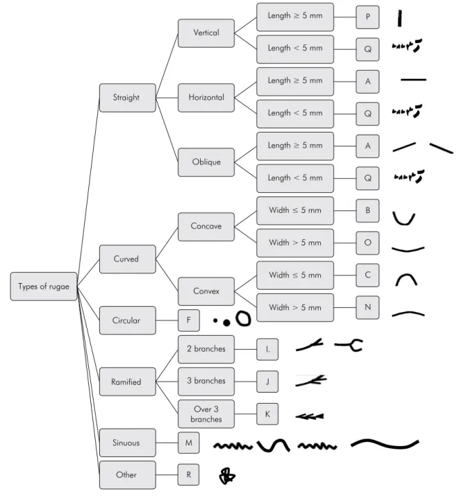
e. Some metric parameters were incorporated into the morphologic parameters in order to make interpretation of certain palatal rugae objective.
These parameters are illustrated in Figure 2.
Each group of rugae, in numerical order, continued
to be separated in the formula by the minus sign (“−”).
Classiication of the incisive papillae also remained
the same.
A total of 100 new readouts were performed using
the improvements proposed. The reproducibility
of the improved method, assessed by intra-examiner agreement, was 88%. When comparing readouts with the RP, of the 100 classifications obtained, 30% were in disagreement. Table 1
Figure 2. Proposed improvements to the 2011 Briñón classification.
P
Q
A
Q
A
Q
B
O
C
N
I.
J
K
M F
R Sinuous
Ramified Circular Curved
Oblique
Concave
Convex
2 branches
3 branches
Over 3 branches
Width > 5 mm Width ≤ 5 mm Width > 5 mm Width ≤ 5 mm Length < 5 mm Length ≥ 5 mm Length < 5 mm Length ≥ 5 mm Length < 5 mm Length ≥ 5 mm
Horizontal Vertical
Straight
Types of rugae
shows a comparison of results obtained with the improved method versus those obtained with the
method originally proposed by Briñón in 2011 (method “B”).
When the improved method was applied to a new sample of singleton subjects, its reproducibility for both
examiners was 93.75%, whereas the inter-examiner agreement was 25%.
We observed that no individual presented the
same classiication, even between pairs of twins.
Furthermore, the patterns found on the left side were not equal to those observed the right side.
Table 2 shows the total number of rugae and
repeated occurrences when the pairs were compared.
There was no statistically signiicant difference in
frequency of repetitions of palatal rugae between
groups (twins and singletons).
Table 3 presents the number of rugae, stratiied by
type, in twin and singleton subjects. No association was observed between types of rugae and groups
(p > 0.1). In this sample, the most prevalent types of
rugae were A, M, and Q.
Discussion
Rugoscopy represents a scientiically established auxiliary method of human identiication. However, many classiication systems are available15, and some
are still very dificult to use.
In this study, palatal rugae were classiied according to the schemes proposed by Briñón.9,10 To the best of
our knowledge, this was the irst report to scientiically
evaluate these methods.
We considered the intra-examiner agreement obtained with method “A” as insuficient (70%). For method “B”, intra-examiner agreement was even worse at 13%, indicating that the same trained observer was
unable to adequately adjust his agreement.
One of the key essential properties of any method of human identiication is classiiability, i.e., it must
be easy to record, amenable to iling, and allow data
to be retrieved later.16 The intra-examiner agreement results obtained for our updated methodology of
palatal rugae classiication were not consistent with the proposal of Briñón, who restructured the original 1982 method9 to clarify and speed
up classiication of palatal rugae for purposes of human identiication.10
An explanation for the better reproducibility of method “A” is the fact that the classiication generates a single inal numerical code. Therefore, the types of rugae are not shown in the classiication, only the number of rugae on the right and left maxillary hemiarches. For method “B”, on the other hand, the
description of all types of rugae in the formulas for both sides produced a greater number of variables,
improving examiner error. Furthermore, classiication
of rugae on the basis of a chart of figures was considered highly subjective.
The subjectivity of intra- and inter-examiner observations and of the methods of classiication
of palatal rugae was reported as a problem by Caldas et al.15 Gondivikar et al.11 also reported subjectivity in ascertaining the shape of palatal
rugae, although this parameter is extremely simple
to record. The subjectivity found in this study may be due to the variations in the shape and dimension of the same type of palatal rugae in the same individual and between individuals.
The important role played by palatal ridge anatomy in human identification should not be ignored. However, as two individuals may present with the same number of rugae in the palate, method
“B” (which includes different shapes) would have potential for use as a classiication scheme if some
changes were made to reduce subjectivity and increase reproducibility.
Palatal rugae are classiiable structures. However, the schemes proposed by Briñón are complex and,
as shown in this study, preclude precise reproducibility.
Table 1. Reproducibility of rugoscopic methods and classification of intra-examiner agreement.
Method Readouts (n) Intra-examiner agreement (%) Classifications in disagreement with reference pattern (%)
Briñón (2011) 100 13 93
We proposed a series of changes seeking to simplify
the updated Briñón classiication10 and obtain better
intra-examiner agreement.
The results were deemed more satisfactory in
terms of reproducibility between the two examiners, as the rate of code repetition was higher than 90%.
Uniformity between examiners could not be ensured, as demonstrated by the inter-examiner agreement of 25%. However, when comparing
classifications, the differences were perceived
to be small (up to three rugae per subject). This
was due to the fact that, our proposed changes notwithstanding, the number of variables was still very high. Given the subjectivity of classifying the shape of palatal rugae, this may create a sense of
insecurity in the examiner.
Although there was a substantial difference
between examiners, we cannot state that rugoscopy is not an effective method for identiication. This
is because, in forensic practice, identification is
performed by comparison between antemortem and postmortem records of the palate, not by comparison
of palatal rugae classiications.
Several authors1,5,6,7,17,18,19 have afirmed that the palatal rugae are unique in each individual, and the present study supports this statement. No subject presented the same set of palatal rugae, not even monozygotic twins.
As far as the uniqueness of palatal rugae in twins is concerned, these same findings were described in few studies, such as that of Ritter
(apud English et al.20), who concluded that the
pattern of palatal rugae between twins was very
similar, but not identical; and Lysell,4 who veriied
differences and similarities in rugoscopic patterns in monozygotic and fraternal twins. Kamala et al.,14 after analyzing a pair of twins and their parents, concluded that twins do not present the same palatal rugae patterns. Indira et al.6 evaluated palatal rugae patterns in ive pairs of dizygotic twins and found
these patterns to be different and unique in all subjects of the sample, despite some similarities
at speciic locations in two pairs of twins.
In the present study, on pairwise comparison, the degree of repeated occurrences of palatal rugae was
similar in twins and singletons; that is, monozygotic twin pairs did not exhibit any set of repetitions that
could differentiate them from the general population.
Table 3. Distribution of types of palatal rugae in twins and singletons.
Type Number of rugae in twins (n = 241) Number of rugae in singletons (n = 248) Chi-square
Present Absent Present Absent
A 62 179 53 195 1.05*
B 1 240 3 245
-C 5 236 3 245
-F 23 218 34 214 1.67*
I 25 216 19 229 0.79*
J 3 238 1 247 -
K 0 241 0 248
-M 43 198 57 191 1.68*
N 18 223 18 230
-O 16 225 8 240
-P 0 241 1 247
-Q 45 196 51 197 0.17*
R 0 241 0 248
-*p > 0.1 (not significant).
Table 2. Occurrence of coincident (repeated) palatal rugae in twins and singletons.
Variable Twins Singletons
Coincident 194 197
Non-coincident 47 51
Total 241 248
Moreover, the equivalence in types of rugae between the twin and singleton groups made it clear that both groups belonged to the same population.
The palatal rugae types A, M, and Q were the most prevalent in both groups. These results are similar to those reported in previous studies. Gondivkar et al.11, analyzing a western Indian population, found that the
most common type of palatal rugae was the “sinuous” type, corresponding to type “M” in the present
study. In a Portuguese sample studied by Santos and Caldas1, the most prevalent types of rugae were the
“straight” and “wavy”, corresponding to types “A” and “M” in the classiication used herein. Ibeachu
et al.,12 in a study of Nigerians, found that the most
frequent types were “wavy” rugae (corresponding to type “M” in our classiication) in the Igbo tribe and “curved” and “straight” rugae (corresponding to types “B/C/N/O” and “A” in our classiication, respectively) in the Ikwerre people.
Some authors have suggested that the predominance of certain palatal rugae types in different population
groups can be used for identiication purposes.1,11
Our study does not bear out this proposal, probably due to the heterogeneity in classiications used in
different studies, which make it impossible to perform a more faithful comparison.
Conclusion
A degree of subjectivity was observed when the
Briñón methods9,10 were used for classiication of palatal
rugae. The methods were not reproduced precisely.
However, the more recent version (2011) demonstrated potential for use as a classiication reference, as it takes
into account type rather than number of rugae. The satisfactory percentage of repetitions of palatal
rugae classifications (intra-examiner agreement) suggests that the improvements to the 2011 Briñón
classification10 proposed in this study, although preliminary, do contribute to the achievement of better results. Further investigation is needed to ascertain the effectiveness of the improved method
as compared with other palatal rugae classiications
proposed in the literature.
Palatal rugae are unique to each individual, even in
monozygotic twins, who, in this study, did not exhibit
any special rugae patterns that could differentiate them from other groups. Nevertheless, studies in larger samples are suggested.
Acknowledgments
The authors thank the volunteers for their participation in the study and the reviewers for their observations.
References
1. Santos C, Caldas IM. Palatal rugae pattern in a Portuguese population: a preliminary analysis. J Forensic Sci. 2012;57(3):786-8. doi:10.1111/j.1556-4029.2011.02016.x 2. Tornavoi DC, Silva RHAD. Rugoscopia palatina e a
aplicabilidade na identificação humana. Saúde Ética Justiça. 2010;15(1):28-34. doi:10.11606/issn.2317-2770.v15i1p28-34 3. Santos KC, Fernandes CMS, Serra MC. Evaluation of
a digital methodology for human identification using palatal rugoscopy. Braz J Oral Sci. 2011;10(3):199-203. 4. Lysell L. Plicae palatinae transversae and papilla incisiva
in man: a morphologic and genetic study. Acta Odontol Scand. 1955;13(suppl 18):5-137.
5. Saraf A, Bedia S, Indurkar A, Degwekar S, Bhowate R. Rugae patterns as an adjunct to sex differentiation in forensic identification. J Forensic Odontostomatol. 2011;29(1):14-9.
6. Indira A, Gupta M, David MP. Usefullness of palatal rugae patterns in establishing identity: preliminary results from Bengaluru city, India. J Forensic Dent Sci. 2012;4(1):2-5. doi:10.4103/0975-1475.99149
7. Dawasaz AA, Dinkar AD. Rugoscopy: predominant pattern, uniqueness, and stability assessment in the Indian Goan population. J Forensic Sci. 2013;58(6):1621-7. doi:10.1111/1556-4029.12190
8. Kesri R, Das G, Tote J, Thakur P. Rugoscopy: science of palatal rugae: a review. Int J Dent Med Res. 2014;1(4):103-7. 9. Briñón EN. Rugas palatinas. In: Briñón EN. Odontología
legal y práctica forense. Buenos Aires: Purinson; 1982. p. 291-317.
11. Gondivkar SM, Patel S, Gadbail AR, Gaikwad RN, Chole R, Parikh RV. Morphological study of the palatal rugae in western Indian population. J Forensic Leg Med. 2011;18(7):310-2. doi:10.1016/j.jflm.2011.06.007
12. Ibeachu PC, Didia BC, Arigbede AO. A comparative study of palatal rugae patterns among Igbo and Ikwerre Ethnic Groups of Nigeria: aUniversity of Port Harcourt Study. Anat Res Int. 2014;2014:ID123925. doi:10.1155/2014/123925
13. Fahmi FM, Al-Shamrani SM, Talic YF. Rugae pattern in a Saudi population sample of males and females. Saudi Dent J. 2001;13(2):92-5.
14. Kamala R, Gupta N, Bansal A, Sinha A. Palatal Rugae Pattern as an Aid for Personal Identification: a forensic study. J Indian Acad Oral Med Radiol. 2011;23(3):173-8.
15. Caldas IM, Magalhães T, Afonso A. Establishing identity using cheiloscopy and palatoscopy. Forensic Sci Int. 2007;165(1):1-9. doi:10.1016/j.forsciint.2006.04.010
16. Turano LM, Silva M. Noções de dactiloscopia. In: Silva M. Compêndio de odontologia legal. Rio de Janeiro: Medsi; 1990. p. 161-6.
17. De Angelis D, Riboli F, Gibelli D, Cappella A, Cattaneo C. Palatal rugae as an individualising marker: reliability for forensic odontology and personal identification. Sci Justice. 2012;52(3):181-4. doi:10.1016/j.scijus.2011.09.002 18. Shetty D, Juneja A, Jain A, Khanna KS, Pruthi N, Gupta A
et al. Assessment of palatal rugae pattern and their reproducibility for application in forensic analysis. J Forensic Dent Sci. 2013;5(2):106-9. doi:10.4103/0975-1475.119775 19. Barbieri AA, Scoralick RA, Naressi SC, Moraes ME,
Daruge E Jr, Daruge E. The evidence of the rugoscopy effectiveness as a human identification method in patients submitted to rapid palatal expansion. J Forensic Sci. 2013;58(S1):S235-8. doi:10.1111/j.1556-4029.2012.02263.x 20. English WR, Robison SF, Summitt JB, Oesterle LJ, Brannon