596
1. Service of Pediatric Cardiovascular Surgery of São José do Rio Preto - Hospital de Base -São José do Rio Preto Medical School, SP, Brazil.
Correspondence address: Ulisses Alexandre Croti
Hospital de Base – Faculdade de Medicina de São José do Rio Preto –
Ulisses Alexandre CROTI1, Domingo Marcolino BRAILE1, Daniela Patini ESPADA1, Betsy Maria VILLEGAS1 Rev Bras Cir Cardiovasc 2010; 25(4): 596-597
CLINICAL-SURGICAL CORRELATION
RBCCV 44205-1235
Correção de coarctação de aorta com interposição de tubo em criança de 12 anos
Correction of aortic coarctation with tube
interposition in a 12 years-old child
FAMERP – Avenida Brigadeiro Faria Lima, 5544 – São José do Rio Preto, SP, Brasil – CEP 15090-000
E-mail: [email protected]
Article received on October, 25th, 2010 Article accepted onNovember, 15th, 2010 CLINICAL DATA
12-year-old female child, 53 kg, brown, from Palestina, SP. She was referred to our hospital’s pediatric service presenting hypertension (240/140 mmHg) during surgery to remove a synovial cyst in the right hand.
The anamnesis of admission to the Pediatric Cardiology Service indentified holocranial headache identified as single symptom, not using any medication.
Physical examination showed the pressure difference between upper and lower limbs, and 190/140 mmHg in the left arm, 190/140 mmHg in the right arm, 130/100 mmHg in the left lower limb and 130/90 mmHg in the right leg, in addition to lower pulse diminished. The heart sounds were normal with systolic murmur +/6+ in the left lower sternal border. Murmur was not noted on the back.
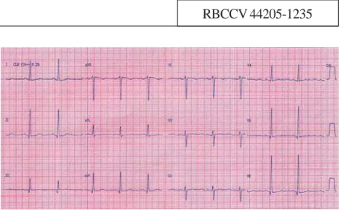
ELECTROCARDIOGRAM
Sinus rhythm, frequency of 70 beats/min, SÂP + 60º, SÂQRS + 60°, PR 0,16 s, QRS 0,10 s, QT 0,40, QTc 0,42. Left ventricular overload and alterations of ventricular repolarization (Figure 1).
RADIOGRAPHY
Visceral situs solitus in levocardia. Coastal erosion in the lower edges. Slightly increased cardiac area, reduced transparency in the right lung base due to possible injuries of airspace.
ECHOCARDIOGRAPHY
Situs solitus in levocardia, aortic coarctation of segment with hemodynamic repercussion, minimal muscular
trabecular interventricular communication detected only by Doppler echocardiography and without bicuspid aortic valve dysfunction [1].
Ascending aorta with 29 mm, arch before the innominate artery 22 mm between the brachiocephalic trunk and left subclavian artery 7 mm. The ejection fraction was 68.1%.
MULTI-DETECTOR COMPUTED TOMOGRAPHY Reduction target size of aortic arch in the descending portion of about 2.2 cm long. It is also noted collateral vessels distal to the affected region (Figure 2 / Video 1).
DIAGNOSIS
Fundamentally, it should be noted that the diagnosis could easily have been performed in the city of origin with the simple blood pressure measurement in the four limbs. It should also be emphasized the fact that a serious disease was present with little or no symptoms until adolescence [2].
597 CROTI, UA ET AL - Correction of aortic coarctation with tube
interposition in a 12 years-old child
Rev Bras Cir Cardiovasc 2010; 25(4): 596-597
The additional tests confirmed the diagnosis, and appropriate drug treatment and medical operation to correct the defect were indicated.
OPERATION
Children in the lateral position, lateral-posterior thoracotomy, opening of the pleura, wide dissection of the aortic arch, the coarcted area, descending aorta, left subclavian artery and careful isolation of the intercostal arteries.
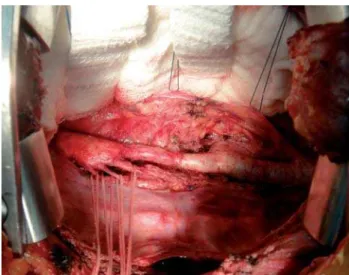
Observed long segment of the aorta with reduction in size between the left subclavian artery and descending aorta (Figure 3).
Lateral clamping of the descending aorta below the affected region, lateral incision and end-to-side anatomosis of polytetrafluoroethylene (PTFE) tube no.16. Sectioning the other end of the tube in bezel, this was also anastomosed (end-to-side) in the aorta near the origin of the LSA, which was also clamped laterally. The ischemic times for the anastomoses were respectively 10 and 12 minutes, without cardiopulmonary bypass [3]. The sutures were performed using polypropylene 5-0 yarn (Figure 4 / Video 2).
The tremor observed in the tube after the anastomoses indicated high blood flow, which was confirmed by postoperative echocardiography.
The patient was discharged after 35 days of hospital stay due to suture dehiscence, need for antibiotic therapy and hyperbaric oxygen therapy.
REFERENCES
1. Perloff JK. The variant associations of aortic isthmic coarctation. Am J Cardiol. 2010;106(7):1038-41.
2. Tanous D, Benson LN, Horlick EM. Coarctation of the aorta: evaluation and management. Curr Opin Cardiol. 2009;24(6):509-15.
3. Carvalho MVH, Pereira WL, Gandra SMA, Rivetti LA. Coarctação de aorta no adulto: a respeito de um caso e sobre desvios extra-anatômicos. Rev Bras Cir Cardiovasc. 2007;22(4):501-4.
Fig. 3 – Aortic segment compromised after extensive aortic dissection. It is noted the presence of intercostal arteries repaired with cotton thread
Fig 4/Video2 - Polytetrafluoroethylene tube no. 16 implanted between the aorta near the left subclavian artery and descending aorta below the compromised segment
Fig 2/Video 1 (http://www.rbccv.org.br/video/v25n4b/)