419 1. São José do Rio Preto Pediatric Cardiovascular Surgery Service
-Hospital de Base - São José do Rio Preto Medical School, SP, Brazil.
Fabiana Nakamura AVONA1, Ana Carolina Leiroz Ferreira Botelho MAISANO KOZAK1, Ulisses Alexandre CROTI1, Domingo Marcolino BRAILE1
Rev Bras Cir Cardiovasc 2009; 24(3): 419-421
RBCCV 44205-1112
Correção total de tetralogia de Fallot em criança com agenesia da artéria pulmonar esquerda
Total correction of tetralogy of Fallot in child with
left pulmonary artery agenesis
Correspondence address: Ulisses Alexandre Croti
Hospital de Base - FAMERP - Avenida Brigadeiro Faria Lima, 5544. ZIP Code: 15090-000 - São José do Rio Preto, SP, Brazil.
Fone (Fax): 17 - 3201 5025 / 3222 6450 / 9772 6560 E-mail: [email protected]
Article received on August 6th, 2009 Article accepted on September 8th 2009
CLINICAL-SURGICAL CORRELATION
CLINICAL DATA
2-year-old full-term male child, born by c-section. Born in Piracicaba-SP, at 3 months of life when heart murmur was auscultated. An echocardiogram was performed that diagnosed tetralogy of Fallot with agenesis of the left pulmonary artery (LPA) and indicated use of propranolol 5mg a day. The diagnosis was confirmed by cardiac catheterization performed at 8 months of life. Evolved with low weight gain and, when reaching 1 year of age, he started presenting frequent episodes of cyanosis, but none required hospitalization. Clinical follow-up was maintained follow-up to 2 years, when surgical correction was chosen. Prior to the operation, he underwent multiple detector computed tomography (MDCT).
On physical examination, the patient was in good general condition, ruddy, hydrated, cyanotic +++/+ 4 and eupneic. Normodynamic precordium without bulging, with a regular rhythm of two clicks, hyperphonesis of the second sound, ejection systolic murmur ++/6+ in left upper sternal border and a regurgitant systolic murmur +++/6+ in mid left sternal border. The abdomen was flat with the liver palpable at 1 cm below the right costal margin, spleen not palpable. The peripheral pulses were symmetrical.
420
AVONA, FN ET AL - Total correction of tetralogy of Fallot in child with left pulmonary artery agenesis
Rev Bras Cir Cardiovasc 2009; 24(3): 419-421
with a rare congenital heart defect, which implies a challenge in the postoperative period, which requires other examinations to detail anatomy and elucidate diagnosis [1]. The hemodynamic study confirmed the suspicion of the echocardiogram and showed a continuation of the pulmonary artery only with the right branch, in addition to reject the possibility of the presence of systemic-pulmonary ELECTROCARDIOGRAM
Sinus rhythm, heart rate of 120 beats/min, SÂP +60°, SÂQRS+120°, PR interval of 0.12s. Right ventricular overload evidenced by the qR complex in V1.
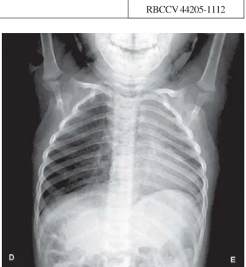
RADIOGRAM
Visceral situs solitus in levocardia, cardiac area with difficult delimitation due to deviation of the mediastinal structures to the left hemithorax. Thoracic skeleton with preserved aspect. Presence of lesion in the right interstitial-alveolar basement and left upper lobe. Free diaphragmatic domes (Figure 1).
ECHOCARDIOGRAM AND HEMODYNAMIC STUDY Situs solitus in levocardia. Normal venoatrial and atrioventricular connections. Normal ventriculoarterial connection. At Doppler, turbulent and accelerated flow in the right ventricle outflow tract and pulmonary artery was found, consistent with infundibular-valvar stenosis, with peak gradient of 97 mmHg. Continuous flow of descending aorta to the left lung was revealed. Diameter of the pulmonary valve annulus of 8.7 mm, pulmonary artery trunk of 5.9 mm, right pulmonary artery of 7.1 mm and left pulmonary artery (LPA) was not visualized. Thus, the echocardiogram showed tetralogy of Fallot of unfavorable anatomy, severe hypoplasia or agenesis of LPA, small systemic-pulmonary multiple arteries and aortic arch at right.
Hemodynamic study during right ventriculography identified severe infundibular stenosis, with thick pulmonary valve stenosis and pulmonary trunk connected directly with the right branch. It was not visualized the left pulmonary artery branch. During injection performed in the descending aorta, it was not observed the presence of systemic pulmonary circulation developed, and the injection into the left subclavian artery showed that the ductus arteriosus was occluded at its origin.
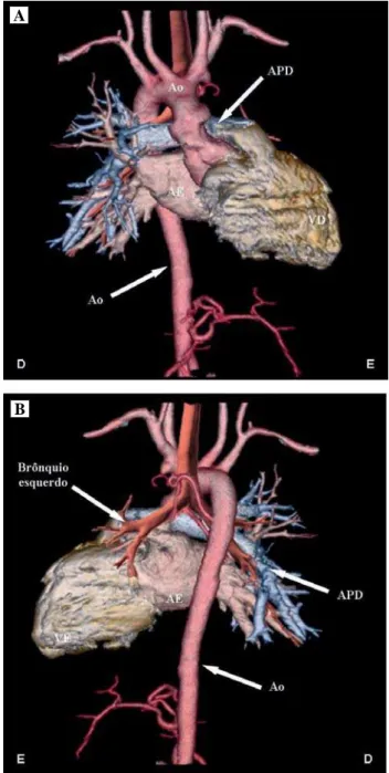
MULTIPLE DETECTOR COMPUTED TOMOGRAPHY The chest MDCT showed that the transverse, descending and ascending aorta, were at right. The absence of LPA was confirmed by complete agenesis. The pulmonary trunk and its right branch have normal caliber. Reduction of the left lung volume with deviation of mediastinal structures to this side, with the appearance of hypoplastic lung. There was only one pulmonary vein of small size that drained this lung. The right lung presented increased volume and normal pulmonary venous drainage (Figures 2A and 2B).
DIAGNOSIS
The diagnosis of tetralogy of Fallot with agenesis of the LPA was first detected by echocardiography. It deals
Fig. 2 - A: Multiple detector computed tomography in anterior view showing clearly the agenesis of the left pulmonary artery. B: Rear view of the descending aorta to the right. AE: left atrium, VE: left ventricle, VD: right ventricle, APD: right pulmonary artery, Ao: aorta
A
421 REFERENCES
1. Bockeria LA, Podzolkov VP, Makhachev OA, Zelenikin MA, Alekian BG, Ilyin VN, et al. Surgical correction of tetralogy of Fallot with unilateral absence of pulmonary artery. Ann Thorac Surg. 2007;83(2):613-8.
2. Halit V, Olguntürk R, Erer D, Kula S. Tetralogy of Fallot and absence of left pulmonary artery. Thorac Cardiovasc Surg. 2008;56(7):430-2.
Fig. 3 – Anterior mediastinum occupied by the right lung (pulmão direito) immediately after sternotomy
Fig. 4 - General aspect of the heart and great vessels after opening of the pericardial sac. It is clear the absence of left pulmonary artery. VSC: superior vena cava, AD: right atrium, VD: right ventricle, TP: pulmonary trunk, APE: left pulmonary artery collateral arteries, which could influence the surgical
approach. The MDCT sealed the final diagnosis of agenesis of the left pulmonary artery branch, which is consistent with the literature, indicating that agenesis of the LPA is more common than the RPA agenesis [2]. Clinical follow-up could be performed up to 2 years, because the patient remained stable, with appropriate weight and height gain and without cyanosis.
OPERATION
Sternotomy, by observing the right lung expanding into the mediastinum anterior to the pericardial sac, consistent with the image of the chest radiograph (Figure 3). Momentary stopping of mechanical ventilation and opening of the pericardial sac.
Complete agenesis of the LPA was noted, because the main pulmonary artery continued only to the right (Figure 4).
The surgery was performed as usual for tetralogy of Fallot with heparinization of 4 mg/kg, introduction of cannulas in the aorta and vena cavae, cardiopulmonary bypass (CPB) with moderate hypothermia at 28ºC. Aortic clamping, intermittent and anterograde hypothermic blood cardioplegia at 4ºC, every 20 minutes.
Opening of the right atrium and pulmonary trunk, atrial infundibular resection and by the pulmonary trunk through the pulmonary valve. The pulmonary valve was trileaflet, with normal appearance as for the mobility and thickness, but commissurotomy was necessary due to commissural fusion between two valves.
The ventricular septal defect was closed via atrial with U-shaped polypropylene sutures anchored in bovine pericardium. The points were passed around the defect and using a bovine pericardial patch of appropriate diameter the communication has been corrected.
The pulmonary trunk was sutured and a small residual atrial septal defect was purposely left if the patient presents pulmonary hypertension in later life.
The operation was finished as usual, with hemostasis, insertion of drains, temporary pacemaker wires and the chest was closed using layer technique. The duration of CPB was 93 minutes and ischemia 75 minutes.
Postoperatively, the patient evolved uneventfully and was discharged from hospital on the sixth day under use of furosemide.
Three months later, the patient is in excellent clinical condition without medication, with echocardiography showing defect corrected with satisfactory results and minimal atrial septal defect with flow from left to right. AVONA, FN ET AL - Total correction of tetralogy of Fallot in child
with left pulmonary artery agenesis