Subjects susceptible to chronic periodontitis (CP) show a high risk for the development of peiimplantitis (PI). Both diseases are multifactorial, presenting similarities in their pathophysiology and polygenic profile. MMP-13 (matrix metalloproteinases 13/ collagenase 3) is a collagenolytic enzyme, which expression is induced by TGF beta 3 (transforming growth factor type 3) in human gingival fibroblasts and inhibited by TIMP-2 (tissue inhibitor of metalloproteinase type TIMP-2). The aim of this study was to investigate the occurrence of peiimplantitis (PI) in subjects with history of chronic periodontitis (CP) and polymorphisms frequency in MMP13, TIMP2 and TGFB3 genes. One hundred and sixty-three volunteers received dental implant placement were submitted to oral and radiographic examination in order to identify past history of CP or presence of PI. Volunteers were divided into 4 groups: Control (without PI and CP, n=72), CP (with CP and without PI, n=28), PI (with PI and without CP, n=28) and diseased (with CP and PI, n=35). The chi-square test correlated genotypes in specific regions of MMP13 (rs2252070), TIMP2 (rs7501477) and TGFB3 (rs2268626) genes, considering the interaction between CP and PI. The results showed that volunteers with CP had 3.2 times more susceptibility to develop PI (p=0.0004) compared to those without CP. No significant association was observed in MMP13, TIMP2 and TGFB3 genes with CP or PI. CP is a risk factor to develop PI, however, there is no association of both diseases with polymorphisms in the MMP13, TIMP2 and TGFB3 genes.
M M P 1 3 , T I M P 2 a n d T G F B 3
G e n e P o l y m o r p h i s m s i n
Brazilian Subjects with Chronic
Periodontitis and Periimplantitis
Roberto Gonçalves Junior1, Aristides da Rosa Pinheiro2,3, José Jorge Schoichet3, Carlos Henrique Ramirez Nunes3, Rackel Gonçalves3, Leticia Ladeira Bonato1, Valquiria Quinelato1, Leonardo Santos Antunes4, Erika Calvano Küchler5, Julie Lobo6, Ricardo de Mello Villas-Bôas1, Alexandre Rezende Vieira7,8, José Mauro Granjeiro9,6, Priscila Ladeira Casado1,10
1Postgraduate Program in Dentistry, UFF - Universidade Federal Fluminense, Niterói, RJ, Brazil 2Dental Clinical Department, UFF - Universidade Federal Fluminense, Niterói, RJ, Brazil 3Oral Implantology Post-graduation, UFF - Universidade Federal Fluminense, Niterói, RJ, Brazil 4Departament of Specific Formation, School of Dentistry, UFF -
Universidade Federal Fluminense, Nova Friburgo, RJ, Brazil
5Department of Pediatric Dentistry, School of Dentistry of Ribeirão Preto, USP - University of São Paulo, Ribeirão Preto, SP, Brazil 6Clinical Research Unit and Biology Institute, UFF - Universidade Federal Fluminense, Niterói, RJ, Brazil 7Department of Oral Biology, School of Dental Medicine, University of Pittsburgh, Pittsburgh, PA, USA 8Center for Craniofacial and Dental Genetics, Department of Human Genetics, University of Pittsburgh, Pittsburgh, PA, USA 9National Institute of Metrology, Quality and Technology, Rio de Janeiro, RJ, Brazil
10Department of Periodontology, UFF - Universidade Federal Fluminense, Niterói, RJ, Brazil
Correspondence: Profa. Dra. Priscila Ladeira Casado. Rua Mario Santos Braga, 28 Centro, Niterói Campus do Valonguinho. CEP :24020-140. Tel: 2629-9901 E-mail: [email protected]
Key Words: dental implants, chronic periodontitis, genetic polymorphism.
Introduction
The increased failure rate in implant dentistry is associated with the development of periimplant disease (PID) (1). The main causes of PID have been attributed to biomechanical failure resulting from bacterial infection and occlusal overload, in which the main risk factors are poor oral hygiene, smoking, diabetes mellitus, as well as periodontitis. The implant success rate in patients without a history of periodontitis is 96.5%, whereas in patients with a history of periodontitis it is 90.5% (1,2).
Microbial etiology of PID is closely related to the microbiota associated with chronic periodontitis (CP). However, the individual’s immune response to bacterial insult is responsible for the development or cessation of the disease, including a profile of the immune response regulated by different cytokines (3). Increased levels of
immunoregulatory molecules, such as IL-1-b (interleukin-1 beta), IL-6 (interleukin-6), TNF (tumor necrosis factor) and PGE2 (prostaglandin E2), have been observed in periodontitis and periimplantitis sites compared with healthy sites (4-6).
Overexpression of these cytokines may result in the destruction of mineralized and/or non-mineralized tissues through autocrine or paracrine induction. This stimulates other biological mediators such as matrix metalloproteinases (MMPs) (7) family, which is composed of 23 enzymes able of degrading most extracellular matrix (ECM) proteins, including native and denatured collagen (7,8).
P
olymorphisms in periodontitis/periimplantitis
endothelial cells (9,10).
The activity of these MMPs is primarily controlled by tissue inhibitors, known as TIMPs, which have an N-terminal domain able to inhibit MMPs. The balance between MMPs and TIMPs determines the degree of ECM degradation, together with hormones, oncogenic products, pro- and anti-inflammatory cytokines and growth factors (10,11).
TGF-beta is among the main growth factors involved in the regulation of ECM in the periodontal tissue and it has shown to block or initiate cell differentiation and migration, becoming overexpressed after placement of endosseous implants. TGF-beta 3 has a central role in fibroblastic proliferation and differentiation, stimulating collagen deposition (12).
TGF-beta is able to induce the expression of MMP-13 in human gingival fibroblasts in wound healing, by a cascade of mitogen-activated protein kinases (MAPK), p38 and complex AP-1 (activating protein-1). This suggests that MMP-13 plays an important role in the rapid remodeling of the extracellular matrix collagen during repair of periodontal lesions (9). In addition, the inflammatory process is controlled by genetic factors. Recent studies have focused on the individual genetic pattern to explain the common etiology of periodontitis and periimplantitis, but have yielded conflicting results and limited analyses of specific regions in a single gene (5).
Taking into account that homeostasis is regulated by MMP-13 and considering its regulators in the mineralized and non-mineralized tissues, the aim of this study was to evaluate the occurrence of periimplantitis (PI) in subjects with history of chronic periodontitis (CP) and its interaction with polymorphisms in MMP13, TIMP2 and TGFB3 genes.
Material and Methods
Volunteer Selection
One hundred and sixty-three individuals, presenting 587 osseointegrated endosseous implants, were selected for this cross-sectional study over the course of one year at the Dental Implantology Clinic of the Dental School at the Universidade Federal Fluminense, Niterói, RJ, Brazil. The clinical procedures were conducted in accordance with the recommendations from the University Research Ethics Board (Registration number 286.354). Informed consent was obtained from all participants. Volunteers answered a personal, medical and dental history anamnesis. The exclusion criteria were: implant failure (pathologic bone loss, implant mobility or implant loss) before the osseointegration period (3 months for mandible and six months for maxilla), bisphosphonate use, pregnancy and/ or lactation in female volunteers, lack of preoperative radiography, one stage or immediate implant placement,
concurrent bone grafting required, early implant exposure during osseointegration period, non-treated periodontitis and non-compliance with study protocol. The inclusion criteria were as follows: at least one osseointegrated endosseous implant, immediate postoperative radiography showing the vertical bone level around implant to compare bone level after the osseointegration period, periapical radiography showing periodontal status before implant placement, and annual follow-up clinical and radiographic examinations. All implants were placed in a submerged healing modality (two-stage concept) in sites that had previously shown favorable bone quality and quantity.
Diagnosis of Periimplantitis
All periimplant regions were clinically and radiographically evaluated. Clinical examination of the periimplant sites consisted of visual inspection and palpation, analysis of mucosa inflammation, probing pocket depth, bleeding on probing and spontaneous bleeding on four surfaces (mesial, buccal, distal and lingual/palatine), presence of plaque, periimplant phenotype, implant mobility (any mobility during percussion test), osseointegration period and implant platform configuration. Conventional periapical radiography, using the paralleling technique, was used to assess the vertical bone loss around the implants measuring the height of periimplant bone and comparing it with the initial radiography taken immediately after implant placement. According to the clinical and radiographic characteristics of the periimplant sites, volunteers were characterized as having healthy sites - no clinical signs of inflammation in the periimplant mucosa and no signs of bone loss – or periimplantitis with radiographic signs of pathologic bone loss in at least one region. Physiological bone loss was characterized considering as normal bone loss of 1 mm during the first year following implant placement and 0.2 mm for subsequent years. According to the implant osseointegration period, the total amount of bone loss was calculated based on the difference between immediate postoperative radiography and diagnosis radiography (at the moment of periimplant examination). If total bone loss was more than 1 mm and 0.2 mm per year, the volunteer was diagnosed with periimplantitis.
Diagnosis of Chronic Periodontitis
Volunteers were characterized as healthy (without history of chronic periodontitis) or having chronic periodontitis (with a history of chronic periodontitis).
R. Gonçalves Jr et al.
periodontitis.
Diagnosis and classification of generalized chronic periodontitis was based on Armitage (13). All volunteers diagnosed with chronic periodontitis have been treated and were under regular follow-up care.
Based on periimplant and periodontal status, volunteers were divided into the following groups: healthy – control group (no history of chronic periodontitis and periimplantitis, n=72); and test-case groups: disease (with a history of chronic periodontitis and periimplantitis, n=35); chronic periodontitis (chronic periodontitis history without periimplantitis, n=28) and periimplantitis (with periimplantitis without chronic periodontitis, n=28).
Single Nucleotide Polymorphism Genotyping
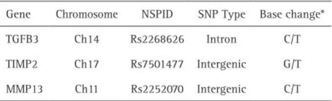
Genomic DNA was obtained from saliva samples, as previously described (14).The amount and purity of the DNA was determined by the spectrophotometer (Nanodrop® 1000; Thermo Scientific, Wilmington, DE, USA). Only DNA samples presenting A260nm/A280nm ratio greater than 1.8 were used. All examiners at the laboratories were blinded to the group assignment. Analyses for the presence of polymorphism in the MMP13 (rs2252070), TIMP2 (rs7501477) and TGFb3 (rs2268626) (Table 1) genes were performed from the reactions of real-time polymerase chain reactions using the Taqman assay (Stratagene Mx3005P; Agilent Technologies, Santa Clara, CA, USA). Primers, probes and the universal master mix were provided by Applied Biosystems (Foster City, CA, USA).
Statistical Analyses
Nominal variables were expressed as frequencies and percentages. To access the significance of nominal variables among the groups, the chi-square (χ2) test was applied. Continuous variables were expressed as means and standard deviations. After the Shapiro-Wilk test evaluated the distribution among variables, analysis of variance (ANOVA), t-test or Mann-Whitney tests were used to compare means among the groups, when the variables presented normal or non-normal distribution. Differences in the prevalence of genotypes and alleles among the groups were analyzed using the Pearson chi-square (χ2) test after fitting for Hardy-Weinberg equilibrium. Values of p<0.05 were considered statistically significant. Statistical analyses were performed using Stata 11.1 (StataCorp, College Station, TX, USA).
Results
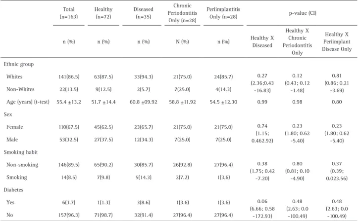
One hundred sixty-three volunteers were evaluated over the course of one year. The sample consisted of 110 (67.5%) females and 53 (32.5%) males, with a mean age of 55.4 ±13.2 years. No difference was found among the test
groups (diseased; chronic periodontitis only; periimplantitis only) compared with the control group regarding the ethnic group, age, gender and smoking habits. In contrast, comparing with the control volunteers, the prevalence of diabetes showed a higher incidence (8.6%) in the diseased group (p=0.06) (Table 2).
Taking into account the periimplant status (Table 3), plaque accumulation around implant was highly prevalent in all test groups compared with control (p<0.01). In addition, spontaneous bleeding was significantly higher in the diseased (p=0.01) and the periimplantitis (p=0.004) groups, as well as bleeding on probing (p<0.0001). A tendency for higher probing pocket depth was also observed in the diseased group (p=0.05). Considering the platform type of implant, there was no difference among groups. However, volunteers with history of CP showed 3.2 times more chance of developing PI than volunteers without CP history (Table 4).
Genotype distributions were within Hardy-Weinberg equilibrium analyses (data not shown). All genotypes and allele frequencies were compared between control group and each test group to further clarify the results. Genotypes and allele frequencies for MMP13 (rs2252070), TIMP2 (rs7501477) and TGFB3 (rs2268626) showed no association with test groups (Table 5). When adjusted by age (data not shown), the results were similar for all genotype associations.
Discussion
Based on the previously established relationship between periimplantitis and chronic periodontitis (2), the aim of this study was to investigate the association between genetic information contained in the DNA, considering the MMP13, TIMP2 and TGFB3 genes, involved in the homeostasis of ECM, with the development of periimplantitis in volunteers with a history of chronic periodontitis. The results showed that, despite the clinical differences observed among groups, polymorphisms in the MMP13, TIMP2 and TGFB3 genes have no association with both diseases.
These results also showed the association among several risk factors for periodontitis, already established in the literature, like diabetes (15)and plaque accumulation (1) in volunteers with periimplantitis. This association may
Table 1. Characteristics from polymorphic regions
Gene Chromosome NSPID SNP Type Base change*
TGFB3 Ch14 Rs2268626 Intron C/T
TIMP2 Ch17 Rs7501477 Intergenic G/T
P
olymorphisms in periodontitis/periimplantitis
indicate that, as in periodontitis, external factors have direct influence in the spread of periimplantitis, irrespective the history of chronic periodontitis.
There is a predominance of anaerobic bacteria in periimplant plaque accumulation, as well as CP, such as Porphyromonas gingivalis, Aggregatibacter actinomycetemcomitans, Prevotella intermedia, Treponema denticola and Tannerella forsythenses (16,17), which cause apical spread of infection and culminate in the loss of surrounding bone through mechanisms similar to the ones that destroy ECM. This exaggerated immune response, mediated by T helper cells 1 (Th1) or Th2 cells, will determine the stability or progression of the lesion.However, considering that the periimplant tissue has anatomical differences and particularities that anatomically distinguish it from the periodontium, the presence of pathogens in the periimplant tissue results in the disruption of the mucosal seal, considered as the only physical barrier against bacterial penetration, leading to the rapid extension of the pathological pocket to the bone tissue (18).
The periimplant mucosa is less capable of recovering from lesions associated with plaque than the gingiva (18,19). However, most of the volunteers who received implants have a history of periodontitis and, therefore, a destructive inflammatory tissue response as consequence
of microbiota changes. This immune response may lead to a higher incidence of periimplant bone loss (periimplantitis) observed in subjects with CP, as demonstrated in the present research.
This excessive destruction of connective tissue components plays a role in the loss of functional tissue architecture (7). Within this context, the equilibrium in the activity of MMPs, controlled primarily by protein tissue inhibitors (TIMPs), represent a crucial aspect for the maintenance of the ECM homeostasis (20) in the periimplant soft tissue, as response to the presence of pathogens. MMP-13 has a high ability to digest type II collagen and its gelatinase activity is 40-fold greater than MMP-1 and MMP-8 (20).The presence of protein derived from P. gingivalis is able to decrease the amount of TIMP-2, a potential regulator of MMP-13, suggesting that this may be a pathological mechanism that promotes tissue destruction. Furthermore, TIMP-2 has erythroid- and mitogenic-potentiating activity, but its overexpression reduces the growth of tumor cells (21), which justifies its reduction to be associated with ECM tissue destruction by MMPs.
On the other hand, TGF beta may induce the expression of MMP-13 in human gingival fibroblasts reducing the expression of TIMP-2 (22). Therefore, changes in the
Table 2. Baseline characteristics from volunteers included in the study
Total (n=163)
Healthy (n=72)
Diseased (n=35)
Chronic Periodontitis Only (n=28)
PeriimpIantitis
Only (n=28) p-value (CI)
n (%) n (%) n (%) N (%) n (%) Healthy X
Diseased
Healthy X Chronic Periodontitis
Only
Healthy X Periimplant Disease Only
Ethnic group
Whites 141(86.5) 63(87.5) 33(94.3) 21(75.0) 24(85.7) 0.27
(2.36;0.43 -16.83)
0.12 (0.43; 0.12
-1.48)
0.81 (0.86; 0.21
-3.69)
Non-Whites 22(13.5) 9(12.5) 2(5.7) 7(25.0) 4(14.3)
Age (years) (t-test) 55.4 ±13.2 51.7 ±14.4 60.8 ±09.92 58.8 ±11.92 54.5 ±12.30 0.99 0.98 0.80
Sex
Female 110(67.5) 45(62.5) 23(65.7) 21(75.0) 21(75.0) 0.74
(1.15; 0.462.92)
0.23 (1.80; 0.62
-5.40)
0.23 (1.80; 0.62
-5.40)
Male 53(32.5) 27(37.5) 12(34.3) 7(25.0) 7(25.0)
Smoking habit
Non-smoking 146(89.5) 65(90.2) 30(85.7) 26(92.8) 27(96.4) 0.38
(1.75; 0.42 -7.20)
0.80 (0.81; 0.10
-4.90)
0.37 (0.39; 0.023.56)
Smoking 14(8.5) 7(9.8) 5(14.3) 2(7,2) 1(3,6)
Diabetes
Yes 6(3.7) 1(1.3) 3(8.6) 1(3.6) 1(3.6) 0.06
(6.66; 0.58 -172.93)
0.48 (2.63; 0.0
-100.49)
0.48 (2.63; 0.0
-100.49)
No 157(96.3) 71(98.7) 32(91.4) 27(96.4) 27(96.4)
R. Gonçalves Jr et al.
levels of these molecules may offer greater protection or susceptibility to periimplantitis. Genetic modifications may be associated to a greater or lesser degree with the production of these molecules, or interfere in their physiology (5).
Polymorphism in the MMP13 gene was previously associated with the development of colorectal cancer,
aneurysm of the abdominal aorta, tooth agenesis and caries (11,23). In addition, polymorphisms in the TGFB3 gene were associated with non-syndromic cleft lip and development of tooth agenesis (24). Some studies have shown a clear association between various genetic polymorphisms, including IL-6 gene with PID (5) and periodontitis (25), but so far no studies have correlated the presence of
Table 3. Periimplant status of the studied population
Periimplant status
Healthy (n=72)
Diseased (n=35)
Chronic periodontitis
Only (n=28)
Periimplant
Only (n=28) p value
n (%) n (%) n (%) n (%)
Healthy X Diseased
Healthy X Chronic Periodontitis
Only
Healthy X Peri-ImpIant
Disease Only
Bleeding on probing
*Presence 9 (12.5) 20 (57.1) 5 (17.9) 14 (50.0) <0.0001
(9.33; 3.23 -27.82)
0.48 (1.52; 0395.71)
<0.0001 (7.00; 2.27
-2.14)
Absence 63 (87.5) 15 (42.9) 23 (82.1) 14 (50.0)
Spontaneous bleeding
*Presence 0 (00.0) 3 (8.6) 0 (00.0) 3 (10.7) 0.01
(RR 3.25; 2.44-4.34)
0
0.004 (RR 3.88; 2.77-5.44)
Absence 72 (100.0) 32 (91.4) 28 (100.0) 25 (89.3)
Probing Pocket
Depth (mm) 1.64 ±0.77 2.27 ±1.19 1.44 ±0.52 2.28 ±0.75 0.05 0.36 0.17
Periimplant plaque
*Presence 2 (2.8) 10 (28.6) 6 (21.4) 5 (17.9) <0.0001
(14.00; 2.59-99.93)
0.002 (9.55; 1.56
-74.38)
0.007 (7.61; 1.18
-61.37)
Absence 70 (97.2) 25 (71.4) 22 (78.6) 23 (82.1)
Periimplant phenotype
*Thin 29 (40.2) 16 (45.7) 17 (60.7) 11 (39.2) 0.57
(1.26; 0.513.10)
0.06 (2.32;
0.87-6.27)
0.94 (0.97; 0.36
-2.60)
Thick 43 (59.8) 19 (54.3) 11 (39.3) 17 (60.8)
Implant Mobility 0 (00.0) 1 (02.8) 0 (00.0) 2 (7.1)
0.14 (RR 3.12; 2.36-4.11)
0.0
0.04 (RR 3.18; 2.40-4.22)
Implant-platform type
External Hexagon 35 (48.6) 19 (54.3) 14 (50.0) 16 (57.1)
0.49 0.99
0.85
Internal Hexagon 5 (6.9) 4 (11.4) 2 (7.1) 1 (3.6)
Morse Cone 29 (40.3) 12 (34.3) 11 (39.3) 10 (35.7)
Others 3 (4.2) 0 (00.0) 1 (3.6) 1 (3.6)
Implant region
*Upper 41 (56.9) 17 (48.6) 9 (32.1) 17 (60.7) 0.41
(0.71; 0.29 -1.73)
0.02 (0.36;
0.13-0.98)
0.73 (1.17; 0.44
-3.13)
Lower 31 (43.1) 18 (51.4) 19 (67.9) 11 (39.3)
Edentulism
*Partial 62 (86,1) 27 (77,1) 21 (75,0) 25 (89,3) 0.24
(0.54; 0.17-1.72)
0.18 (0.48 0.14
-1.63)
0.67 (1.34; 0.30
-6.76)
Total 10 (13,9) 8 (22,9) 7 (25,0) 3 (10,7)
Osseointegration
P
olymorphisms in periodontitis/periimplantitis
Table 4. Correlation between history of chronic periodontitis and periimplantitis
Healthy Periimplant
tissue (n)
Periimplantitis
(n) p-value Odds ratio (CI)
Without
history of CP 72 28
0.0004 3.21 (1.58-6.59) With history
of CP 28 35
CI: confidence interval.
Table 5. Allele and genotype frequencies of SNP markers
Healthy Diseased
Chronic periodontitis
Only
Periimplantitis Only
p value (OR; CI)
(n=72) (n=35) (n=28) (n=28) Healthy X
Diseased
Healthy X Chronic Periodontitis Only
Healthy X Periimplant Disease Only
n (%) n (%) n (%) n (%)
MMP13
rs2252070
CC 7 (9.72) 5(14.28) 2(07.14) 1(3.57)
0.42 0.63 0.59
CT 26(36.11) 12(34.28) 9(32.14) 11(39.28)
TT 36(50.00) 18(51.42) 17(60.71) 15(53.57)
CT+TT 62(86.11) 30(85.71) 26(92.85) 26(92.85) 0.53
(0.68; 0.17-2.72)
0.64 (1.47; 0.25-11.01)
0.30 (2.94;0.33-66.6)
C 40(27.77) 22(31.42) 13(23.21) 13(23.21) 0.71
(1.12; 0.57-2.19)
0.41 (1.35;0.62-2.96)
0.49 (1.29;0.59-2.83) T 98(68.05) 48(68.57) 43(76.78) 41(73.21)
TGFB3
rs2268626
0.58 (0.77;0.26-2.19)
CC 0(00.00) 0(00.00) 0(00.00) 0(00.00)
1.0 (1.00;0.39-2.55)
0.21 (0.52;0.16-1.61) CT 46(63.88) 23(65.71) 20(71.42) 22(78.57)
TT 24(33.33) 12(34.28) 8(28.57) 6(21.42)
C 46(31.94) 23(32.85) 20(35.71) 22(39.28) 1.0
(1.00;0.52-1.93)
0.70 (0.88;0.44-1.78)
0.39 (0.76;0.38-1.51) T 94(65.27) 47(67.14) 36(64.28) 34(60.71)
TIMP2
rs7501477
GG 51(70.83) 27(77.14) 19(67.85) 21(75.00)
0.58 0.67 0.66
GT 17(23.61) 8(22.85) 7(25.00) 7(25.00)
TT 2(2.77) 0(00.00) 0(00.00) 0(00.00)
GT+TT 19(26.38) 8(22.85) 7(25.00) 7(25.00) 0.63
(0.80;0.28-2.25)
0.98 (0.99;0.32-3.02)
0.82 (0.89;0.29-2.70)
G 119(82.62) 62(88.57) 45(86.5) 49(87.50) 0.47
(0.73;0.28-1.87)
0.07 (0.88;0.32-2.38)
0.65 (0.81;0.29-2.18)
T 21(14.58) 8(11.42) 7(13.5) 7(12.50)
comorbid history of CP with PID and polymorphisms in genes associated with ECM homeostasis
This study evaluated for the first time the biological relationship between CP and PI considering polygenic
profile in MMP3, TIMP2 and TGFB3. Despite having found no genetic predisposition associated with both diseases, the authors state that CP is a risk factor for PI development. In order to investigate the central role of this pathway in periodontitis and periimplantitis pathogenesis, further studies analyzing gene expression and protein levels from gingival and bone tissues in a bigger sample size should be encouraged. The findings of this study leads to the belief that novel methods, such as gene therapy (with local or systemic applications) and tissue engineering, may be developed for implant dentistry based on genetic response.
R. Gonçalves Jr et al.
periodontitis is a risk factor for periimplantitis. However, polymorphisms in MMP13, TIMP2 and TGFB3 are not associated with the development of periimplantitis.
Acknowledgements
The authors gratefully thank the assistance of all Department of Oral Biology at the University of Pittsburgh and Fundação Carlos Chagas Filho de Amparo à Pesquisa do Estado do Rio de Janeiro.
Resumo
Indivíduos susceptíveis à periodontite crônica (CP) apresentam alto risco para o desenvolvimento de periimplantite (PI). Ambas doenças são multifatoriais e apresentam similaridades na patofisiologia e perfil poligênico. A MMP-13 (metaloproteinase da matriz tipo 13) é uma enzima colagenolítica cuja expressão é induzida por TGF beta 3 (fator transformador do crescimento tipo 3) nos fibroblastos gengivais humanos e inibida por TIMP-2 (inibidor tecidual de metaloproteinase tipo 2). O objetivo deste estudo foi investigar a ocorrência de periimplantite em sujeitos com periodontite crônica e a frequência dos polimorfismos nos genes MMP13, TIMP2 e TGFB3. Cento e sessenta e três voluntários submetidos à instalação de implantes endósseos foram analisados clínica e radiograficamente quanto à presença de histórico de CP e PI, sendo divididos em 4 grupos: Controle (sem história de CP e PI, n=72), CP (com CP e sem PI, n=28), PI (com PI e sem CP, n=28) e Doentes (com CP e PI, n=35). O teste do qui-quadrado correlacionou os genótipos nas regiões dos genes MMP13 (rs2252070), TIMP2 (rs7501477) e TGFB3 (rs2268626), considerando a interação entre CP e PI. Os resultados mostraram que voluntários com CP possuem 3.2 vezes mais chances de desenvolver PI (p=0.0004) comparados aos sem CP. Nenhuma associação significativa foi observada entre os genes MMP13, TIMP2 e TGFB3 e CP ou PI. A CP é um fator de risco ao desenvolvimento de PI, no entanto, não há associação entre ambas as doenças com polimorfismos nos genes MMP13, TIMP2 e TGFB3.
References
1 Lindhe J, Meyle J. Peri-implant diseases: Consensus Report of the Sixth European Workshop on Periodontology. J Clin Periodontol 2008;35(Suppl. 8):282–285.
2 Casado PL, Pereira MC, Duarte MEL, Granjeiro JM. History of chronic periodontitis is a high risk factor for peri-implant disease. Braz Dent J 2013;24:134-141.
3. Heitz-Mayfield LJA. Peri-implant diseases: diagnosis and risk indicators. J Clin Periodontol 2008;35:292-304.
4. Casado PL, Canullo L, Filardy AA, Granjeiro JM, Barboza EP, Duarte MEL. Intreleukins 1B and 10 expressions in the periimplant crevicular fluid from volunteers with untreated periimplant disease. Implant Dent 2013;22:1-8.
5. Casado PL, Villas-Boas R, Mello W, Duarte MEL, Granjeiro JM. Peri-implant disease and chronic periodontitis: is interleukin-6 gene promoter polymorphism a common risk factor in a Brazilian population? Int J Oral Maxillofac Implant 2013;28:35-43.
6. Gorska R, Gregorek H, Kowalski J, Laskus-Perendyk A, Syczewska M, Madalinski K. Relationship between clinical parameters and cytokine profiles in inflamed gingival tissue and serum samples from volunteers with chronic periodontitis. J Clin Periodontol 2003;30:1046-1052. 7. Graves DT, Cochran D. The contribution of interleukin-1 and tumor
necrosis factor to periodontal tissue destruction. J Periodontol 2003;74:391-401.
8. Knäuper V, López-Otín C, Smith B, Knight G, Murphy G. Biochemical characterization of human collagenase-3. J Biol Chem 1996;271:1544-1550.
9. Ravanti L, Häkkinen L, Larjava H, Saarialho-Kere U, Foschi M, Han J, et al.. Expression of human collagenase-3 (MMP-13) by fetal skin fibroblasts is induced by transforming growth factor beta via p38 mitogen-activated protein kinase. FASEB J 2001;15:1098-1100. 10. Visse R, Nagase H. Matrix metalloproteinases and tissue inhibitors of
metalloproteinases: structure, function, and biochemistry. Circ Res 2003;92:827-839.
11. Van Nguyen S, Skarstedt M, Lofgren S, Zar N, Andersson RE, Lindh M, et al.. Gene polymorphism of matrix metalloproteinase-12 and -13 and association with colorectal cancer in Swedish volunteers. Anticancer Res 2013;33:3247-3250.
12. Papakonstantinou E, Aletras AJ, Roth M, Tamm M, Karakiulakis G. Hypoxia modulates the effects of transforming growth factor-beta isoforms on matrix-formation by primary human lung fibroblasts. Cytokine 2003;24:25-35.
13. Armitage GC. Periodontal diagnoses and classification of periodontal diseases. Periodontol 2000 2004;34:9-21.
14. Küchler EC, Tannure PN, Falagan-Lotsch P, Lopes TS, Granjeiro JM, Amorim LMF. Buccal cells DNA extraction to obtain high quality human genomic DNA suitable for polymorphism genotyping by PCR-RFLP and Real-Time PCR. J Appl Oral Sci 2012;20:467-471.
15. Ferreira S, Silva G, Cortelli J, Costa J, Costa F. Prevalence and risk variables for peri-implant disease in Brazilian volunteers. J Clin Periodontol 2006;33:929–935.
16. Hultin M, Gustafsson A, Hallstrom H, Johansson LA, Ekfeldt A, Klinge B. Microbiological findings and host response in volunteers with peri-implantitis. Clin Oral Impl Res 2002;13:349-58.
17. Mayanagi G, Sato T, Shmauchi H, Takahasho N. Detection frequency of periodontitis-associated bacteria by polymerase chain reaction in subgingival and supragingival plaque of periodontitis and healthy volunteers. Oral Microb Immunol 2004;19:379-385.
18. Ericsson L, Berglundh T, Marinello C, Lijenberg, B, Lindhe J. Long-standing plaque and gingivitis at implants and teeth in the same dog. Clin Oral Implants Res 1992;3:99-103.
19. Lindhe J, Berglundh T, Ericsson J, Lijenberg B, Marinello C. Experimental breakdown of peri-implant and periodontal tissues. Clin Oral Implants Res 1992;3:9-16.
20. Uchida M, Shima M, Shimoaka T, Fujieda A, Obara K, Suzuki H, et al.. Regulation of matrix metalloproteinases (MMPs) and tissue inhibitors of metalloproteinases (TIMPs) by bone resorptive factors in osteoblastic cells. J Cell Physiol 2000;185:207-214.
21. Nagase H, Woessner JF Jr. Matrix metalloproteinases. J Biol Chem 1999;274:21491-21494.
22. Eickelberg O, Kohler E, Reichenberger F, Bertschin S, Woodtli T, Erne P, et al.. Extracellular matrix deposition by primary human lung fibroblasts in response to TGF-beta1 and TGF-beta3. Am J Physiol 1999;276:L814-L824.
23. Antunes LS, Kuchler EC, Tannure PN, Dias JB, Ribeiro VN, et al.. Genetic variations in MMP9 and MMP13 contribute to tooth agenesis in a Brazilian population. J Oral Science 2013;55:281-286.
24. Antunes LS, Küchler EC, Tannure PN, Lotsch PF, Costa MC, Gouvêa CV, et al.. TGFB3 and BMP4 polymorphism are associated with isolated tooth agenesis. Acta Odontol Scand 2012;70:202-206.
25. Shih YS, Fu E, Fu MM, Lin FG, Chiu HC, Shen EC, et al.. Association of CCL5 and CCR5 gene polymorphisms with periodontitis (in Taiwanese). J Periodontol 2014;14:1-14.