This study evaluated the effect of matrix metalloproteinase (MMP) inhibitors - 2% (CHX) and sodium fluoride (NaF) (5000 ppm) - on microtensile bond strength (μTBS) of composite resin to Er:YAG laser-irradiated dentin after chemical degradation of the bond interface. The occlusal surface of forty sound human molars was removed exposing the dentin surface (n=10), which was polished, irradiated with Er:YAG laser, acid etched and dried. Twenty specimens were rewetted with 2% CHX (control group) and 20 were rewetted with NaF (5000 ppm). The adhesive system was applied and a 4-mm-high plateau of light-cured composite resinwas built up. Resin-dentin sticks were obtained with a rectangular cross-sectional area (0.8-1 mm2) and were either stored in water at 37 °C for 24 h or submitted to chemical degradation. For chemical degradation, they were immersed in 10% NaOCl aqueous solution for 5 h and rinsed in water for 1 h. The sticks were submitted to microtensile test in a mechanical testing machine at 0.5 mm/min until failure. Fracture pattern was analyzed using SEM. μTBS values were calculated in MPa and submitted to analysis of variance ANOVA (α=0.05). The variance analysis showed that the ‘MMP inhibitor’ and ‘degradation’ factors (p=0.214 and p=0.093, respectively) and interaction between the factors were not statistically significant (p=0.143). Mixed failure predominated in all groups. In conclusion, the 2% CHX and NaF 5000 ppm presented similar μTBS of composite resin to laser-irradiated dentin before and after chemical degradation
E f f e c t o f M e t a l l o p r o t e i n a s e
I n h i b i t o r s o n t h e M i c r o t e n s i l e
Bond Strength of Composite Resin
to Er:YAG Laser-Irradiated Dentin
Beatriz Carlos Correa1, Rodrigo Galo1, Camila Scatena2, Maria Cristina Borsatto1 Aloísio Oro Spazzin3, Silmara Aparecida Milori Corona1,Daniel Galafassi3
1Department of Pediatric Dentistry,
School of Dentistry of Ribeirão Preto, USP - Universidade de São Paulo, Ribeirão Preto, SP, Brazil
2Department of Pediatric Dentistry,
FSG - Faculdade da Serra Gaúcha, Caxias do Sul, RS, Brazil
3Department of Restorative Dentistry,
School of Dentistry, IMED – Faculdade Meridional, Passo Fundo, RS, Brazil
Correspondence: Daniel Galafassi, Rua Senador Pinheiro, 304, 99070-220 Passo Fundo, RS, Brasil. Tel: +55-54-3045-6100. e-mail: [email protected]
Key Words: Er:YAG laser, dentin, bond strength, degradation.
Introduction
Preparation with Er:YAG laser has been proposed as an alternative technique for removing carious tissue and preparation of micro-cavities (1). Several advantages have been related to the use of laser irradiation in operative dentistry compared to conventional rotary instruments, such as a more conservative cavity design (2), antibacterial activity (3), decrease of enamel solubility (prevention of recurrent caries) (4), and mainly laser use provides more comfort to the patient due to the absence of vibration and a lower pain sensation with consequent decrease for local anesthesia compared to the use of conventional rotary instruments. A limitation of the laser use is the long time required for cavity preparation, generally, twice than with rotary instruments (5).
The bonding mechanism for irradiated dentin and also the impact of the Er:YAG lasers on the collagen fibers have not been completely clarified. Previous studies showed microstructural changes, microruptures and denaturation of collagen fibers (6,7), which may affect the bonding mechanism between the resin materials and dentin, due the bond direct dependence on the interaction of resin monomers with the exposed collagen fibers (8).
The degradation of the bonding interface has a considerable influence on the clinical performance and survival of the resin composite restorations (9,10). Studies have shown that matrix metalloproteinase (MMP) inhibitors, such as chlorhexidine (CHX) and more recently sodium fluoride (NaF), are able to preserve the structural integrity of the hybrid layer and consequently improve the longevity of restorations (11-14).
The research was proposed due the lower bond stability of irradiated dentin to the adhesive systems and limited information concerning the effect of the MMP inhibitors to stabilize the degradation of this hybrid layer. Therefore, the aim of this laboratory study was to evaluate the effect of two MMP inhibitors - 2% CHX and NaF (5000 ppm) - on microtensile bond strength (μTBS) of a composite resin to Er:YAG laser-irradiated dentin after chemical degradation of the bonded interface. The null hypothesis tested was that NaF (5000 ppm) is similar to CHX in the preservation of bonded interface.
Material and Methods
Experimental Design and Tested Groups
This laboratory study was designed to test the effect of ISSN 0103-6440 Brazilian Dental Journal (2016) 27(4): 442-445
Braz Dent J 27(4) 2016
443
Bond strength of laser irradiated dentin
the MMP inhibitors 2% CHX (Clorhexidina S, FGM, Joinville, SC, Brazil) (control group) and NaF (5000 ppm) (DaTerra, Ribeir̃o Preto, SP, Brazil) as well as degradation processes (24 h water storage and chemical degradation with 10% NaOCl for 5 h) on the μTBS between a composite resin and dentin treated with Er:YAG laser. The sample of the study comprised 40 human third molars assigned to 4 groups (n=10). The response variable was μ-TBS (MPa). Failure mode analysis of the bonded interface after μTBS testing was carried out by scanning electron microscopy (SEM).
Specimen Preparation
Forty extracted intact human third molars were used according to the guidelines of the local Ethics Committee, under protocol number (31160914.6.0000.5419). The teeth roots were included in polyester resin, by a polyvinyl chloride (PVC) cylinder. The occlusal surface was removed with a diamond saw (Isomet 1000; Buehler, Lake Bluff, IL, USA) exposing a coronal dentin surface, which was ground in a polishing machine (Beta; Buehler) with wet #600 sandpaper.
The dentin surfaces of all teeth were irradiated with Er:YAG laser (260 mJ/4 Hz) (Twin Light, Fotona Medical Lasers, Ljubljana, Slovenia) with 0.81 W power, 47 J/cm2 energy density and 163.5 W/cm2 irradiance, scanning transversally and horizontally across the surface of the exposed dentin using a device that holds the laser pen in place. The laser beam was applied using a noncontact method, focused at a distance of 12 mm from the substrate with water spray of 1.5 mL/min (9).
The specimens were etched with 35% phosphoric acid (Etching gel, 3mas M ESPE, St. Paul, MN, USA) for 15 s, rinsed for 30 s and dried for 30 s with air. Half of the specimens were rewetted with 1.5 mL of 2% CHX (Clorhexidina S, FGM, Joinville, SC, Brazil) with a micropipette (Labmate Soft, PZ HTL S.A., Warsaw, Poland) for 60 s and the excess removed with absorbent paper. The remaining specimens were rewetted with NaF 5000 ppm in the same way as previously reported. The adhesive system (Single Bond 2; 3M ESPE) was applied following the manufacturer’s instructions and cured with halogen light (Curing Light XL-3000; 3M Dental Products, St. Paul, MN, USA) for 10 s. A 4-mm-high plateau of composite resin (Filtek Z350; 3M ESPE) was built
on adhesive-coated dentin surface using the incremental technique in four 1-mm layers.
Degradation and μTBS Test
The specimens were vertically serially sectioned (Isomet 1000, Buehler Ltd.) into approximately 1-mm thick dentin-resin slabs, and then rotated 90° and sectioned again to obtain resin-dentin sticks with a rectangular cross-sectional area of 0.8–1 mm2. Four sticks were randomly selected from the central area of each sample. Half of the sticks groups were stored for 24 h in 37 °C water before μ-TBS. The other half were submitted to chemical degradation immersed in 10% NaOCl aqueous solution (da Terra) for 5 h and rinsed in water for 1 h (15). The bonded surface area was calculated before each test using a digital caliper. The sticks were individually fixed to a jig using cyanoacrylate glue (Super Bonder Gel; Loctite, S̃o Paulo, SP, Brazil) in a mechanical testing machine at 0.5 mm/min (EMIC, S̃o José dos Pinhais, PR, Brazil) and tested until failure. μTBS values were calculated in MPa. The teeth were considered the specimen numbers (n) per group (n=10). Therefore, the mean value of μTBS of the 4 sticks was considered the μTBS of the specimen. Data were subjected to analysis of variance ANOVA (α=0.05).
All fractured sticks were dehydrated in increasing alcohol concentrations, as follows: 25% for 20 min; 50% for 20 min; 75% for 20 min; 95% for 20 min; and 100% for 60 min and mounted on aluminum stubs with carbon adhesive tape. The specimens were sputter-coated (SCD 050 sputter coater, BAL-TEC, Balzers, Liechtenstein) with pure gold and observed with a SEM, to examine the mode of failure of the debonded interfaces. Failure modes were classified as adhesive (in the bonding agent), cohesive in the composite resin, cohesive in dentin and mixed (remnants of composite
Table 1. Mean (standard deviation) of microtensile bond strength (MPa) as function of the metalloproteinase inhibitor and degradation
Group Water stored
(24 h)
Chemistry degradation
2% chlorhexidine (control) 20.5 (5.2) 16.5 (5.0)
Sodium fluoride (5000 ppm) 15.7 (4.0) 15.3 (4.5)
Braz Dent J 27(4) 2016
444
B
. C. Correa et al.
and/or bonding agent on the dentin surface). The frequency of each mode was calculated as percentage.
Results
Mean values and standard deviation of the μTBS are presented in Table 1. The variance analysis showed that the ‘MMP inhibitor’ and ‘degradation’ factors (p=0.214 and p=0.093, respectively) and interaction between the factors were not statistically significant (p=0.143). The MMP inhibitor 2% CHX and NaF 5000 ppm presented similar μTBS before and after the chemical degradation.
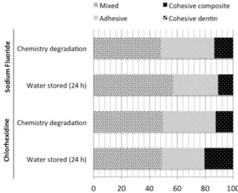
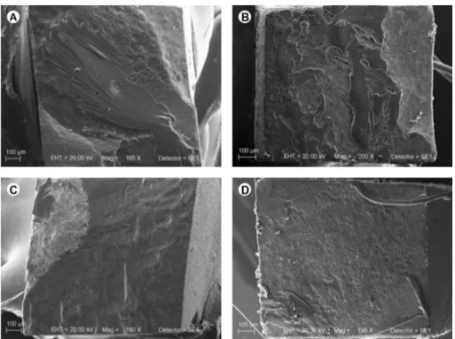
Distribution of failure modes is presented in Figure 1. Mixed failures predominated in all groups. The number of cohesive failures was slightly higher in the group where 2% CHX was used and the specimens were stored in water. Representative SEM images of mixed failure in all groups are illustrated in Figure 2.
Discussion
Most of the experiments designed to evaluate stability of the resin-dentin bond by enzyme inhibition have been performed with CHX, a potent antimicrobial agent. CHX effectively inhibits MMP-2, -8 and -9 (13,16,17) and cysteine cathepsins (18). In addition, many studies have demonstrated that CHX may improve the structural stability of collagen matrix in the hybrid layer and decrease time-dependent reduction in resin-dentin bond strength (14,17,19,20). Therefore, CHX was used as control group due to a large
number of studies demonstrating its efficacy to decrease the degradation of the resin-dentin bonding interface.
Another MMP inhibitor presented in the literature is sodium fluoride. However, the mechanism of MMPs inhibition by NaFis not yet fully understood. Kato et al. (11) showed the inhibition of MMP-2 and -9 by sodium fluoride, suggesting that the high electronegativity of the fluorine and heavy concentration of this mineral could release free cations to participate in the catalytic process. However, the study showed that irreversible inhibition was observed only in the fluoride at high concentration (5000 ppm); at lower concentrations (250, 500, 1500 ppm) MMP inhibitory effect was considered to be reversible. In the present study the groups using different MMP inhibitors for rewetting the dentin presented similar results after water storage for 24 h.
Accelerated aging is a relevant procedure to evaluate the long-term stability of the bonded interface (15). To simulate in the laboratory aging of adhesive restorations, a commonly used procedure is storing in 10% NaOCl. This is a nonspecific deproteinizing agent with tendency of creating superoxide radicals in aqueous solutions, therefore inducing oxidation phenomena, fragmenting the protein peptide chains (15,21,22). The 10% NaOCl may affect the resin-dentin bond by two different ways: first, degradation of the etched and non-resin-infiltrated layer; and second, by the collagen fibrils that were not properly resin-infiltrated and/or later exposed because of the resin dissolution by 10% NaOCl (23). Therefore, collagen fibrils in the etched
Braz Dent J 27(4) 2016
445
Bond strength of laser irradiated dentin
dentin and the resin adhesive may have been affected by 10% NaOCl action, leading to a rapid degradation of the resin-dentin layer (15,24). In the current study, the results demonstrated that the MMP inhibitors, 2% CHX and NaF(5000 ppm) were able to prevent chemical degradation of the adhesive interface, confirming the tested hypothesis. The predominance of mixed failures, especially in dentin irradiated with Er:YAG agree with the results of a previous study (25). Overall, no relevant change was observed in the failure mode among the groups.
It is possible to conclude that the MMP inhibitors, 2% CHX and NaF 5000 ppm presented similar μTBS between Er:YAG laser irradiated dentin and composite resin after the chemical degradation. Further studies are suggested to evaluate longer times and different degradation forms to confirm the effectiveness of these inhibitors in the Er:YAG laser irradiated dentin.
Resumo
Este estudo avaliou o efeito dos inibidores de metaloproteinase, clorexidina 2% e fluoreto de sódio (5000 ppm), na resistência de unĩo entre a dentina irradiada por laser Er:YAG e a resina composta após a degradaç̃o química da interface de unĩo. A superfície oclusal de quarenta molares humanos hígidos (n=10) foi removida expondo uma superfície de dentina, que foi polida, irradiada com laser Er:YAG, condicionada com ácido e seca. Vinte espécimes foram re-umedecidos com clorexidina 2% (Grupo controle) e 20 com fluoreto de sódio (5000 ppm). O sistema adesivo foi aplicado e um platô de resina compostafotopolimerizável de 4 mm de altura foi construído. Palitos de resina-dentina foram obtidos com secç̃o transversal retangular (0,8-1 mm2). Eles foram armazenados em água (24 h a 37 °C) ou submetidos a degradaç̃o química. Para a degradaç̃o química, foram imersos em soluç̃o aquosa de hipoclorito de sódio a 10% durante 5 horas e lavados em água durante 1 h. Os palitos foram submetidos ao teste de microtraç̃o em uma máquina de ensaios mecânicos a 0,5 mm/min até a fratura. O padr̃o de fratura foi analisado em MEV. Os valores de resistência de unĩo foram calculados em MPa e submetidos à análise de variância ANOVA (α=0,05). A análise de variância mostrou que os fatores inibidor de metaloproteinases e degradaç̃o (p=0,214 e p=0,093, respectivamente), e a interaç̃o entre os fatores ño foram estatisticamente significantes (p=0,143). A predominância de falha mista foi detectada para todos os grupos. Em conclus̃o, a clorexidina a 2% e fluoreto de sódio (ppm 5000) apresentaram resistência de unĩo entre dentina irradiada e resina composta semelhante antes e após a degradaç̃o química.
Acknowledgements
The authors would like to acknowledge the S̃o Paulo State Research Foundation - FAPESP for the financial support (Grant #18868-7).
References
1. Buyukhatipoglu I, Secilmis A. The use of erbium: yttrium-aluminum-garnet laser in cavity preparation and surface treatment: 3-year follow-up. Eur J Dent 2015;9:284-287.
2. de Almeida Neves A, Coutinho E, Cardoso MV, Lambrechts P, Van Meerbeek B. Current concepts and techniques for caries excavation and adhesion to residual dentin. J Adhes Dent 2011;13:7-22.
3. Turkun M, Turkun LS, Celik EU, Ates M. Bactericidal effect of Er,Cr:YSGG laser on Streptococcus mutans. Dent Mater J 2006;25:81-86. 4. Cecchini RC, Zezell DM, de Oliveira E, de Freitas PM, Eduardo C de P.
Effect of Er:YAG laser on enamel acid resistance: morphological and
atomic spectrometry analysis. Lasers Surg Med 2005;37:366-372. 5. Keller U, Hibst R, Geurtsen W, Schilke R, Heidemann D, Klaiber B, et al..
Erbium:YAG laser application in caries therapy. Evaluation of patient perception and acceptance. J Dent 1998;26:649-656.
6. Ceballo L, Toledano M, Osorio R, Tay FR, Marshall GW. Bonding to Er-YAG-laser-treated dentin. J Dent Res 2002;81:119-122.
7. Aranha AC, De Paula Eduardo C, Gutknecht N, Marques MM, Ramalho KM, Apel C. Analysis of the interfacial micromorphology of adhesive systems in cavities prepared with Er,Cr:YSGG, Er:YAG laser and bur. Microsc Res Tech 2007;70:745-751.
8. Schein MT, Bocangel JS, Nogueira GE, Schein PA. SEM evaluation of the interaction pattern between dentin and resin after cavity preparation using ER:YAG laser. J Dent 2003;31:127-135.
9. Amaral FL, Colucci V, Palma-Dibb RG, Corona SA. Assessment of in vitro
methods used to promote adhesive interface degradation: a critical review. J Esthet Restor Dent 2007;19:340-353; discussion 354. 10. Frassetto A, Breschi L, Turco G, Marchesi G, Di Lenarda R, Tay FR, et al..
Mechanisms of degradation of the hybrid layer in adhesive dentistry and therapeutic agents to improve bond durability-A literature review. Dent Mater 2016;32:e41-e53.
11. Kato MT, Bolanho A, Zarella BL, Salo T, Tjaderhane L, Buzalaf MA. Sodium fluoride inhibits MMP-2 and MMP-9. J Dent Res 2014;93:74-77. 12. Carrilho MR, Geraldeli S, Tay F, de Goes MF, Carvalho RM, Tjaderhane L,
et al.. In vivo preservation of the hybrid layer by chlorhexidine. J Dent Res 2007;86:529-533.
13. Pashley DH, Tay FR, Yiu C, Hashimoto M, Breschi L, Carvalho RM, et al.. Collagen degradation by host-derived enzymes during aging. J Dent Res 2004;83:216-221.
14. Carrilho MR, Carvalho RM, de Goes MF, di Hipolito V, Geraldeli S, Tay FR, et al.. Chlorhexidine preserves dentin bond in vitro. J Dent Res 2007;86:90-94.
15. Toledano M, Cabello I, Yamauti M, Giannini M, Aguilera FS, Osorio E, et al.. Resistance to degradation of resin-dentin bonds produced by one-step self-etch adhesives. Microsc Microanal 2012;18:1480-1493. 16. Gendron R, Grenier D, Sorsa T, Mayrand D. Inhibition of the activities of
matrix metalloproteinases 2, 8, and 9 by chlorhexidine. Clin Diagn Lab Immunol 1999;6:437-439.
17. Hebling J, Pashley DH, Tjaderhane L, Tay FR. Chlorhexidine arrests subclinical degradation of dentin hybrid layers in vivo. J Dent Res 2005;84:741-746.
18. Scaffa PM, Vidal CM, Barros N, Gesteira TF, Carmona AK, Breschi L, et al.. Chlorhexidine inhibits the activity of dental cysteine cathepsins. J Dent Res 2012;91:420-425.
19. Breschi L, Mazzoni A, Nato F, Carrilho M, Visintini E, Tjaderhane L, et al.. Chlorhexidine stabilizes the adhesive interface: a 2-year in vitro study. Dent Mater 2010;26:320-325.
20. Breschi L. Chlorhexidine application to stabilize the adhesive interface: why and how? J Adhes Dent 2013;15:492.
21. Osorio R, Toledano M, Osorio E, Aguilera FS, Tay FR. Effect of load cycling and in vitro degradation on resin-dentin bonds using a self-etching primer. J Biomed Mater Res A 2005;72:399-408.
22. Monticelli F, Osorio R, Pisani-Proenca J, Toledano M. Resistance to degradation of resin-dentin bonds using a one-step HEMA-free adhesive. J Dent 2007;35:181-186.
23. Toledano M, Osorio R, Albaladejo A, Aguilera FS, Osorio E. Differential effect of in vitro degradation on resin-dentin bonds produced by self-etch versus total-self-etch adhesives. J Biomed Mater Res A 2006;77:128-135.
24. Yoshida E, Hashimoto M, Hori M, Kaga M, Sano H, Oguchi H. Deproteinizing effects on resin-tooth bond structures. J Biomed Mater Res B Appl Biomater 2004;68:29-35.
25. Galafassi D, Scatena C, Colucci V, Rodrigues-Junior AL, Campos Serra M, Corona SA. Long-term chlorhexidine effect on bond strength to Er:YAG laser irradiated-dentin. Microsc Res Tech 2014;77:37-43.