MUSCLE INVOL
MUSCLE INVOL
MUSCLE INVOL
MUSCLE INVOL
MUSCLE INVOLVEMENT IN LEPROSY
VEMENT IN LEPROSY
VEMENT IN LEPROSY
VEMENT IN LEPROSY
VEMENT IN LEPROSY
STUDY OF THE ANTERIOR TIBIAL MUSCLE IN 40 PATIENTS
LINEU CESAR WERNECK*, HÉLIO A. G. TEIVE*, ROSANA HERMINIA SCOLA*
ABSTRACT - The involvement of skeletal striated muscle in leprosy is considered secondary due to peripheral neuropathy, but some studies point it to a primary muscle lesion. In order to investigate the muscle involvement in leprosy, we studied 40 patients (lepromatous 23, tuberculoid 13, borderline 2 and indeterminate 2). The motor nerve conduction of the peroneal nerves had a reduction of the velocity, decreased compound muscle action potential and sometimes absence of potentials. The electromyographic study of the anterior tibial muscle showed signs of recent and chronic denervation in 77.5% of the cases and no myopathic potentials. The anterior tibial muscle biopsy revealed denervation in 45% of the cases, interstitial inflammatory myopathy in 30% and mixed (myopathic and neuropathic) pattern in 12.5%. Acid fast bacillus was detected in 25% of the cases, always in the interstitial tissue. Inflammatory reaction was present in the interstitial space and in patients with the lepromatous type. The histological findings clearly defined the presence of the so-called “Leprous Interstitial Myositis” on the top of denervation signs.
KEY WORDS:leprosy, skeletal muscle, muscle biopsy, myositis.
Alterações musculares na lepra: estudo do músculo tibial anterior em 40 pacientes
RESUMO - O envolvimento do músculo estriado na lepra é considerado secundário à lesão dos nervos periféricos, mas alguns estudos relataram acometimento muscular primário. A fim de verificar esta controvérsia estudamos 40 pacientes com lepra, sendo 23 da forma lepromatosa, 13 da tuberculoide, 2 borderline e 2 indeterminada. Realizamos a neurocondução do nervo peroneiro, junto com eletromiografia e biópsia do músculo tibial anterior. Encontramos redução de velocidade de condução, da amplitude e algumas vezes ausência de potenciais no nervo peroneiro. A eletromiografia do tibial anterior mostrou sinais de desinervação recente e crônica em 77,5% dos casos e não foi encontrada evidência de padrão “miopático”. A biópsia do músculo tibial anterior revelou desinervação em 45% dos casos, miopatia inflamatória intersticial em 30% e padrão misto (miopático e neuropático) em 12,5%. Bacilos alcool-ácido resistentes foram encontrados em 25% dos casos, sempre localizados no perimisio e endomisio. Na forma lepromatosa, a reação inflamatória estava presente no espaço intersticial. Os dados histológicos claramente definiram a presença de “Miosite Lepromatosa Intersticial” sobreposta às alterações histológicas encontradas em desinervação.
PALAVRAS-CHAVES: lepra, músculo esquelético, biópsia muscular, miosite.
Leprosy is a chronic infection illness caused by theMyobacterium leprae(M.leprae) that
preferably attacks peripheral nerves. This disease is endemic all over the world, specifically in Brazil, and it is considered the commonest cause of treatable peripheral neuropathy1-3.
*Neuromuscular Disorders Service, Neurology Division, Internal Medicine Department, Hospital de Clínicas, Federal University of Paraná, Curitiba, Brazil.Supported in part by a grant from CNPq (Brazil). Aceite: 12-julho-1999.
The involvement of the skeletal striated muscle in leprosy has been characterized as a
consequence of the peripheral neuropathy with subsequent muscular denervation2,4,5. However, some
experimental studies with inoculation of the M.leprae in animals point to a primary muscle
involvement6-10. This primary muscle involvement has also been reported11-15and an inflammatory
reaction was described, being called lepromatous myositis16-18, leprous interstitial myositis19or leprous
nodular interstitial myositis20. However, these studies do not give the incidence of this abnormality
and histological/histochemical details are lacking.
We undertook this study in order to verify the histologic and electromyographic alterations in the muscle of patients with leprosy, as well as to look for their possible relationship.
METHOD
METHOD
METHOD
METHOD
METHOD
We studied 40 patients with an established diagnosis of leprosy through clinical examination, skin biopsy and bacilloscopy; 34 (85%) were male and 6 (15%) female. Their mean age was 45.6 years and the average disease duration was 3.85 years. Of these patients, 57.5% had the lepromatous form (Virchow form), 32.5% the tuberculoid form, 5% the borderline form (dimorphous) and 5% an indeterminate form (Table 1). None of the patients had any clinical symptoms suggesting polymyositis or any other myopathy; four had mononeuropathy, 24 had multiple mononeuropathy and 10 had polyneuropathy (Table 1). Four patients (10%) were studied before treatment and 36 (90%) under drug therapy (36 with dapsone, 6 with rifampin, 5 with clofazimine, 6 with thalidomide and 5 with prednisone). The mean treatment time was 3.48 years (one month to 18 years).
White and red blood cell counts, erythrocyte sedimentation rate (ESR), serum glucose, creatinine, syphilis Venereal Disease Research Laboratory test (VDRL), creatinekinase (CK), aldolase, lactic dehydrogenase (LDH), aspartate aminotransferase (SGOT) and alanineamino transferase (SGPT) were done in all patients.
We studied the motor conduction velocity of the peroneal nerve with cutaneous electrodes on both sides. We stimulated below the head of the fibula and just above the ankle with recording electrodes placed over the extensor digitorum brevis. We recorded the proximal and distal latencies, the amplitude and duration of the compound motor muscle potential and calculated the conduction velocity after measuring the distance between the stimulation points22.
Electromyography (EMG) was performed on the anterior tibial muscle with a coaxial needle electrode. We chose to study the anterior tibial muscle because it is a distal and superficial one, possible with lower temperature. Also the peroneal nerve enervates the anterior tibial muscle, the second most common nerve involved in leprosy. At rest we registered spontaneous activity like insertion activity, fibrillation, fasciculation, positive waves, high frequency and myotonic discharges. At voluntary contraction, we evaluated the duration, amplitude, degree of polyphasic potentials, as well as the recruitment pattern during weak and strong contraction21,22. We called it recent denervation when we found only spontaneous potentials with all the other parameters being normal; chronic denervation, when we had increased duration, large amplitude, excess of polyphasic potentials and decreased recruitment; recent and chronic with the combination of both, and borderline when we had only few or one type of abnormality present.
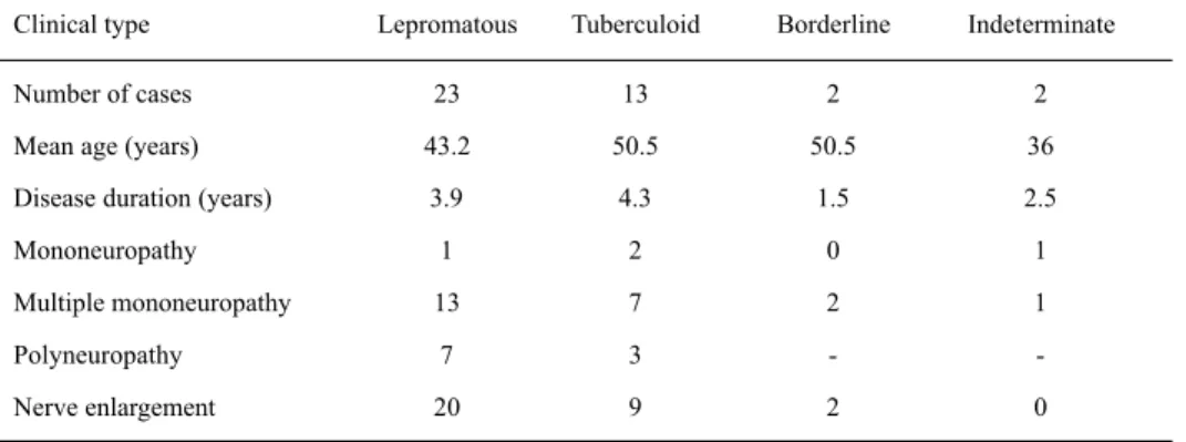
Table 1. Clinical findings.
Clinical type Lepromatous Tuberculoid Borderline Indeterminate
Number of cases 23 13 2 2
Mean age (years) 43.2 50.5 50.5 36
Disease duration (years) 3.9 4.3 1.5 2.5
Mononeuropathy 1 2 0 1
Multiple mononeuropathy 13 7 2 1
Polyneuropathy 7 3 -
All the patients were submitted to an anterior tibial muscle biopsy, always on the opposite side of the electromyography. The muscle fragment was studied with fresh-frozen sections and submitted to the following staining and histochemical reaction: hematoxilin-eosin, modified Gomori trichrome, oil red O, PAS, cresyl violet, sirius red, NADH-tetrazolium reductase, ATPases pH 4.3, 4.6, 9.4, myophosphorylase, non-specific esterase, alkaline phosphatase, acid phosphatase, succinic dehydrogenase and cytochrome c-oxidase23.
The final histological diagnoses were reported as normal (no alterations found), interstitial inflammatory myopathy (inflammatory infiltrate in the perimisium, diffuse or in the perivascular space, necrosis, phagocytosis, excess of internal nuclei, basophilic fibers, increased acid phosphatase in the fibers and in the interstitial cells), active denervation (atrophic angulated fibers in the NADH-tetrazolium reductase and non-specific esterase and rare necrotic fibers), chronic denervation (when found target fibers, type grouping, absence of necrosis and phagocytosis, type 1 and 2 atrophy, type 1 and 2 hypertrophy), active and chronic denervation (combination of all elements of chronic and active denervation) and mixed (combination of elements of interstitial inflammatory myopathy, active and chronic denervation). Ziehl-Neelsen coloration was done on all specimens using Fite-Faraco method. Each specimen had several serial slides section and an extensive search for acid fast bacilli (AFB) was conducted21.
RESUL
RESUL
RESUL
RESUL
RESULTS
TS
TS
TS
TS
Laboratory investigation found an increase of ESR in 17 cases, hypochromic and microcytic anemia in 12, eosinophilia and intestinal parasites in 8, positive VDRL with negative FTA-ABS in 7. Two cases had a slight elevation of the SGOT and SGPT, but they had also liver involvement (Hepatic granuloma on liver biopsy). All the cases had normal CK, aldolase and LDH.
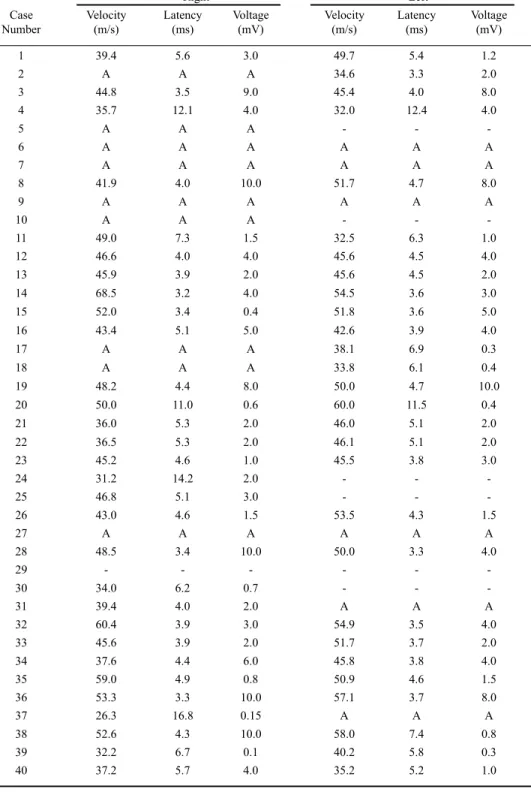
The nervous conduction of the right peroneal showed reduction of the velocity in 28.2% of the cases, normal conduction in 48.7% and absence of conduction in 23%. The distal latency was increased in 36.6% and there was a reduction in the amplitude in 60% of the cases. The left peroneal nerve had reduction of the velocity in 17% of the cases, normal conduction in 64% and absence of conduction in 17.6%. The distal latency was increased in 32.1% and there was a reduction in the amplitude of motor response in 60% of the cases (Table 2).
The electromyographic examinations showed denervation signs in 31 (77.5%) cases, borderline signs of denervation in 3 (7.5%) and were normal in 6 cases (15.0%) (Table 3).
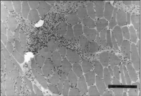
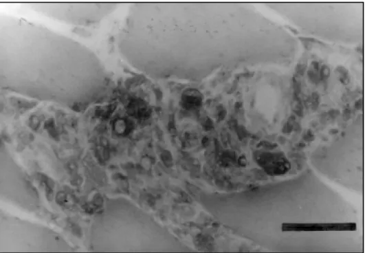
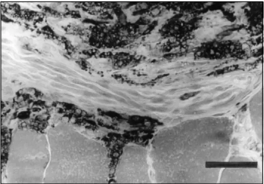
The most frequent abnormalities found in the muscle biopsy of all the cases were: variation of muscle fiber diameter, type 1 fiber predominance, atrophic rounded fibers, type 2 fiber atrophy, type 1 fiber atrophy, type grouping, atrophic dark angulated fibers in the non-specific esterase and NADH-tetrazolium reductase. There were some differences in the incidence of abnormalities regarding the type of leprosy, but no statistical difference was found between the tuberculoid and lepromatous types (Chi-Square test) (Table 4). The inflammatory infiltrate was more prominent in the perimysium and less in the endomysium, with rare fibers with necrosis and phagocytosis (Figs 1,2,3,4). The inflammatory reaction of the perimysium was also present into the nerve branches, which could be responsible for the denervation found in the muscle (Fig 5). The AFB were found exclusively in the lepromatous type and they were located in the interstitial space, inside macrophages, vessels walls and intramuscular nerves (Fig 6) (Table 4).
The muscle biopsy abnormalities permited the following histological diagnosis: denervation in 45% of the cases, inflammatory myopathy in 30%, (17.5% with the presence of AFB), mixed abnormalities with myopathic and neuropathic findings in 12.5% of the cases, and non specific abnormalities in 15% of the cases. The AFB in the muscle was detected in 25% of the cases (Tables 4 and 5).
Table 2. Nervous conduction velocity: peroneal nerve.
Right Left
Case Velocity Latency Voltage Velocity Latency Voltage
Number (m/s) (ms) (mV) (m/s) (ms) (mV)
1 39.4 5.6 3.0 49.7 5.4 1.2
2 A A A 34.6 3.3 2.0
3 44.8 3.5 9.0 45.4 4.0 8.0
4 35.7 12.1 4.0 32.0 12.4 4.0
5 A A A - -
-6 A A A A A A
7 A A A A A A
8 41.9 4.0 10.0 51.7 4.7 8.0
9 A A A A A A
10 A A A - -
-11 49.0 7.3 1.5 32.5 6.3 1.0
12 46.6 4.0 4.0 45.6 4.5 4.0
13 45.9 3.9 2.0 45.6 4.5 2.0
14 68.5 3.2 4.0 54.5 3.6 3.0
15 52.0 3.4 0.4 51.8 3.6 5.0
16 43.4 5.1 5.0 42.6 3.9 4.0
17 A A A 38.1 6.9 0.3
18 A A A 33.8 6.1 0.4
19 48.2 4.4 8.0 50.0 4.7 10.0
20 50.0 11.0 0.6 60.0 11.5 0.4
21 36.0 5.3 2.0 46.0 5.1 2.0
22 36.5 5.3 2.0 46.1 5.1 2.0
23 45.2 4.6 1.0 45.5 3.8 3.0
24 31.2 14.2 2.0 - -
-25 46.8 5.1 3.0 - -
-26 43.0 4.6 1.5 53.5 4.3 1.5
27 A A A A A A
28 48.5 3.4 10.0 50.0 3.3 4.0
29 - - -
-30 34.0 6.2 0.7 - -
-31 39.4 4.0 2.0 A A A
32 60.4 3.9 3.0 54.9 3.5 4.0
33 45.6 3.9 2.0 51.7 3.7 2.0
34 37.6 4.4 6.0 45.8 3.8 4.0
35 59.0 4.9 0.8 50.9 4.6 1.5
36 53.3 3.3 10.0 57.1 3.7 8.0
37 26.3 16.8 0.15 A A A
38 52.6 4.3 10.0 58.0 7.4 0.8
39 32.2 6.7 0.1 40.2 5.8 0.3
40 37.2 5.7 4.0 35.2 5.2 1.0
Table 3. Electromyographyc abnormalities (%).
Type of abnormality present Total lepromatous Tuberculoid
Number of cases 40 23 13
Insertion activity
Increased 5.0 - 15.4
Decreased 2.5 4.3
-Fibrillation potentials 20.0 21.7 23.1
Positive sharp waves 25.0 21.7 30.8
Fasciculation potentials 5.0 - 7.7
Complex repetitive discharges 7.5 4.3 15.4
Duration of motor unit potentials
Increased 47.5 47.8 38.5
Decreased 2.5 - 7.7
Amplitude of motor unit potentials
Increased 5.0 4.3
-Decreased 2.5 - 7.7
Short polyphasic motor units 10.0 13.0 7.7
Long polyphasic motor units 90.0 95.7 84.6
Recruitment pattern
Increased 2.5 - 7.7
Decreased 80.0 91.3 61.5
Table 4. Anterior tibial muscle biopsy abnormalities (%).
Histological/histochemical alteration Total Lepromatous Tuberculoid
Number of cases 40 23 13
HE and modified Connective tissue proliferation 15.0 13.0 15.4 Gomiri trichrome Adipose tissue infiltration 17.5 17.3 15.4
Necrosis 15.0 8.7 23.1
Phagocytosis 12.5 8.7 15.4
Diffuse inflammatory infiltrate 32.5 43.5 23.1 Perivascular inflammatory infiltrate 32.5 43.5 15.4
Internal nuclei 25.0 8.7 46.2
Basophilic fibers 5.0 4.3 0
Hypercontractile fibers 5.0 4.3 7.7
Inflammatory infiltrate excessive 27.5 39.1 15.4 Variation of muscular fibers diameter 95.0 91.4 100.0 Large agglomerations of atrophic fibers 7.5 0 15.4 Small agglomerations of atrophic fibers 15.0 4.3 30.8 Atrophic rounded fibers dispersed 60.0 65.2 53.9 Hypertrophic fibers dispersed 30.0 21.7 53.9
Nuclear clumps 22.5 13.0 30.8
Granular fibers (ragged reds) 2.5 0 7.7
Vacuoles 10.0 4.3 15.4
Cytoplasmatic inclusion body 2.5 4.3 0
Splitting fibers 2.5 0 7.7
NADH- Atrophic angulated fibers 42.5 43.5 30.8
tetrazolium reductase Target fibers 5.0 0 7.7
Moth-eaten fibers 5.0 0 7.7
Ring fibers 10.0 4.3 23.1
Focal increase NBT granules 47.5 52.2 46.2
Type grouping 45.0 47.8 30.8
ATPase Type 1 fiber atrophy 50.0 60.9 38.5
pH 9.4,4.3,4.6 Type 2 fiber atrophy 57.5 60.9 53.8
Type 1 fiber hypertrophy 25.0 13.0 46.2 Type 2 fiber hypertrophy 27.5 21.7 38.5
Type 2 fiber predominance 2.5 4.3 0
Type 2 deficiency fibers 12.5 8.7 23.1
Non-specific Motor plate - Increased size 5.0 4.3 7.7
esterase Atrophic angulated fibers 42.5 34.8 46.2
Interstitial activity in mononuclears 37.5 52.2 15.4 Acid phosphatase Focal increased into fibers 37.5 21.7 46.2
Positive fibers 7.5 4.3 15.4
Interstitial isolated mononuclear cells 35.0 43.4 23.1 Phagocytes near or in necrotic fibers 10.0 8.7 7.7 Nerves (Mononuclear/myelin sheath) 17.5 26.1 0
Alkaline phosphatase Positive fibers 7.5 0 15.4
Interstitial increase 37.5 47.8 15.4
Nerves (Mononuclear/myelin sheath) 5.1 8.7 0
Oil Red O Type 1 fiber increased 5.0 0 7.7
Succinic dehydrogenase Sub-sarcolemmal accumulation 50.0 52.2 38.5 Cytochrome c-oxidase Sub-sarcolemmal accumulation 32.5 39.1 15.4
Sirus red Interstitial tissue increase 10.0 0 23.1
Fite-Faraco for acid Endomysium and perimysium 17.5 30.4 0
Fast bacilli Inside macrophage 20.0 34.8 0
Around blood vessel 7.5 13.0 0
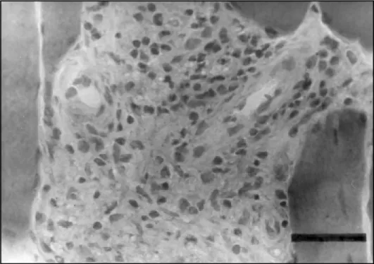
Fig 2. Inflammatory infiltrate in the perimysium with mononuclear cells. Haematoxylin & Eosin. Bar 50µ.
Table 5. Histological diagnosis of anterior tibial muscle biopsy.
Types of leprosy
All types Lepromatous Tuberculoid Borderline Indeterminate
Normal 3 1 1 - 1
Inflammatory myopathy 5 2 2 1
-Inflammatory myopathy + AFB 6 6 - -
-Active denervation 3 1 1 - 1
Active denervation + AFB 1 1 - -
-Chronic denervation 8 5 2 1
-Chronic denervation + AFB 1 1 - -
-Active + chronic denervation 4 2 2 -
-Mixed 4 2 2 -
-Mixed + AFB 1 1 - -
-Non specific 4 1 3 -
-Total 40 23 13 2 2
Fig 3. Inflammatory cells (macrophages) in the interstitial, spreading into the endomysium through the vasculo-nervous space. Acid phosphatase. Bar 50µ.
Fig 5. Lymphomononuclear cells near a nervous branch in the interstitial space. Non specific esterase. Bar 50µ.
Table 6. Relation between anterior tibial muscle biopsy and electromyography.
Histological diagnosis EMG
Denervation
Normal Borderline Active Chronic Active and
Chronic
Normal 1 - - 2
-Interstitial inflammatory myopathy - 1 1 8 1
Active denervation 1 - 1 2
-Chronic denervation 1 - - 5 3
Active and chronic denervation 1 - - 3
-Mixed 1 1 1 - 2
Non specific 1 1 - 1 1
DISCUSSION
DISCUSSION
DISCUSSION
DISCUSSION
DISCUSSION
The neurophysiological evaluation of these patients demonstrated that the study of nervous conduction in the peroneal nerves brought out meaningful results. Some patients had no conduction or very large latencies due to the severity of the disease. These abnormalities were asymmetric, according to the nature of the disease. These findings are in agreement with the literature, which points to the peroneal nerve as the most frequently compromised one, after the ulnar nerve24-27.
The electromyographic study of the tibialis anterior muscle showed alterations in most of the cases; 77.5% showed signs of denervation. These denervation potentials are due to peroneal nerve
or its branch involvement2.The electromyographic examination allows the detection of precocious
alterations in leprosy, even when clinical examination gives no evidence of it18. Electromyographic
alterations can be detected in up to 98% of the cases when several muscles are studied25,28. We did
not find electromyographic potentials compatible with primary muscle involvement (myopathic), as it had rarely been described18,29-31. The scope of this work was the study of the involvement of the
anterior tibial muscle in leprosy and for this reason we did not study other muscles with EMG, even knowing that we could find abnormalities in other muscles, if a complete examination was done. Also, even studying only one muscle, as in this paper, a good correlation can be obtained, according previously published papers21,32.
As expected, we found a histological pattern of denervation, with angulated atrophic fibers,
piknotic nuclear clumps, large agglomerations of atrophic fibers and type grouping21,23. We were
able to record the presence of an inflammatory infiltrate superimposed to the denervation, preponderant
in the interstitial and peri-fascicular area. This infiltration was of mononuclear cells, monocytes, lymphocytes and macrophages. Sometimes, it was very severe, with the formation of a granuloma with giant cells. These findings were described previously, with some variation as “Leprous Interstitial Myositis”12-14,16-19,33,34.
The inflammatory infiltration was present in the perimysium, near the blood vessels, and infiltrated the muscle following the vascular tree inside de muscle fascicle. In reality, it does not damage the muscle fibers and fiber necrosis was rare. The inflammatory infiltration among the striated muscle fibers, muscular fascicle atrophy as well as the presence of AFB was previously
called “Myositis Interstitialis Leprosa”16,19. Most of our patients who presented inflammatory
interstitial myopathy were carriers of leprosy in its lepromatous form, as it had already been observed in the literature. The fact that, in this group of patients, there is anergy to M.leprae, with systemic dissemination of the bacillus, somehow explains the characterization of a real bacillary septicemia13,14.
The presence of AFB was observed in 7 cases with inflammatory reaction, being detected in the interstitial area of muscle fascicles, together with inflammatory cells, granulomas, inside the macrophages, around vessels, nerves, in the arterioles, in nerve fascicles and inside foam cells. The presence of AFB, compatible with M. leprae in the smooth muscle and in the striated muscle of patients with leprosy has been demonstrated4,11-14,16,17,19,20,34,35.
Job et al. described alterations in the striated muscle, defining three levels of lesions: on the first, there is an invasion and proliferation of M.leprae in the muscular fibers; on the second there is a degeneration of the muscular fibers, an infiltration of lymphocytes, macrophages, polymorphonuclear cells and bacillus fragmentation; on the third, a destruction of the muscle fibers occurs, with the presence of fibrous tissues, but with absence of bacilli17.
Pearson et al. propose that Hansen’s bacillus can survive and multiply in the striated muscle more easily than it does in the skin, and that it is less attacked by the body’s immune system, disseminating itself systemically afterwards. The muscle would, then, have an exponential role in the pathogenesis of the leprosy, functioning as a bacillary reservoir14.
We did not find AFB inside muscle fibers in our material, even after a long search and several serial slides section for each case, as it was previously described by several authors14-18,36,37and in
experimental animal studies6,7,9,10. Also, some authors doubt the muscle fiber invasion by AFB12,38,
and the absence of AFB in some of our cases could be due to treatment.
In concluding, we found, besides the denervation process due to peroneal nerve involvement, an interstitial inflammatory myopathy with AFB, the so-called “Leprous Interstitial Myositis” in 45% of the cases. This interstitial myositis was predominantly observed in patients with the lepromatous form. The muscle inflammatory reaction has no electromyographic representation, since the examination only detected denervation, suggesting that the lesion takes place in the perivascular, perineural and interstitial area between the muscle fibers.
Acknowledgments- We would like to thank Miss Sumico Nakagawa and Angela Chiquito, for their technical assistance.
REFERENCES
REFERENCES
REFERENCES
REFERENCES
REFERENCES
1. Adams RD, Victor M. Principles of Neurology. 2Ed. New York: McGraw-Hill, 1981:960-1015.
2. Sabin TD, Swift TR, Jacobson RR.. Leprosy. In: Dyck PJ, Thomas PH, Griffin JW, Low PA, Poduslo JF. Peripheral neuropathy 3Ed. Philadelphia: W.B.Saunders 1993:1354-1379.
3. Talhari S, Neves RG. Hansenologia. Manaus, Calderaro-Funcomiz, 1984.
4. Slotwiner P, Song SK, Anderson PJ. Skeletal muscle changes in leprosy: their relationship to changes in other neurogenic diseases affecting muscle. J Pathol 1969;97:211-218.
5. Dastur DK. Pathology and pathogenesis of predilective sites of nerve damage in leprous neuritis: nerves in the arms and the face. Neurosurg Rev 1983;6:139-152.
7. Rees RJW, Weddell AGM. Experimental models for studying leprosy. Ann NY Acad Sci 1968;154:214-236.
8. Esiri MM, Weddell GM, Rees RJW. Infection of murine striated muscle with Mycobacterium leprae: study by light and electron microscopy. J Pathol 1972;106:73-80.
9. Lancaster RD, Hilson GRF, McDougall AC, Colston MJ. Mycobacterium leprae infection in nude mice: bacteriological and histological responses to primary infection and large inocula. Infect Immun 1983;39:865-872.
10. Job CK, McCormick GT, Hastings RC. Intracellular parasitism of parenchymal cells by Mycobacterium leprae. Int J Leprosy 1989;57:659-670.
11. Dastur DK, Daver SM. Striated muscle in four categories of leprosy: II.-Fine structural changes. Int J Leprosy 1980;48:149-152. 12. Daver SM, Dastur DK, Revankar CR, Shah JS. Striated muscle in four categories of leprosy.I-Histology and histochemistry.
Int J Leprosy 1980;48:140-148.
13. Gupta JC, Jesupadam T, Gupta MC, Gupta DK. A histopathologic study of striated muscle biopsies in leprosy. Int J Leprosy 1975;43:348-355.
14. Pearson JMH, Rees RJW, Weddell AGM. Mycobacterium leprae in the striated muscle of patients with leprosy. Lepr Rev 1970;41:155-166.
15. Mansour S-E, Mehasen A, El-Ariny AF. Muscular changes in lepromatous leprosy. Trans R Soc Trop Med Hyg 1970;64:918-920. 16. Convit J, Arvelo JJ, Mendonze S. Lepromatous myositis. Int J Leprosy 1960;28:417-422.
17. Job CK, Karat ABA, Karat S, Mathan M. Leprous myositis: a histopathological and electron-microscopic study. Lepr Rev 1969; 40:9-16.
18. Sebille A, Gray F. Electromyographic recording and muscle biopsy in lepromatous leprosy. J Neurol Sci 1979;40:3-10. 19. Ishihara S. A study of myositis interstitialis leprosa. Int J Leprosy 1959;27:341-346.
20. Jopling WH, Metha HD. A case of leprous nodular interstitial myositis. Lepr Rev 1972;43:39-43.
21. Werneck LC, Lima JGC. Muscle biopsy correlated with electromyography. Arq Neuropsiquiatr 1988;46:156-165. 22. Kimura J. Electrodiagnosis in diseases of nerve and muscle. Philadelphia: F.A.Davis, 1983.
23. Werneck LC. The value of muscle biopsy in neurology. Rev Bras Clin Terap 1981;10(Supl) 2-22.
24. Baccaredda-Boy AK, Mastropaoli C, Pastorino P, Sacco G, Farris G. Electromyographic findings in leprosy. Int J Leprosy 1963;31:531-532.
25. DeFaria CR, Silva IM. Electromyographic diagnosis of leprosy. Arq Neuropsiquiatr 1990;48:403-413.
26. Hackett ER, Shipley DE, Livengoos R.. Motor nerve conduction velocity studies of the ulnar nerve in patients with leprosy. Int J Leprosy 1968;36:282-287.
27. Karat S, Karat ABA, Potchandy P. Electromyographic findings in conduction velocities of ulnar nerves in leprosy. Lepr India 1970;42:77-82.
28. Brasil-Neto JP. Electrophysiologic studies in leprosy. Arq Neuropsiquiatr 1992;50:313-318.
29. Magora A, Sheskin J, Chaco J, Adler E. An electrodiagnostic study of the lower motor unit in leprosy. Int J Leprosy 1965;33:829-864.
30. Oderiz A, Reyes O. Convit J. Miositis lepromatosa. Dermatol Venez 1966;5:50-55.
31. Mansour S, Mehasen A. El-Eriny AF. Muscular changes in lepromatous leprosy. Trans R Soc Trop Med Hyg 1970;64:918-920. 32. Buchthal F, Kamieniecka Z. The diagnostic yield of quantified electromyography and quantities muscle biopsy in
neuromuscular disorders. Muscle Nerve 1982;5:265-280. 33. Job CK. Nerve damage in leprosy. Int J Leprosy 1989;57:532-539.
34. Shiemy SE, Hefnawy HE, Fattah AA, Hawary MFE, Fares R.. Muscle involvement in leprosy and its correlation with serum aldolase activity. Int J Dermatol 1977;16:587-593.
35. Esiri MM. Studies of intramuscular leprosy bacilli. Thesis, University of Oxford. Oxford,1969.
36. Koranne RV, Singh R, Yvengar B. Mycobacterium leprae in the striated muscle of tuberculoid leprosy patient. Lepr India 1978;50:375-380.