Original Article
RADIOLOGICAL REVIEW OF 173 CONSECUTIVE CASES OF
PARACOCCIDIOIDOMYCOSIS*
Henrique Simão Trad1, Clovis Simão Trad2, Jorge Elias Junior2, Valdair Francisco Muglia2
* Study developed at Clinics Hospital of the Faculty of Medicine of Ribeirão Preto,
Universidade de São Paulo, Ribeirão Preto, SP, Brazil.
1. M.D., Resident at Clinics Hospital of the Faculty of Medicine of Ribeirão Preto,
Universidade de São Paulo, Ribeirão Preto, SP.
2. Doctor Professor at Department of Medical Clinics of the Faculty of Medicine of Ribeirão
Preto, Universidade de São Paulo, Center of Imaging Sciences and Medical Physics.
Mailing address: Dr. Henrique Simão Trad. Avenida Sumaré, 690. Ribeirão Preto, SP, Brazil,
14025-450. E-mail: [email protected]
Received January 28, 2005. Accepted after revision August 3, 2005.
Abstract
OBJECTIVE: To determine the incidence of most significant radiological findings of
paracoccidioidomycosis and to verify its possible variants. MATERIALS AND METHODS: One
hundred and seventy-three cases of paracoccidioidomycosis presented between 1970 and 1980 were
reviewed, including their radiological workup reanalysis by at least two experienced radiologists.
RESULTS: Ninety-four cases were pulmonary only and 38 were pulmonary associated with
ganglial, visceral and osseous lesions or in association with tuberculosis. There was no pulmonary
involvement in 41 cases, with small bowel, viscera, bone lesions, or a combination of these. Most
significant radiological findings in cases of pulmonary involvement were bilateral, diffuse reticular
and nodular interstitial infiltrate, followed by the diffuse bilateral alveolar form. Visceral and
gastrointestinal tract lesions presented predominantly with liver, jejunum and ileum involvement.
Lymph nodal involvement was predominantly diffuse, abdominal or peripheral. In bones, osteolytic
lesions affected predominantly long bones and clavicle. CONCLUSION: Paracoccidioidomycosis is
a granulomatous disease commonly found in Brazil, primarily affecting lungs, caused by inhalation
of fungus spores. Other rare or less frequent forms of the disease should be taken into consideration
Keywords: Paracoccidioidomycosis; Granulomatous disease.
INTRODUCTION
Paracoccidioidomycosis is a systemic fungal disease caused by Paracoccidioides
brasiliensis, a dimorphic fungus that grows like yeast at 37ºC. It is found in Central and South
Americas and is a common cause of the disease in Brazil, Venezuela, Colombia, Ecuador, Peru and
Paraguay. This fungus has not been isolated in other regions of the planet and, invariably, patients
presenting this disease have a history of residence in or visit to endemic areas(1–4).
Paracoccidioidomycosis affects predominantly adult males from rural areas, in the age range
between 30 and 50 years(2,3). It is rare in children, but, when it occurs, affects both sexes equally. The disease may affect urban residents or any individual in contact with the fungus. The
Paracoccidioides brasiliensis has been isolated from soil for many times, so presently the soil is
considered as its most probable natural habitat.
The infection is presumably acquired via inhalation of the fungus in the mycelial phase(1–3),
followed by primary infection of the lungs with subsequent lymphahematogenic dissemination. This
primary pulmonary lesion may be clinically significant or remain latent, asymptomatic and
non-diagnosed.
The disease affects principally the lungs and the reticuloendotelial, tegmental, digestive and
osseous systems. Two main forms of the disease are described: the acute or subacute and
generalized, with lymphadenopathy and hepatosplenomegaly, affecting youngsters, principally up
to the third decade of life(1–5), and another, chronic and progressive, presenting typical
oropharyngeal ulcerations and affecting adult patients (1,2,4).
The literature on paracoccidioidomycosis is plentiful, showing a great concern in determining
its form of contagion and dissemination(6–17), the oropharyngeal mucosa being considered for years
as the portal of entry of the agent(18). However, we have not found significant reports regarding to the incidence of the several presentations of such as heterogeneous disease, even in great series(9,10).
MATERIALS AND METHODS
Between 1970 and 1980, 173 consecutive confirmed cases treated in the Clinics Hospital of
the Faculdade de Medicina de Ribeirão Preto, Universidade de São Paulo, were selected for this
study. Cases confirmed by histological analysis of different lesions were included. In cases suspect
Imaging examinations were performed according to the needs of each patient. In all of the
cases, a chest X-ray was request. More specific studies, like gastrointestinal series, linear CT,
lymphographies, bronchographies and other were performed in patients with specific clinical
suspicions. Later reevaluation of films was performed by at least two experimented radiologists.
RESULTS
One hundred and seventy-three patients were studied, of whom 93.7% were male and 6.3%
female. Ages ranged between 6 and 75 years, with a mean age of 40.8 years.
Sites of affection in this study were: lungs, ganglia, tegmentum, bowel, viscera and bones.
Percentage of affection for each site in relation to the total of cases is demonstrated in Table 1.
Different associations were found and divided into 15 groups with their respective incidences
demonstrated in Table 2.
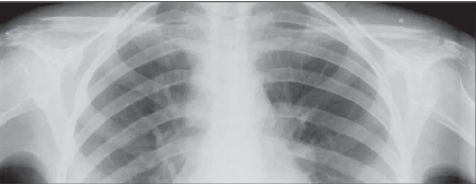
Among pulmonary presentations, predominance was as follows: reticular interstitial opacities
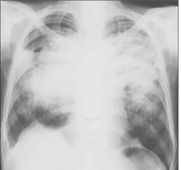
in 89.3% of cases (Figure 1), nodular opacities in 54.5%, bilateral alveolar opacities in 45.4%
(Figure 2) and mixed opacity with “butterfly wing” pattern in 44.7% (Figure 3). Emphysema was
found in 34.1%, Kerley’s septal lines were seen in 25.7%, retraction was observed in 18.2%, pleural
thickening was found in 9.1% and pleural effusion in 7.5%. Nine cases (6.8%) of cavitary lesions
were found at chest X-ray (Figure 4). Six percent of cases presented as unilateral alveolar lesion and
only 2.2% (three cases) as giant nodules. Increased mediastinal lymphadenopathy was found at
chest X-ray in 25 cases (14.4%), 15 of them related only to pulmonary disease. In five cases (2.9%
of total, 3.8% of pulmonary cases) there was concomitant infection by Micobacterium tuberculosis.
Main sites of generalized ganglial involvement were periphery and abdomen, followed by
ganglial mediastinal involvement.
At plain abdominal X-ray, intestinal forms presented as distention in 22% of cases, mass in
10%, calcification in 6% and occlusion in only 2%. In the digestive tract, lesions of jejunum, ileum
and colon predominated, presenting as thickening in 74% of cases (Figure 5); as flocculation in
60% (Figure 6), as segmentations in 48% and stricture in 44% (Figure 5). There was no case of
intestinal fistula. Three cases of gallbladder lesion were found in this series. No case of gastric
lesion was evidenced.
In the visceral form, the liver was involved in 100% of cases (24% of total) and spleen in
54.7%. Two cases of suprarenal involvement and one case with affection of central nervous system
Bone lesions presenting lytic aspect and without sclerotic reaction were evidenced in seven
cases, involving long bones (Figure 7) and clavicle (Figure 8) (five cases each), flat bones in three
cases and the pelvis in two. No case of involvement of spine was found.
DISCUSSION
With up to 3,000 cases diagnosed each year in Brazil, the paracoccidioidomycosis is an
endemic disease especially in the states of São Paulo, Rio de Janeiro, Minas Gerais, Rio Grande do
Sul e Mato Grosso(2). Due to its systemic nature, practically all the systems and organs of the human
body can be affected. Its predominance in male individuals was evidenced also in this study, with a
mean age of 40 years, demonstrating predominance of a chronic and progressive presentation of this
disease.
In this study, similarly to others, there was a clear predominance of pulmonary
involvement(9,10,19,20). Among pulmonary patterns and in concordance with the literature(21–27), the interstitial lesion was predominant, followed by bilateral alveolar lesions and the “butterfly-wing”
pattern. Emphysema, retraction as well as Kerley’s septal lines were significant findings in this
study(28), although in lower proportion than that previously described. Some authors have
emphasized the presence of pulmonary functions alterations as a result of paracoccidiodomycosis(29– 31)
. Some studies have been describing this disease alterations at chest high resolution CT(32). Pleural
lesions, effusion and thickening showed a similar proportion to that reported in a previous study by
Machado & Miranda(33). Association with pulmonary tuberculosis was a less frequent finding.
In the present study, the ganglial lesions proportion was lower than that previously
described(9). Preferential sites are the periphery and abdomen, followed by mediastinum Ganglial
alterations at lymphography have already been described(34–37) and presently have been practically
abandoned due to the availability of other better methods of evaluation. The laryngeal form of this
disease was radiologically documented in only seven cases and, in this study, we could not evaluate
statistically its incidence. The easy evaluation of this lesion by means of direct examination
dispenses with the imaging evaluation. Lauand(38) and Lauand et al.(39–41) had significantly
contributed to the study of these lesions.
Regarding to intestinal involvement, its incidence was higher than that reported in the
previous great series(9,10). Lesions characteristics had already been described(42–46), although without evaluation of their distribution. The predominance of lesions in jejunum, ileum and colon has been
the total of patients. The visceral involvement has predominated in the liver and spleen and lesions
of suprarenal and central nervous system have been interpreted as rare complications. Some authors
suggest that the central nervous system may occur more frequently(48).
The bone lesions found in 4% of cases in this study were those already previously described
as lytic lesions without sclerotic reaction(48–52). They have predominated in long bones and clavicle. Symmetric lesions on clavicles distal extremities are highly suggestive although being
non-pathognomonic findings, therefore other imaging findings and clinical data must be taken into
consideration.
CONCLUSION
Paracoccidioidomycosis is a systemic disease and does not spare any system of the human
body, presenting radiological findings suffice to a diagnosis definition. When findings are
non-specific, the possibility of paracoccidioidomycosis should be taken into consideration for
differential diagnosis.
REFERENCES
1. Dismukes WE. Paracoccidioidomycosis. In: Bennett JC, Plum F, editors. Cecil Textbook of
medicine. Philadelphia: Saunders, 1996;1822–1823.
2. Kwon-Chung KJ, Bennett JE. Paracoccidioidomycosis. In: Kwon-Chung KJ, Bennett JE,
editors. Medical mycology. Philadelphia: Lea & Febiger, 1992;594–619.
3. Fraser RS, Müller NL, Colman N, Paré PD. South American blastomycosis. In: Fraser RS,
Müller NL, Colman N, Paré PD, editors. Fraser & Paré Diagnosis of diseases of the chest. 4th ed.
Philadelphia: Saunders, 1999;902–904.
4. Corrin B. Paracoccidioidomycosis (South American blastomycosis). In: Corrin B, editor.
Pathology of the lungs. London: Churchill Livingstone, 2000;220–221.
5. Londero AT, Gonçalves AJR, Cruz MLS, et al. Paracoccidioidomicose disseminada
“infanto-juvenil” em adolescentes. Arq Bras Med 1987;61:5–12.
6. Guimarães FN, Macedo DG. Contribuição ao estudo das blastomicoses na Amazônia
(blastomicose queloidiana e blastomicose sul-americana). O Hospital 1950;38:223–253.
7. Guimarães N. Micose de Lutz na Bahia (a propósito de um novo caso). O Hospital
1950;38:693–698.
9. Machado Filho J, Miranda JL. Considerações relativas à blastomicose sul-americana.
Localizações, sintomas iniciais, vias de penetração e disseminação em 313 casos consecutivos. O
Hospital 1960;58:99–137.
10. Machado Filho J, Miranda JL. Considerações relativas à blastomicose sul-americana. Evolução,
resultados terapêuticos e moléstias associadas em 394 casos consecutivos. O Hospital 1961;60:375–
412.
11. Lopes CF. Contribuição ao estudo da blastomicose sul-americana (doença de Lutz). J Bras Med
1962;6:539–548.
12. Portugal H. Anatomia patológica da micose de Lutz. J Bras Med 1962;6:489–496.
13. Sampaio SAP. Tratamento da blastomicose sul-americana. J Bras Med 6:516–521.
14. Gonçalves AO. Tratamento da micose de Lutz (blastomicose sul-americana). J Bras Med
1962;6:522–530.
15. Furtado TA. Tratamento da blastomicose brasileira. J Bras Med 1962;6:531–538.
16. Silva JR. Blastomicose sul-americana. O Hospital 1964;65:53–65.
17. Lacaz CS, Ferri RG, Netto AF, et al. Blastomicose queloidiana associada à blastomicose
sul-americana. Registro de um caso. O Hospital 1967;71:1–11.
18. Azulay RD, Feldman J, Azulay JD. Caso de micose de Lutz (blastomicose sul-americana) de
localização ganglionar. Sua importância no diagnóstico diferencial das linfopatias. O Hospital
1955;48:309–317.
19. Magalhães A. Paracoccidioidomicose (blastomicose sul-americana). Aspectos radiológicos.
Rev Hosp Clin Fac Med São Paulo 1980;35:147–155.
20. Gonçalves AP, Bardy C. Aspectos clínicos e radiológicos da blastomicose brasileira pulmonar.
O Hospital 1946;30:1021–1041.
21. Paula A. O pulmão na blastomicose brasileira. J Bras Med 1962;6:480–483.
22. Bardy C. Sinais radiológicos pulmonares da blastomicose sul-americana. J Bras Med
1962;6:484–488.
23. Passos Filho MCR. Blastomicose sul-americana. Comentários em torno de 83 casos de
localização pulmonar. Classificação radiológica. O Hospital 1966;70:127–152.
24. Ferreira LC. O comprometimento pulmonar na blastomicose sul-americana. Arq Bras Tuberc
Doenças Tórax 1966;25:41–60.
25. Simão C, Moraes CR. O comprometimento brônquico na blastomicose sul-americana. Estudo
broncográfico. Rev Inst Med Trop São Paulo 1971;13:252–256.
26. Sequeira OF, Júdice LF, Gabetto JM, Silva LASR, Lima OAS. Blastomicose sul-americana.
27. Valle ACF, Guimarães RR, Lopes DJ, Capone D. Aspectos radiológicos torácicos na
paracoccidioidomicose. Rev Inst Med Trop São Paulo 1992;34:107–115.
28. Moraes CR, Simão C. Linhas septais de Kerley (linhas B) na blastomicose sul-americana. Rev
Inst Med Trop São Paulo 1968;10:214–218.
29. Machado Filho J, Lisboa RM, Matos AD, Januzzi A, Miranda JL. Considerações relativas a
blastomicose sul-americana. As repercussões cardiovasculares das lesões pulmonares. Dados
hemodinâmicos, oximétricos e angiopneumográficos. O Hospital 1961;60:241–259.
30. Simão AT, Tavares W, Tomassini MCC, Krakowski D, Silva JR. Alterações da função
ventilatória na blastomicose pulmonar. Correlação radiológica e espirográfica. Rev Soc Bras Med
Trop 1967;1:79–89.
31. Lemle H, Vieira LOBD, Milward GAF, Miranda JL. Lung function studies in pulmonary South
American blastomycosis. Am J Med 1970;48:434–442.
32. Muniz MAS, Marchiori E, Magnago L, Moreira LBM, Almeida Jr JG. Paracoccidioidomicose
pulmonar: aspectos na tomografia computadorizada de alta resolução. Radiol Bras 2002;35:147–
154.
33. Machado Filho J, Miranda JL. Considerações relativas à blastomicose sul-americana. Da
participação pulmonar em 338 casos consecutivos. O Hospital 1960;58:23–43.
34. Kalaf JM. Linfografia. Técnica de exame e indicações clínicas. Rev Paul Med 1969;74:131–
144.
35. Arruda PRB, Castro RM. Linfografia na blastomicose sul-americana. An Bras Dermatol
1967;40:7–14.
36. Simão C. Alterações radiológicas ganglionares na blastomicose sul-americana. Rev Inst Med
Trop São Paulo 1975;17:242–246.
37. Brandão PP. Calcificações ganglionares em blastomicose sul-americana. Rev Imagem
1979;1:79–80.
38. Lauand F. Contribuição para o estudo da morfologia do Paracoccidioides brasiliensis nos
tecidos orais. Rev Inst Med Trop São Paulo 1966;8:69–78.
39. Lauand F, Lia RCC, Paino MAS. Blastomicose sul-americana. Estudo clínico das lesões bucais.
Rev Fac Farm Odont (Araraquara) 1975;9:243–251.
40. Lauand F, Lia RCC, Paino MAS. Paracoccidioidomicose: evolução clínica e histopatológica
dos tecidos bucais após terapêutica sulfamídica. Rev Fac Farm Odont (Araraquara) 1976;10:145–
41. Lauand F, Lia RCC, Paino MAS. Emprego de uma técnica para evidenciação do
Paracoccidioides brasiliensis nos tecidos bucais. Rev Fac Farm Odont (Araraquara) 1976;10:53–
64.
42. Cunha MAR, Gouveia OF. Forma linfático-abdominal da doença de Lutz. Arq Bras Med
1961;51:269–276.
43. César HC, Cariani A, Lauand F, Lia N. Abdome agudo de etiologia blastomicótica. O Hospital
1962;61:229–241.
44. Cunha MAR, Pereira AA, Gouveia OF, et al. Contribuição ao estudo radiológico da
blastomicose sul-americana no aparelho digestivo. O Hospital 1966;69:195–200.
45. Moraes CR, Fiorillo AM, Costa JC. Lesões radiológicas intestinais na blastomicose
sul-americana. O Hospital 1967;71:145–156.
46. Avritchir Y, Perroni AA. Radiological manifestations of small intestinal South American
blastomycosis. Radiology 1978;127:607–609.
47. Forattini OP. Blastomicose da região pancreática. Rev Paul Med 1947;31:165–172.
48. Costa MAB, Carvalho TN, Araújo Jr CR, et al. Manifestações extrapulmonares da
paracoccidioidomicose. Radiol Bras 2005;38:45–52.
49. Maffei WE, Hungria Filho JS. Tumor blastomicótico do fêmur. Arq Hosp Santa Casa São Paulo
1956;2:41–54.
50. Mello Filho A, Vilela MP, Giannini SD, Sandreschi ET. Um caso de blastomicose óssea com
lesões múltiplas clinicamente primitivas. Rev Paul Med 1967;70:246–254.
51. Vasconcellos LPWC, Lazzareschi M. Relato de um caso de blastomicose sul-americana de
localização óssea. Rev Bras Ort R Janeiro 1974;9:117–124.
52. Kurok JK, Cury LA, Palmieri IT, et al. Blastomicose sul-americana com localização óssea
REVISÃO RADIOLÓGICA DE 173 CASOS
Tabelas e Figuras
Figure 1. Reticular interstitial and bilateral pattern.
Figure 2. Bilateral alveolar pattern associated with cavitary formations in both lungs.
Table 1 Percentage by site of affection. Site of affection
Toracic (pulmonary + pleural) Ganglial
Tegmental Intestinal Visceral Bone
No. of cases
132 34 7 50 43 7 Percentage 76.3% 19.6% 4.0% 28.9% 24.8% 4.0%
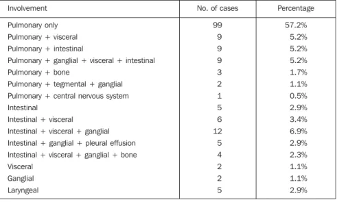
Table 2 Percentage of different associated affections. Involvement
Pulmonary only Pulmonary + visceral
Pulmonary + intestinal
Pulmonary + ganglial + visceral + intestinal Pulmonary + bone
Pulmonary + tegmental + ganglial
Pulmonary + central nervous system Intestinal
Intestinal + visceral
Intestinal + visceral + ganglial
Intestinal + ganglial + pleural effusion Intestinal + visceral + ganglial + bone Visceral
Ganglial
Laryngeal
No. of cases
Figure 4. Cavitary lesions in both lungs.
Figure 3. Mixed lesion in “butterfly-wing” pattern, with predominating confluence.
Figure 6. Flocculations in intestinal loops.
Figure 7. Multiple osteolytic lesions in three patients, without reactional sclerotic bone on tibia, fibula, humerus, radium and ulna, with pathological humerus fracture.