The expression of the aminoacid transporters ASCT2
(SLC1A5) and LAT1 (SLC7A5) in astrocytomas
Maria José Ferreira AlvesI,II, Miyuki UnoI,II, Roseli da SilvaI, Sueli Mieko Oba-ShinjoI, Suely Kazue Nagahashi MarieI,III
DOI: 10.5935/MedicalExpress.2016.06.05
I Universidade de São Paulo, Faculdade de Medicina, Departamento de Neurologia, Laboratório de Biologia Molecular e Celular (LIM 15), São Paulo, SP, Brasil. II Instituto do Câncer do Estado de São Paulo (ICESP), Centro de Investigação Translacional em Oncologia, São Paulo, SP, Brasil.
III Universidade de São Paulo, Faculdade de Medicina, Centro de Estudos de Terapia Celular e Molecular (NETCEM), São Paulo, SP, Brasil
OBJECTIVE: ASCT2 and LAT1 are aminoacid transporters involved in glutamine transport and play a role in tumor growth. Previous studies have shown an association of ASCT2 to cell proliferation through the mechanistic Target of Rapamycin (mTOR) translational machinery; LAT1 has been shown as a prognostic marker due to its relation to tumor invasion, microscopic vascular invasion and metastasis. This study analyzed the gene expression of ASCT2 and LAT1 in astrocytomas of diferent grades and how this correlates to clinical outcome in glioblastoma patients.
METHOD: This is an observational study with ASCT2 and LAT1 mRNA expression analysis in 153 samples of human astrocytomas, distributed in diferent World Health Organization (WHO) grades of malignancy (23 at grade I or pilocytic astrocytoma, 26 at grade II or low-grade astrocytoma, 18 at grade III or anaplastic astrocytoma, 86 at grade IV astrocytoma or glioblastoma (AGIV or GBM)); these were compared to 22 non-neoplastic brain samples.
RESULTS: Signiicant hyperexpression of both genes was observed particularly in malignant astrocytomas (GIII & GBM). Moreover, LAT1 hyperexpression impacted negatively in the overall survival in a subset of GBM patients.
CONCLUSION: LAT1 is more expressed in higher grade astrocytomas. It leads to a poorer prognosis among GBM patients and may be a potential therapeutical target.
KEYWORDS: ASCT2, LAT1, astrocytomas, glutamine, gene expression.
Alves MJF, Uno M, Silva R, Oba-Shinjo SM, Marie SKN. The expression of the aminoacid transporters ASCT2 (SLC1A5) and LAT1 (SLC7A5) in astrocytomas, MedicalExpress (São Paulo, online). 2016;3(6):M160605
Received for Publication on November 16, 2016; First review on November 22, 2016; Accepted for publication on December 5, 2016; Online on December 16, 2016
E-mail: [email protected]
■
INTRODUCTIONAmino acids are the primary source of cellular nitrogen, used for the synthesis of nucleotides, glutathione, amino sugars, and proteins. The carbon skeletons of amino acids are often used, in addition to glucose and fatty acids, as an oxidative fuel source for ATP generation, and may also contribute to sterol and lipid biosynthesis.1ASCT2 is a Na+ dependent,
broad-scope neutral amino acid exchanger that belongs to the solute carrier family-1 (SLC1, the high-affinity glutamate and neutral amino acid transporter family).2Expression of ASCT2, also known as SLC1A5, has been reported as essential for cell growth and viability of human hepatoma
cells through the mTOR signaling pathway.3LAT1, also
known as SLC7A5, codes a L-system transporter; it preferentially transports large, neutral amino acids, such as leucine, isoleucine, valine, phenylalanine, tyrosine, tryptophan, methionine, and histidine.4 Cumulative
evidence suggest that LAT1 is implicated in growth and proliferation of tumor cells, and also normal cells during tissue development. Very few observations have been reported about these glutamine transporters in central nervous system tumors. Astrocytomas are the most common primary brain tumors. The World Health
Organization (WHO) classifies astrocytomas into four
grades: grade I or pilocytic astrocytoma, grade II or low-grade astrocytoma (AGII), grade III or anaplastic astrocytoma (AGIII) and grade IV or glioblastoma (AGIV or GBM).5 Diffusely infiltrative astrocytomas (AGII-GBM)
of 60°C and were synthesized by IDT (Integrated DNA
Technologies, Coralville, USA) as follows (5′ to 3′):
ASCT2 F: TCCGCTTCTTCAACTCCTTCA;
ASCT2 R: CACATCCTCCATCTCCACGA;
LAT1 F: ATCCTCTCCATGATCCACCCA;
LAT1 R: GCGTAGAGCAGCGTCATCAC;
HPRT F: TGAGGATTTGGAAAGGGTGT; HPRT R: GAGCACACAGAGGGCTACAA; GUSB F: GAAAATACGTGGTTGGAGAGCTCATT; GUSB R: CCGAGTGAAGATCCCCTTTTTA; TBP F: AGGATAAGAGAGCCACGAACCA; TBP R: CTTGCTGCCAGTCTGGACTGT.
The minimum primer concentrations necessary were determined to give the lowest threshold cycle (Ct) and
maximum amplification efficiency, while minimizing non-specific amplification. A standard curve was established to ensure amplification efficiency and analysis of melting
curves demonstrated a single peak for all PCR products. Additionally, agarose gel electrophoresis was employed to
check the size of the PCR product amplified. SYBR Green I amplification mixtures (12 µl) contained 3 µl of cDNA, 6 µl of 2X Power SYBR Green I Master Mix (Life Technologies,
Carlsbad, USA) and forward and reverse primers. PCR reactions were run on an ABI Prism 7500 sequence detector (Life Technologies, Carlsbad, USA) as follows: 2 min at 50°C, 10 min of polymerase activation at 95°C, and 40 cycles of 15 s at 95°C and 1 min at 60°C. All the reactions were performed in duplicate. The following equation was applied to calculate gene expression in tumor and non-neoplastic tissue samples: 2-∆Ct, where DCt = Ct specific gene – mean Ct of housekeeping. For statistical analysis, gene expression status was scored as “hyper” or “hypo expressed” in relation to the value calculated by the receiver operator characteristic (ROC) curve as described below.
Ethical statement. Written informed consent was obtained from all patients per the ethical guidelines approved by the Department of Neurology, School of Medicine, University of São Paulo (case # 0599/10).
Statistical analysis. The statistical analysis of relative gene expression in different grades of astrocytoma was assessed using the Kolmogorov-Smirnov normality test, and the non-parametric Kruskal-Wallis and Dunn tests. Correlation between relative gene expression values was assessed using the non-parametric Spearman-rho correlation test and the parametric Pearson’s correlation test. Categorical variables were compared by Pearson Chi-square. Discrimination of variables was calculated by the receiver operator characteristic (ROC) curve utilizing
area under curve and asymptotic significance. Among the
continuous variables categorized through the ROC curve the
value with the best sensitivity and specificity was chosen as the cut-off value. The gene expressions were classified
as hyper or hypoexpression based on this cut-off value. The Kaplan-Meier survival curve was analyzed using the
tissue, hampering tumor resection; glioblastoma is the most aggressive adult brain tumor with a usually dismal outcome.6,7 The aim of the present study is to analyze the
gene expression of ASCT2 and LAT1 in astrocytomas of different grades and correlate to clinical outcome among GBM patients.
■
MATERIAL AND METHODSTissue samples. The studied series consisted of 23 AGI, 26 AGII, 18 AGIII, 86 GBM, and 22 non-neoplastic (NN) brain anonymized cases from epilepsy patients subjected to temporal lobectomy. Tumor samples were obtained during therapeutic surgery of patients treated by the Neurosurgery Group of the Department of Neurology at Hospital das Clínicas at the School of Medicine of the University of São Paulo, in the period of 2000 to 2007. The cases were categorized according to the WHO grading system by a neuropathologist from the Division of Pathological Anatomy of the same institution. Samples were macrodissected; immediately following surgical removal
they were snap-frozen in liquid nitrogen. A 4µm-thick
cryosection of each sample was analyzed under a light microscope after hematoxylin-eosin staining for assessment of necrotic, cellular debris and non-neoplastic areas (in tumor samples); this was followed by removal from the frozen block by microdissection prior to RNA extractions.
Sample preparation. Total RNA was extracted from frozen tissues (tumor and non-neoplastic) using an RNeasy Mini Kit (Qiagen, Hilden, Germany). Evaluation of RNA concentration and purity were carried out by measuring absorbance at 260 and 280 nm. Ratios of 260/280 measures ranging from 1.8 to 2.0 were considered satisfactory for purity standards. Denaturing agarose gel electrophoresis was used to assess the quality of the samples. A conventional reverse transcription reaction was performed to yield
single-stranded cDNA. The first strand of cDNA was synthesized from 1 µg of total RNA previously treated with 1 unit of DNase
I (FPLC-pure, GE Healthcare, Uppsala, Sweden) using random and oligo (dT) primers, RNase inhibitor, and SuperScript III reverse transcriptase according to the manufacturer’s recommendations (Life Technologies, Carlsbad, USA). The resulting cDNA was subsequently treated with 1 unit of RNase H (GE Healthcare, Uppsala, Sweden), diluted with TE buffer, and stored at -20°C until later use.
Quantitative real time PCR (qRT-PCR). The relative expression levels of ASCT2 and LAT1 were analyzed by
qRT-PCR, using the SYBR Green approach. Quantitative data
were normalized in relation to the geometric mean of three reference genes, suitable for the analysis: hypoxanthine phosphoribosyltransferase (HPRT), glucuronidase beta (GUSB) and TATA box binding protein (TBP), as previously demonstrated by our group.8,9 The primers were designed to
log-rank parameter. Statistical analysis was carried out with SPSS for Windows, version 20.0 (IBM Corporation, Armonk, USA). Differences were considered statistically
significant when p<0.05
■
RESULTSASCT2 and LAT1 were hyperexpressed in malignant astrocytomas
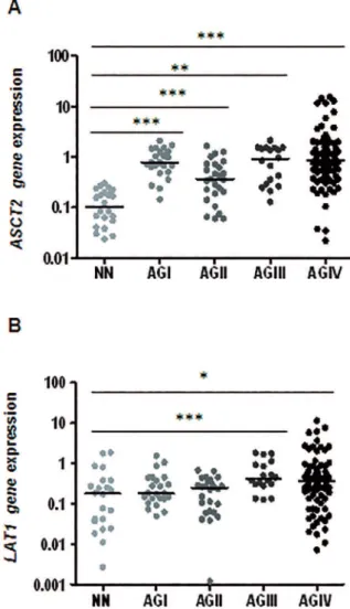
Figure 1A shows that significant ASCT2 hyperex-pression was observed in all grades of astrocytoma in comparison to non-neoplastic brain tissue, and a stepwise increment of ASCT2 expression was detected in diffusely
infiltrative astrocytomas (AGII to GBM). In contrast, Figure 1B shows that a significant LAT1 hyperexpression was only present in malignant astrocytomas (AGIII and GBM).
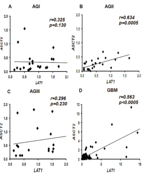
Figure 2 illustrates the correlation between ASCT2 and LAT1 gene expressions for the four grades of
astrocytoma. They correlated significantly for low grade
astrocytomas (AGIII) (r=0.634, p=0.0005) and for GBMs (r=0.563, p<0.0005), as shown in Figures 2B and 2D; but no correlation was found the expression of ASCT2 and LAT1
genes in AGI (Figure 2A) or AGIII (Figure 2C). A cut-off
value for the significant expression level was obtained using
the ROC curve. Considering a best balance of sensitivity
and specificity, a value of 0.315 for ASCT2 expression was
obtained with 0.791 of sensitivity and 1.00 of specificity,
and a value of 0.255 for LAT1 expression with 0.608 of
sensitivity, and 0.682 of specificity.
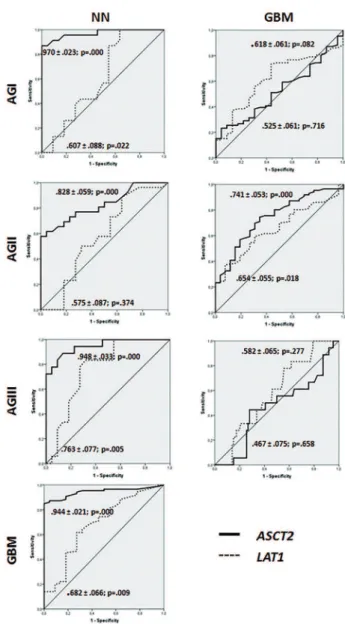
Figure 3 shows that the ROC curves of ASCT2 exhibited
high sensitivity and specificity to differentiate non-neoplastic
vs. neoplastic condition; in contrast LAT1 was found to differentiate the more malignant astrocytomas (AGIII and GBM). When applying these values to the studied population, the calculated odds ratio was 4.12 (p=0.003) for ASCT2 and 3.80 (p=0.007) for LAT1 for GBM cases, meaning high risk of malignant grade of astrocytoma for patients presenting elevated expressions of both ASCT2 and LAT1.
Figure 4 shows a heatmap with an overview of both genes expression levels in different grades of astrocytomas in comparison to non-neoplastic brain tissues: the highest expression levels of both genes occurred in GBM, but the
heterogeneity of the expression profile of the two genes is
evident in GBM cases.
Impact of ASCT2 and LAT1 mRNA expression levels in the outcome of GBM cases.
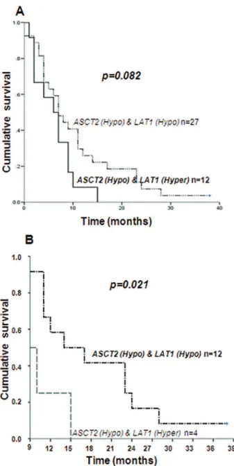
When subsets of GBM patients were compared considering combination of the status of ASCT2 and LAT1
mRNA expression levels (Figure 5), a trend for worse outcome was found for patients presenting only LAT1
hyperexpression (p=0.082), with median overall survival time of 6.2±1.2 months in comparison to 10.5±1.7 months
Figure 1 - Amino acid transporters ASCT2 and LAT1 mRNA expression. Expression
levels of ASCT2 (A) and LAT1 (B) in non-neoplastic tissues (NN) were compared to the expressions on all grades of astrocytoma (AGI: pilocytic astrocitoma, AGII: low grade astrocytoma, AGIII: anaplastic astrocytoma, AGIV: GBM). The expression levels difer signiicantly among the groups for both genes (Kruskal-Wallis test, p<0.0001). The expressions on pair-based comparisons were also signiicant among the group as demonstrated by post-hoc Dunn’s test, where ***p<0.0005, **p<0.005 and *p<0.05. Horizontal bars show the median relative expression in each group. The results are presented in log10 scale for better visualization.
for those presenting hypoexpression of both genes (Figure 5A). However, when we compared these cases after the period of standard of care of 8 months, the difference in overall survival time was statistically different (log rank
p= 0.021): patients presenting hyperexpression of LAT1
presented 10.7±1.4 months, against a mean survival time of 18.4±2.4 months for those presenting hypoexpression for both genes (Figure 5B).
■
DISCUSSIONTo the best of our knowledge, this is the first
Figure 2 - Correlation between ASCT2 and LAT1 mRNA expression levels in diferent grades of astrocytoma. Pearson’s correlation test resulted signiicant among AGII (B) and GBM (D) astrocytomas. The coeicient of corre-lation (r) and p value are shown for each grade. NN: non-neoplastic brain tissues, AGI: pilocytic astrocytoma, AGII: low grade astrocytoma, AGIII: anaplastic astrocytoma, GBM: glioblastoma.
transporters, LAT1 and ASCT1, gene expression in a large series of different grades of astrocytic tumors. Both genes presented higher expression in astrocytomas of more malignant grades. Especially, LAT1 expression impacted in GBM patients outcome.
Although glucose metabolism has been considered as the major source for the bioenergetic necessities of cancerous cells, there is cumulative evidence demonstrating that the highly proliferative cancer cells need additional sources to supplement for biosynthetic precursors of intermediary substrates of Krebs’s cycle.10,11 The
glutaminolysis and pyruvate carboxylation pathways are important alternatives in this context.12-14 Glutamine (Gln)
is a compulsory carbon donator, and it is transported into the cell by different transporting systems. Herein, the gene expression of two major Gln transporters, ASCT2 and LAT1, were analyzed in a series of human astrocytoma cases.
Two functions are attributed to ASCT2: 1) retrovirus receptor during placental development and fusion of endothelial cells in breast cancer,15,16 and 2) neutral
aminoacid transporter with high affinity to Gln.17,18,19
Increased expression of ASCT2 has been previously described in several tumors, including hepatocellular carcinoma,2 colorectal cancer, clear cell renal carcinoma,20
breast cancer,21 laryngeal squamous cell carcinoma,22
prostate carcinoma23 and metastasis,24 suggesting its
relevant role in tumor progression. In the present study,
we demonstrated a significant increase in ASCT2 mRNA expression in a stepwise pattern in parallel to the increase
of malignancy among astrocytomas, confirming previous
data.25 The involvement of ASCT2 in cancer cell proliferation
has been confirmed in vitro assay with C6 mouse glioma cell line,26 and cell line derived from human neuroblastoma.27
Figure 3 - ROC curves for ASCT2 and LAT1 expressions. Curves show sensitivity and speciicity of ASCT2 expression level in all grades of astrocytomas compared to non--neoplastic brain tissues while LAT1 expression levels show better sensitivity and speciicity on more malignant grades of astrocytomas (grade III and IV). When GBM is compared to other grades of astrocytomas, the sensitivity and speciicity is signiicant only in relation to grade II (p=0.000 for ASCT2 and p=0.018 for LAT1). NN: non-neoplastic brain tissues, AGI: pilocytic astrocytoma, AGII: low grade astrocytoma, AGIII: anaplastic astrocytoma,GBM: glioblastoma.
Figure 4: Heatmap of ASCT2 and LAT1 mRNA expression levels in diferent grades of
astrocytoma and non-neoplastic brain tissue. The green color was attributed for gene expression levels below the values determined by ROC curve (0.315 for ASCT2 and 0.255 for LAT1), and red to expression levels equal to or above these values. Hyperex-pression of ASCT2 is observed in all grade of astrocytomas, and hyperexHyperex-pression of LAT1 in more malignant grades of astrocytomas (grades III and GBM). A small set of GBM cases present the highest expression of both genes (in red). NN: non-neoplastic brain tissues, AGI: pilocytic astrocytoma, AGII: low grade astrocytoma, AGIII: anaplastic astrocytoma, GBM: glioblastoma.
may be induced by mTOR and ERK signaling pathways, independently of glutamine metabolism.28-30
LAT1 (isolated in 1998) was the first discovered
transporter of L-system; it transports Gln and asparagine
despite its low affinity (Km of 1.6 and 2.1 μM, respectively). Increased LAT1 expression has also been described in several types of cancer, namely colorectal,31,32 glioma,33,34
esophageal,35-37 ovarian,38,39 neuroendocrine40 and
others.41 Moreover, a high expression of LAT1 has been
associated to bad prognosis in prostate carcinoma,42,43 lung
adenocarcinoma44 and non-small-cell lung carcinoma.45
Additionally, LAT1 hyperexpression in clear cell renal carcinoma was correlated to less differentiated tumor, local invasion, microscopic vascular invasion, and metastasis.46
Of note, it has been demonstrated that LAT1 presents asymmetric intra and extra cellular substrate recognition,47
which may be further exploited for therapeutic purposes.
■
CONCLUSIONOur present findings of LAT1 hyperexpression in more malignant astrocytomas and its impact upon the
■
ACKNOWLEDGMENTSThis study was supported by grants #01/12898-4 and #04/12133-6 from São Paulo Research Foundation (FAPESP), and grant # 483467/2011-1 from National
Council for Scientific Technological Development (CNPq).
We sincerely thank Isac de Castro for the support in the biostatistical analysis.
■
AUTHOR PARTICIPATIONMaria José Ferreira Alves: planning and executing experimental laboratorial work and writing the article
Miyuki Uno: statistical analysis and figure elaboration
Roseli Silva: gene expression analysis
Sueli Mieko Oba-Shinjo and Suely Kazue Nagahashi Marie: experimental design and paper writing
■
CONFLICT OF INTERESTAuthors declare no conflict of interest regarding this project
EXPRESSÃO DOS TRANSPORTADORES DE AMI-NOÁCIDOS ASCT2 (SLC1A5) E LAT1 (SLC7A5) EM
ASTROCITOMAS
OBJETIVO: ASCT2 e LAT1 são transportadores de aminoácidos envolvidos no transporte de glutamina e desempenham um papel no crescimento tumoral. Estudos prévios mostraram uma associação de ASCT2 com proliferação celular através da maquinaria de tradução do mTOR; tem sido mostrado que o LAT1 é um marcador prognóstico devido à sua relação com invasão tumoral, invasão vascular microscópica e metástase. Este estudo analisou a expressão gênica de ASCT2 e LAT1 em astrocitomas de diferentes graus e sua correlação com desfecho clínico em pacientes com glioblastoma.
METODO: Este é um estudo observacional com
análise de expressão de RNAm de ASCT2 e LAT1 em 153 amostras de astrocitomas humanos, distribuídas em diferentes graus de malignidade segundo a OMS (23 astrocitomas de grau I ou astrocitoma pilocítico, 26 de astrocitoma de grau II ou astrocitoma de baixo grau, 18 de astrocitoma de grau III ou astrocitoma anaplásico, 86 de astrocitoma de grau IV ou glioblastoma (AGIV ou GBM); estes foram comparados com 22 amostras cerebrais não neoplásicas.
RESULTADOS: Foi observada uma hiperexpressão de ambos os genes, particularmente nos astrocitomas malignos (GIII & GBM). Além disso, a hiperexpressão LAT1
impactou negativamente na sobrevida global em um grupo
Figure 5 - Survival curves of GBM patients according to ASCT2 and LAT1 gene
ex-pression status. A: The comparison of the median survival time of a subgroup of 27 GBM patients presenting hypoexpression of ASCT2 and LAT1 (10.5±1.7months) to a subgroup of 12 GBM patients with hypoexpression of ASCT2 and hyperexpression of LAT1 (6.2±1.2months) resulted in a trend of worse outcome for those presenting hyperexpression of LAT1 (p= 0.082 by log rank). B: The same comparison after the period of standard care of 8 months resulted in a mean survival time of 18.4±2.4 months for patients presenting hypoexpression of both genes in contrast to the mean survival time of 10.7±1.4 months for those presenting hyperexpression of LAT1, with a log rank p = 0.021.
levels impact more significantly on the clinical outcome of
de pacientes com GBM.
CONCLUSÃO: LAT1 é mais expresso em astrocitomas
de grau maior. Isso leva a um pior prognóstico entre os pacientes com GBM e pode ser um potencial alvo terapêutico.
PALAVRAS-CHAVE: ASCT2, LAT1, astrocItoma,
glutamina, expressão gênica.
■
REFERENCES1. Fuchs BC, Bode BP. Amino acid transporters ASCT2 and LAT1 in cancer: partners in crime? Semin Cancer Biol. 2005; 15(4):254-66.
2. Fuchs BC, Finger RE, Onan MC, Bode BP. ASCT2 silencing regulates mammalian target-of-rapamycin growth and survival signaling in human hepatoma cells. Am J Physiol Cell Physiol. 2007;293(1):55-63. http://dx.doi.org/10.1152/ajpcell.00330.2006
3. Fuchs BC, Perez JC, Suetterlin JE, Chaudhry SB, Bode BP. Inducible antisense RNA targeting amino acid transporter ATB(0)/ASCT2 elicits apoptosis in human hepatoma cells Am J Physiol Gastrointest Liver Physiol. 2004;286(3):467-78. http://dx.doi.org/10.1152/ajp-gi.00344.2003
4. Kekuda R, Prasad PD, Fei YJ, Torres-Zamorano V, Sinha S, Yang-Feng
TL. Cloning of the sodium-dependent, broad-scope, neutral amino acid transporter B-O from a human placental choriocarcinoma cell line. J Biol Chem. 1996; 271(31):18657-61.5. http://dx.doi.org/10.1074/ jbc.271.31.18657
5. Louis DN, Ohgaki H, Wiestler OD, Cavenee WK, Burger PC, Jouvet A.
The 2007 WHO classification of tumours of the central nervous system.
Acta Neuropathol. 2007;114(2): 97-109. http://dx.doi.org/10.1007/ s00401-007-0243-4
6. Ohgaki H, Kleihues P. Genetic profile of astrocytic and oligodendro -glial gliomas. Brain Tumor Pathol. 2011;28(3):177-83. http://dx.doi. org/10.1007/s10014-011-0029-1
7. Ohgaki H, Kleihues P. The definition of primary and secondary
glioblastoma. Clin Cancer Res. 2013;19(4):764-72. http://dx.doi. org/10.1158/1078-0432.CCR-12-3002
8. Marie SK, Okamoto OK, Uno M, Hasegawa AP, Oba-Shinjo SM, Cohen T, et al. Maternal embryonic leucine zipper kinase transcript abundance correlates with malignancy grade in human astrocytomas. Int J Cancer. 2008; 122(4):807-15. http://dx.doi.org/10.1002/ijc.23189 9. Valente V, Teixeira SA, Neder L, Okamoto OK, Oba-Shinjo SM, Marie SK
et al. Selection of suitable housekeeping genes for expression analysis in glioblastoma using quantitative RT-PCR. BMC Mol Biol. 2009; 10:17. http://dx.doi.org/10.1186/1471-2199-10-17.
10. DeBerardinis RJ, Mancuso A, Daikhin E, Nissim I, Yudkoff M, Wehrli
S et al. Beyond aerobic glycolysis: transformed cells can engage in glutamine metabolism that exceeds the requirement for protein and nucleotide synthesis. Proc Natl Acad Sci USA. 2007;104(49):19345-50. http://dx.doi.org/10.1073/pnas.0709747104
11. DeBerardinis RJ, Sayed N, Ditsworth D, Thompson CD. Brick by brick: metabolism and tumor cell growth. Curr Opin Genet Dev. 2008;18(1):54-61. http://dx.doi.org/10.1016/j.gde.2008.02.003 12. Fan TW, Lane AN, Higashi RM, Farag MA, Gao H, Bousamra M, Miller
DM. Altered regulation of metabolic pathways in human lung cancer discerned by C-13 stable isotope-resolved metabolomics (SIRM). Mol. Cancer. 2009; 8:41. http://dx.doi.org/10.1186/1476-4598-8-41
13. Gao P., Tchernyshyov I., Chang TC, Lee YS, Kita K, Ochi T, et al. c-Myc
suppression of miR-23a/b enhances mitochondrial glutaminase ex-pression and glutamine metabolism. Nature. 2009;458(7239):762-5. http://dx.doi.org/10.1038/nature07823.
14. Wise DR, DeBerardinis RJ, Mancuso A, Sayed N, Zhang XY, Pfeiffer HK
et al. Myc regulates a transcriptional program that stimulates mito-chondrial glutaminolysis and leads to glutamine addiction. Proc Natl Acad Sci USA. 2008;105(48):18782-7. http://dx.doi.org/10.1073/ pnas.0810199105.
15. Bjerregaard B, Holck S, Christensen IJ, Larsson LI. Syncytin is in-volved in breast cancer-endothelial cell fusions. Cell Mol Life Sci. 2006;63(16):1906-11. http://dx.doi.org/10.1007/s00018-006-6201-9.
16. Blond JL, Lavillette D, Cheynet V, Bouton O, Oriol G, Chapel-Fernandes S et al. An envelope glycoprotein of the human endogenous retrovirus HERV-W is expressed in the human placenta and fuses cells expressing the type D mammalian retrovirus receptor. J Virol. 2000; 74(7):3321-9. http://dx.doi.org/10.1128/JVI.74.7.3321-3329.2000.
17. Bode BP, Kaminski DL, Souba WW, Li AP. Glutamine transport in isolated human hepatocytes and transformed liver cells. Hepatology. 1995;21(2):511-20. http://dx.doi.org/10.1002/hep.1840210236
18. Utsunomiya-Tate N, Endou H, Kanai Y. Cloning and functional charac -terization of a system ASC-like Na+-dependent neutral amino acid transporter. J Biol Chem. 1996;25(271):14883-90. http://dx.doi. org/10.1074/jbc.271.25.14883
19. Hannanein M, Hoeksema MD, Shiota M, Qian J, Harris BK, Chen H et al. SLC1A5 mediates glutamine transport required for lung cancer cell growth and survival. Clin Cancer Res. 2013;19(3):560-70. http:// dx.doi.org/10.1158/1078-0432.CCR-12-2334
20. Liu Y, Yang L, An H, Chang Y, Zhang W, Zhu Y, Xu L, Xu J. High expression
of Solute Carrier Family 1, member 5 (SLC1A5) is associated with poor prognosis in clear-cell renal cell carcinoma. Sci Rep. 2015;5:16954. http://dx.doi.org/10.1038/srep16954
21. van Geldermalsen M, Wang Q, Nagarajah R, Marshall AD, Thoeng A, Gao D et al. ASCT2/SLC1A5 controls glutamine uptake and tu-mour growth in triple-negative basal-like breast cancer. Oncogene. 2016;35(24):3201-8. http://dx.doi.org/10.1038/onc.2015.381 22. Nikkuni O, Kaira K, Toyoda M, Shino M, Sakakura K, Takahashi K, et al.
Expression of Amino Acid Transporters (LAT1 and ASCT2) in Patients with Stage III/IV Laryngeal Squamous Cell Carcinoma. Pathol Oncol Res. 2015; 21(4):1175-81. http://dx.doi.org/10.1007/s12253-015-9954-3 23. Wang Q, Hardie RA, Hoy AJ, van Geldermalsen M, Gao D, Fazli L et al. Targeting ASCT2-mediated glutamine uptake blocks prostate cancer growth and tumour development. J Pathol. 2015;236(3):278-89. http://dx.doi.org/10.1002/path.4518
24. Witte D, Ali N, Carlson N, Younes M. Overexpression of the neutral
amino acid transporter ASCT2 in human colorectal adenocarcinoma. Anticancer Res. 2002; 22(5): 2555-7.
25. Sidoryk M, Matyja E, Dybel A, Zielinska M, Bogucki J, Jaskólski DJ.
Increased expression of a glutamine transporter SNAT3 is a marker of malignant gliomas. Neuroreport. 2004;15(4):575-8. http://dx.doi. org/10.1097/01.wnr.0000117892.81922.b9.
26. Dolinska M, Dybel A, Zablocka B, Albrecht J. Glutamine transport
in C6 glioma cells shows ASCT2 system characteristics. Neuro-chem Int. 2003;43(4-5):501-7. http://dx.doi.org/10.1016/S0197-0186(03)00040-8
27. Wasa M, Wang HS, Okada A. Characterization of L-glutamine transport by a human neuroblastoma cell line. Am J Physiol Cell Physiol. 2002; 282(6):1246-53. http://dx.doi.org/10.1152/ajpcell.00324.2001 28. Fuchs BC, Finger RE, Onan MC, Bode BP. ASCT2 silencing regulates
mammalian target-of-rapamycin growth and survival signaling in human hepatoma cells. Am J Physiol Cell Physiol. 2007;293(1):C55-63. http://dx.doi.org/10.1152/ajpcell.00330.2006
29. Nicklin P, Bergman P, Zhang B, Triantafellow E, Wang H, Nyfeler B,
et al. Bidirectional transport of amino acids regulates mTOR and autophagy. Cell. 2009;136(3):521-34. http://dx.doi.org/10.1016/j. cell.2008.11.044
30. DeBerardinis RJ, Mancuso A, Daikhin E, Nissim I, Yudkoff M, Wehrli
S, et al. Beyond aerobic glycolysis: transformed cells can engage in glutamine metabolism that exceeds the requirement for protein and nucleotide synthesis. Proc Natl Acad Sci USA. 2007;104(49):19345-50. http://dx.doi.org/10.1073/pnas.0709747104
31. Yanagida O, Kanai Y, Chairoungdua A, Kim DK, Segawa H, Nii T, Cha
32. Wolf D, Wang S, Panzica M, Bassily NH, Thompson NL. Expression of a highly conserved oncofetal gene, TA1/ E16, in human colon carcinoma and other primary cancers: Homology to Schistosoma mansoni amino acid permease and Caenorhabditis elegans gene products. Cancer Res. 1996;56(21):5012-22.
33. Nawashiro H, Otani N, Shinomiya N, Fukui S, Ooigawa H, Shima K. L-type amino acid transporter 1 as a potential molecular target in human astrocytic tumors. Int J Cancer. 2006; 119(3): 484-92. http:// dx.doi.org/10.1002/ijc.21866
34. An S, Lu X, Zhao W, Sun T, Zhang Y, Lu Y, et al. Amino Acid Metabolism
Abnormity and Microenvironment Variation Mediated Targeting and Controlled Glioma Chemotherapy. Small. 2016;12(40):5633-45. http:// dx.doi.org/10.1002/smll.201601249
35. Kobayashi H, Ishii Y, Takayama T. Expression of L-type amino acid
transporter 1 (LAT1) in esophageal carcinoma. J Surg Oncol. 2005; 90(4): 233-8. http://dx.doi.org/10.1002/jso.20257
36. Ohshima Y, Kaira K, Yamaguchi A, Oriuchi N, Tominaga H, Nagamori S et al. Efficacy of system l amino acid transporter 1 inhibition as a
therapeutic target in esophageal squamous cell carcinoma. Cancer Sci. 2016; 107(10) 1499-1505. http://dx.doi.org/10.1111/cas.13021
37. Honjo H, Kaira K, Miyazaki T, Yokobori T, Kanai Y, Nagamori S, et al. Clinicopathological significance of LAT1 and ASCT2 in patients with
surgically resected esophageal squamous cell carcinoma. J Surg Oncol. 2016;113(4):381-9. http://dx.doi.org/10.1002/jso.24160
38. Kaji M, Kabir-Salmani M, Anzai N, Yokobori T, Kanai Y, Nagamori S, et
al. Properties of L-type amino acid transporter 1 in epidermal ova-rian cancer. Int J Gynecol Cancer. 2010;20(3):329-36. http://dx.doi. org/10.1111/IGC.0b013e3181d28e13
39. Kaira K, Nakamura K, Hirakawa T, Imai H, Tominaga H, Oriuchi N,
et al. Prognostic significance of L-type amino acid transporter 1
(LAT1) expression in patients with ovarian tumors. Am J Transl Res. 2015;7(15):1161-71.
40. Barollo S, Bertazza L, Watutantrige-Fernando S, Censi S, Cavedon E, Galuppini F et al. Overexpression of L-Type Amino Acid Transporter 1 (LAT1) and 2 (LAT2): Novel Markers of Neuroendocrine Tumors. PLoS One. 2016;11(15):e0156044. http://dx.doi.org/10.1371/journal. pone.0156044.
41. Zhao Y, Wang L, Pan J. The role of L-type amino acid transporter 1
in human tumors. Intractable Rare Dis Res. 2015;4(4):165-9. doi: 10.5582/irdr.2015.01024
42. Sakata T, Ferdous G, Tsuruta T, Satoh T, Baba S, Muto T, et al. L-type aminoacid transporter 1 as a novel biomarker for high-grade malig-nancy in prostate cancer. Pathol Int. 2009;59(1):7-18. http://dx.doi. org/10.1111/j.1440-1827.2008.02319.x
43. Wang Q, Tiffen J, Bailey CG, Lehman ML, Ritchie W, Fazli L, et al. Targe-ting amino acid transport in metastatic castration-resistant prostate cancer: effects on cell cycle, cell growth, and tumor development. J Natl Cancer Inst. 2013;105(19):1463-73. http://dx.doi.org/10.1093/jnci/ djt241
44. Kaira K, Oriuchi N, Imai H, Shimizu K, Yanagitani N, Sunaga N, et al. Prognostic significance of l-type amino acid transporter 1 (LAT1) and
4F2 heavy chain (CD98) expression in stage I pulmonary adenocarci-noma. Lung Cancer. 2009;66(1):120-6. http://dx.doi.org/10.1016/j. lungcan.2008.12.015
45. Kaira K, Oriuchi N, Imai H, Shimizu K, Yanagitani N, Sunaga N, et al. Prognostic significance of L-type amino acid transporter 1 expression
in resectable stage I-III nonsmall cell lung cancer. Br J Cancer. 2008; 98(4):742-8. http://dx.doi.org/10.1038/sj.bjc.6604235
46. Betsunoh H, Fukuda T, Anzai N, Nishihara D, Mizuno T, Yuki H, et al.
Increased expression of system large amino acid transporter (LAT)-1 mRNA is associated with invasive potential and unfavorable prognosis of human clear cell renal cell carcinoma. BMC Cancer. 2013;13:509. http://dx.doi.org/10.1186/1471-2407-13-509
47. Habermeier A, Graf J, Sandhofer BF, Boissel JP, Roesch F, Closs EI.