CT enterography: a preliminary experience in the evaluation
of small bowel diseases*
Enterografia por tomografia computadorizada: experiência inicial na avaliação das doenças do intestino delgado
Luciana Costa-Silva1, Tatiana Martins2, Maria do Carmo Friche Passos3
OBJECTIVE: The present study was aimed at demonstrating the value of computed tomography enterography (CT enterography) and how this imaging method can be useful in the diagnostic elucidation and assessment of patients with small bowel diseases. MATERIALS AND METHODS: Retrospective evaluation of 35 patients submitted to CT enterography in a 16-row multidetector CT equipment from May/2008 to March/2009. All the patients received intravenous and neutral oral iodinated contrast agents (polyethylene glycol). Main indications were: Crohn’s disease, diarrhea of undetermined origin and suspicion of neoplasia. RESULTS: A good correlation was observed between CT enterography findings and clinical, laboratory and endoscopic data related to the disease activity in patients with Crohn’s disease. In 15 cases alterations compatible with Crohn’s disease were identified, nine of them suggesting disease activity. A diagnosis was achieved in the majority of the patients with diarrhea. Carcinoid tumors were identified in two patients. CONCLUSION: CT enterography is a simple and effective method in the evaluation of inflammatory/neoplastic small bowel diseases, particularly in cases of Crohn’s disease, indicating disease activity. One of the main advantages of this method is the possibility of evaluating associated mesenteric and extraintestinal alterations.
Keywords: Enterography; Computed tomography; Small bowel; Crohn’s disease.
OBJETIVO: O objetivo deste estudo é demonstrar a importância da enterografia por tomografia computado-rizada (entero-TC) e como este exame pode contribuir na elucidação diagnóstica e avaliação clínica de pacien-tes com doenças do inpacien-testino delgado. MATERIAIS E MÉTODOS: Análise retrospectiva de 35 pacienpacien-tes submetidos a entero-TC realizadas em aparelho multidetector de 16 canais, entre maio de 2008 e março de 2009. Utilizou-se meio de contraste iodado venoso e oral neutro (polietilenoglicol). As principais indicações foram avaliação de doença de Crohn, diarreia de origem indeterminada e suspeita de neoplasias. RESULTA-DOS: Houve boa correlação dos achados à entero-TC relacionados à atividade da doença em pacientes com doença de Crohn, quando comparados com dados clínicos, laboratoriais e endoscópicos. Em 15 casos iden-tificaram-se alterações compatíveis com doença de Crohn, 9 deles sugerindo atividade. Dos pacientes com diarreia, o exame elucidou a maioria dos casos. Identificaram-se dois casos de tumor carcinoide. CONCLU-SÃO: A entero-TC é método simples e eficaz para estudo das doenças inflamatórias/neoplásicas do intestino delgado, sobretudo na avaliação da doença de Crohn, indicando atividade da doença. Uma de suas princi-pais vantagens é a possibilidade de avaliação de alterações mesentéricas e extraintestinais associadas.
Unitermos: Enterografia; Tomografia computadorizada; Intestino delgado; Doença de Crohn. Abstract
Resumo
* Study developed at the Unit of Radiology and Imaging Diag-nosis, Ecoar Medicina Diagnóstica, Belo Horizonte, MG, Brazil. 1. Master, MD, Radiologist, Assistant Professor at Department of Supplementary Propedeutics, Faculdade de Medicina da Universidade Federal de Minas Gerais (UFMG), Belo Horizonte, MG, Brazil.
2. MD, Radiologist, Specialist in Radiology and Imaging Diag-nosis, Ecoar Medicina Diagnóstica, Belo Horizonte, MG, Brazil. 3. PhD, Associate Professor-2 at Department of Medical Prac-tice, Universidade Federal de Minas Gerais (UFMG), Belo Horizonte, MG, Brazil.
small bowel diseases, being increasingly utilized(2). This is a method that provides high spatial resolution, allowing the visu-alization of the lumen and the mucosal re-lief, with the main advantage of evaluating the parietal thickness, in addition to even-tual association with mesenteric and extraintestinal findings(3).
The most common indications for the examination include the detection and fol-low-up of inflammatory bowel diseases, particularly Crohn’s disease, investigation of small bowel tumors, abdominal pain and diarrhea of unknown origin, and obscure Costa-Silva L, Martins T, Passos MCF. CT enterography: a preliminary experience in the evaluation of small bowel diseases. Radiol Bras. 2010;43(5):303–308.
ventional enteroclysis and small bowel fol-low-through, this later being, for many years, considered the standard imaging method(1). However, as a consequence of advances obtained with the introduction of multidetector computed tomography and the wide variety of available enteric con-trast media, CT enterography has become the method of choice in the evaluation of
Mailing address: Dra. Luciana Costa Silva. Rua Antônio de Albuquerque, 1021/901, Lourdes. Belo Horizonte, MG, Brazil, 30112-011. E-mail: [email protected]
Received February 17, 2010. Accepted after revision Septem-ber 16, 2010.
INTRODUCTION
con-gastrointestinal bleeding. It allows identi-fication of hypervascular lesions, bowel dilatation, stenosis, fistulas, hyperenhancing bowel segments, as well as other abdomi-nal findings. Thus, CT enterography has extensive clinical applicability, particularly in Crohn’s disease, providing data on ac-tivity and possible associated complica-tions(2,4,5).
The aim of this study is to demonstrate how this method can contribute with the di-agnostic elucidation and clinical manage-ment of bowel diseases. The small bowel has always been considered as a structure of difficult propedeutic evaluation, because of its extensive length, presence of folds, its position in the digestive tube between the stomach and the large bowel, its tor-tuosity and loops overlapping(6). CT enterography, with its thin slices and multiplanar reconstruction, minimizes such limitations and many times provides a greater amount of data than other imaging methods such as capsule endoscopy and enteroscopy, enhancing the role of radio-logical studies in the enteral assessment(6,7).
Considering the simplicity and swiftness of this noninvasive method, it is important to disseminate the technique describing its main findings.
MATERIALS AND METHODS
From May 2008 to March 2009, 35 pa-tients (mean age, 43.46 years, standard de-viation: 19.18 years, 62.9% women) were submitted to CT enterography in a private institution for suspicion of small-bowel le-sions and were retrospectively evaluated. The present study was not submitted to the analysis by a Committee for Ethics in Re-search as it did not exist in the institution at the time of data collection.
Patients were instructed to drink 70 g of polyethylene glycol diluted in 1,000 ml of water, divided in two doses: 40 minutes and 20 before the examination. Immediately before the images acquisition, the patients were instructed to drink 350 ml of water to allow an appropriate distention of the stom-ach, duodenum and proximal jejunum.
The studies were performed by using a 16-detector row CT scanner (Philips Medi-cal Systems; Best, The Netherlands). Con-trast-enhanced biphasic CT images (arterial
and enteric phases) were acquired at 30 and 55 seconds by using automatic power in-jector, 1.5 mm section thickness and 1.5– 2.0 ml/kg of non-ionic iodinated contrast medium per body weight at a rate of 3 ml/ sec.
Main indication for the studies included diagnostic evaluation or management of Crohn’s disease (n = 14; 40.0% of the cases) and evaluation of abdominal pain of unknown origin (n = 11; 31.4% of the cases). Other indications included investi-gation of neoplasias in seven patients, chronic diarrhea of unknown origin in two, and management of celiac disease in one patient.
All the images were reviewed at the workstation by a single radiologist with a 13-year experience in abdominal imaging. Several parameters were evaluated at CT enterography: small bowel wall thickness, pattern of enhancement, pattern of distri-bution and aspect of connivent valves, bowel loops diameter, extent and localiza-tion of the disease, besides extraparietal alterations, such as engorged vasa recta, perienteric inflammatory changes and lymphadenomegaly. The presence of fistu-las, abscesses, stenosis and bowel dilata-tion were also investigated.
Bowel loops with a mean diameter of 25.0 mm, achieving 35.0 mm in the je-junum and 30.0 mm in the ileum were con-sidered normal, with the usual parietal thickness ranging from 1.0 to 2.0 mm. It is known that, on average, the jejunum pre-sents four to seven folds every 2.5 cm, and
the ileum, three to five folds in the same length(8). One of the findings in celiac
dis-ease was the reversal jejunoileal fold pat-tern. In the evaluation of bowel tumor, the presence of focal thickening or vegetative lesions was evaluated. Nodes whose small-est axis was over 10 mm were interpreted as pathological. Pathologic mural enhance-ment refers to segenhance-mental hyperattenuation of bowel loops relative to nearby normal-appearing. Hyperenhancement is one of the main criteria indicating active inflamma-tory Crohn’s disease.
Subsequently, CT findings were corre-lated with clinical, laboratory, endoscopic and, whenever present, histopathological data; and in all the cases were discussed with the respective assistant-physician. Previously to the examination, the patients answered a questionnaire on the signs and symptoms. After CT enterography, the pa-tients answered questions on their tolerance and acceptance towards the method.
RESULTS
The examination was well-tolerated by all the patients tolerated, with appropriate ingestion of the neutral oral contrast, which allowed adequate luminal distension (Fig-ure 1). Among the 35 patients included in the present study, CT enterography demon-strated abnormalities in 25. Among the 14 patients whose indication was evaluation of Crohn’s disease, signs of disease activ-ity were observed in eight patients (57%), with identification of mural thickening and
Figure 2. Female, 22-year-old patient. Slightly increased C-reactive protein level. Mild to moderate thick-ening of the terminal ileum walls, in association with parietal stratification and mildly increased contrast-enhancement indicative of Crohn’s disease with mild inflammatory activity.
hyperenhancement in 100% of the cases (Figure 2). In the other six cases, signs com-patible with the presence of Crohn’s dis-ease were observed, although without sug-gesting the presence of inflammatory activ-ity at the moment of the examination. The presence of fistulas was demonstrated in seven patients (87% of the patients with activity) (Figures 3 and 4).
In two cases previously unsuspected carcinoid tumors were identified (Figure 4). The patients were surgically treated, and anatomopathological results confirmed the diagnosis. In a patient whose indication was celiac disease, CT enterography dem-onstrated typical findings of the disease, with the classical aspect of reversed jejunoileal fold pattern, with smooth and regular appearance of jejunal loops related
to villous atrophy and increased number of connivent valves in the ileum, characteriz-ing the “jejunization” pattern (Figure 5).
DISCUSSION
CT enterography is a study based on dis-tension of bowel loops by neutral peroral enteric contrast medium associated with intravenous iodinated contrast injection(9). Such method differs from conventional abdominal and pelvic computed tomogra-phy mainly by the utilization of neutral oral contrast medium(2,3).
While water is the most readily avail-able neutral enteric contrast agent, other agents that distend the small bowel wall to a greater extent – such as methylcellulose solution, polyethylene glycol solution,
lactulosis and low-attenuation barium sus-pensions – are increasingly being em-ployed. Considering that the satisfactory luminal distension is indispensable for the appropriate interpretation of the images, water is not considered as a good contrast medium for enterography, as it is rapidly absorbed along the intestine(4). Thus,
non-absorbable agents such as polyethylene glycol are preferred(10).
Macari et al have considered that appro-priate luminal distension is achieved when the small bowel diameter corresponds to at least 2 cm(9). Other authors consider the
distension ideal in the presence of intralu-minal contrast separating the intestinal walls, allowing the identification of folds, without loops collapsing(11,12). Satisfactory intestinal distension was achieved in all examinations performed in the present study with the oral polyethylene glycol, allowing an appropriate evaluation, with-out masking or simulating alterations that could lead to false-negative or false-posi-tive results. In spite of controversies on the best way to administer the contrast me-dium, recent studies, such as the one devel-oped by Wold et al., have demonstrated that there is no statistically significant differ-ence between enterography and entero-clysis regarding intestinal loops disten-sion(11).
As regards the acceptance of contrast medium ingestion, the authors could ob-serve that all patients included in the present study presented good tolerance to the amount and the interval of administra-tion of the oral contrast medium, which is in agreement with data reported in the
perimposition, providing data on the pari-etal involvement and allowing the evalua-tion of the surrounding mesentery, perien-teric fat and other abdominal structures. The judicious interpretation of the images is based on the analysis of the lesion loca-tion in the small bowel, on the pattern and intensity of the parietal contrast uptake, involvement extent, the degree of parietal thickening, besides other associated find-ings.
The main clinical application of CT enterography is the evaluation of patients with suspected or confirmed(3) Crohn’s
dis-ease. The main advantage of the method in the study of the intestinal transit in Crohn’s disease is the possibility of evaluating bowel wall and perienteric structures with a noticeable higher accuracy in the evalu-ation of fistulas and abscesses, with both intra- and interobserver reproducibility(14). The early recognition of fistulas and ab-scesses is of paramount relevance, in an attempt to reduce hospital stay time and complications in patients with penetrating and stenosing presentations of Crohn’s dis-ease, considering the highest rate of hospi-talization and reoperation along the disease progression among these patients(15)
(Fig-ure 6).
Findings such as mural thickening, mural hyperenhancement, parietal stratifi-cation, increased attenuation of the perien-teric fat, lymphadenomegaly, engorged of the vasa recta (the comb sign), fistulas or abscesses have been associated to active in-flammatory Crohn’s disease(16) (Figure 7).
Parietal thickening and enhancement rep-resent the most sensitive criteria, and were observed in all the patients with active dis-ease in the present series(2,4,17,18). Vasa recta
engorgement and densification of perien-teric fat are the most specific findings in-dicating disease activity(18).
Another important application of CT enterography in patients presenting with Crohn’s disease coursing with abdominal pain is the differential diagnosis of parietal stenosis, whether of fibrotic/cicatricial ori-gin or secondary to edema/spasm in cases of inflammatory activity (Figure 8). It is known that patients with active disease may benefit from the use of steroids, while for those patients with chronic disease, surgery seem to be the best alternative(4,19).
erature, leading to increased use of the technique over enteroclysis(11,13). However,
inpatients were not evaluated in the present study. In such cases, sometimes because of the patients’ poor general condition, the utilization of the technique may not be
fea-sible, and enteroclysis may be necessary, in spite of being more time consuming, un-comfortable and more expensive for the patient.
CT enterography allows the evaluation of each intestinal segment without loop
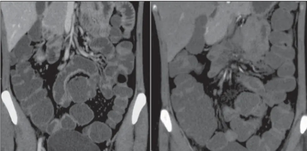
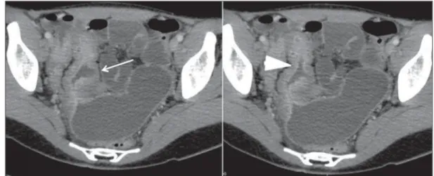
su-Figure 4. 36-year-old patient with Crohn’s disease. CT enterography demonstrates severe thickening of the distal ileum, with stenosis and significant upstream dilation. There is linear pathway originating from the above mentioned segment towards the transverse colon, characterizing enterocolic fistula (arrows).
Figure 6. CT enterography, coronal (A,B) and axial plane (C,D): expansile hypervascular lesion in the terminal ileum. Regional mesenteric lympha-denomegaly. Anatomopatho-logical study: terminal ileum carcinoid with lymph node in-volvement.
In spite of the wide applicability and the excellent results of CT enterography in the evaluation of Crohn’s disease, such method involves the use of ionizing radiation. It is known that the disease affects mainly young adults who, usually, will be submit-ted to several examinations along their lives. Thus, other propedeutic methods such as ultrasonography or MR enterogra-phy may be considered.
Intestinal tract tumors may also be de-tected at CT enterography. Small bowel neoplasias are rare, representing approxi-mately 3% to 6% of all cancers of the di-gestive tract(20). Carcinoid tumors tend to
appear as thickening areas or polyps with
intensely contrast enhancement, most fre-quently in the ileum(2). In the present study,
two cases were identified in this site. Be-cause of technical characteristics related to a more appropriate loops distension and greater capacity to evaluate parietal hyperenhancing, CT enterography can best demonstrate such tumors.
Celiac disease is a self-immune disor-der whose definite diagnosis is achieved by means of intestinal biopsy(21). However,
some imaging findings are suggestive of the disease, particularly in advanced stages, when the usual jejunoileal folds pattern reversal can be identified, most clearly demonstrated on coronal
reconstruc-tions(22). Nonspecific alterations such as
loops dilation, folds separation, intussus-ception and extraintestinal alterations, such as lymphadenomegaly and even eventual complications of the disease may also be observed(2,21).
Another indication for CT enterography reported in some studies is the evaluation of gastrointestinal bleeding, overt or occult. It seems that this method and the endo-scopic capsule play complementary roles and that, because of the inherent difficul-ties of the later such as the identification of the lesion site, visualization impaired by the presence of fluid and intraluminal blood or absence of loops distension, CT
Figure 7. Crohn’s disease with moderate to severe inflamma-tory activity. On A and B, in-creased parietal contrast up-take, thickening and stratifica-tion of the terminal ileum wall (related to mucosal enhance-ment and submucosal edema) can be observe on axial sec-tions; on C, densification of perienteric mesenteric fat; on D, reactional mesenteric lymphadenomegaly; on E, en-gorgement of intestinal vessels (vasa recta) related to the af-fected segment, image in the coronal plane.
A B
enterography may in some cases demon-strate lesions otherwise undetectable by the endoscopic capsule(23,24). Further studies
are necessary to define its role in the evalu-ation of intestinal hemorrhages.
CONCLUSION
The preliminary study allows the obser-vation that CT enterography has demon-strated to be useful in the diagnosis of in-testinal disorders, particularly in the evalu-ation of the inflammatory bowel diseases. It is an image method with a cost simi-lar to that of the conventional abdominal computed tomography, swift and with good acceptance and tolerance by patients. Its applicability has increased and, certainly, it will be increasingly utilized in the clini-cal and radiologiclini-cal practice.
REFERENCES
1. Horsthuis K, Stokkers PCF, Stoker J. Detection of inflammatory bowel disease: diagnostic perfor-mance of cross-sectional imaging modalities. Abdom Imaging. 2008;33:407–16.
2. Paulsen SR, Huprich JE, Fletcher JG, et al. CT enterography as a diagnostic tool in evaluating small bowel disorders: review of clinical experi-ence with over 700 cases. Radiographics. 2006; 26:641–57.
3. Hara AK, Alam S, Heigh RI, et al. Using CT enterography to monitor Crohn’s disease activity: a preliminary study. AJR Am J Roentgenol. 2008;190:1512–6.
4. Hara AK, Swartz PG. CT enterography of Crohn’s disease. Abdom Imaging. 2009;34:289–95. 5. Booya F, Akram S, Fletcher JG, et al. CT
enter-ography and fistulizing Crohn’s disease: clinical benefit and radiographic findings. Abdom Imag-ing. 2009;34:467–75.
6. Hara AK, Leighton JA, Sharma VK, et al. Imag-ing of small bowel disease: comparison of cap-sule endoscopy, standard endoscopy, barium ex-amination, and CT. Radiographics. 2005;25:697– 711.
7. Fidler J. Feature section, small bowel disease: CT imaging. Abdom Imaging. 2009;34:281.
8. Federle MP, Jeffrey RB, Desser TS, et al. Diag-nostic imaging: abdomen. Salt Lake, UT: Amir-sys; 2004.
9. Macari M, Megibow AJ, Balthazar EJ. A pattern approach to the abnormal small bowel: observa-tions at MDCT and CT enterography. AJR Am J Roentgenol. 2007;188:1344–55.
10. Mazzeo S, Caramella D, Battolla L, et al. Crohn disease of the small bowel: spiral CT evaluation
after oral hyperhydration with isotonic solution. J Comput Assist Tomogr. 2001;25:612–6.
11. Wold PB, Fletcher JG, Johnson CD, et al. Assess-ment of small bowel Crohn disease: noninvasive peroral CT enterography compared with other imaging methods and endoscopy – feasibility study. Radiology. 2003;229:275–81.
12. Megibow AJ, Babb JS, Hecht EM, et al. Evalua-tion of bowel distenEvalua-tion and bowel wall appear-ance by using neutral oral contrast agent for multi-detector row CT. Radiology. 2006;238:87–95. 13. Fletcher JG. CT enterography technique: theme
and variations. Abdom Imaging. 2009;34:283–8. 14. Lee SS, Kim AY, Yang SK, et al. Crohn disease of the small bowel: comparison of CT enterogra-phy, MR enterograenterogra-phy, and small-bowel follow-through as diagnostic techniques. Radiology. 2009;251:751–61.
15. Veloso FT, Ferreira JT, Barros L, et al. Clinical outcome of Crohn’s disease: analysis according to the Vienna classification and clinical activity. Inflamm Bowel Dis. 2001;7:306–13.
16. Huprich JE, Fletcher JG. CT enterography: prin-ciples, technique and utility in Crohn’s disease. Eur J Radiol. 2009;69:393–7.
17. Bodily KD, Fletcher JG, Solem CA, et al. Crohn disease: mural attenuation and thickness at con-trast-enhanced CT enterography – correlation with endoscopic and histologic findings of in-flammation. Radiology. 2006;238:505–16. 18. Baker ME, Walter J, Obuchowski NA, et al.
Mu-ral attenuation in normal small bowel and active inflammatory Crohn’s disease on CT enterogra-phy: location, absolute attenuation, relative at-tenuation, and the effect of wall thickness. AJR Am J Roentgenol. 2009;192:417–23.
19. Higgins PD, Caoili E, Zimmermann M, et al. Computed tomographic enterography adds infor-mation to clinical management in small bowel Crohn’s disease. Inflamm Bowel Dis. 2007;13: 262–8.
20. Masselli G, Polettini E, Casciani E, et al. Small-bowel neoplasms: prospective evaluation of MR enteroclysis. Radiology. 2009;251:743–50. 21. Soyer P, Boudiaf M, Fargeaudou Y, et al. Celiac
disease in adults: evaluation with MDCT entero-clysis. AJR Am J Roentgenol. 2008;191:1483– 92.
22. Tomei E, Marini M, Messineo D, et al. Computed tomography of the small bowel in adult celiac disease: the jejunoileal fold pattern reversal. Eur Radiol. 2000;10:119–22.
23. Huprich JE, Fletcher JG, Alexander JA, et al. Obscure gastrointestinal bleeding: evaluation with 64-section multiphase CT enterography – initial experience. Radiology. 2008;246:562–71.
24. Singh V, Alexander JA. The evaluation and man-agement of obscure and occult gastrointestinal bleeding. Abdom Imaging. 2009;34:311–9. Figure 8. Crohn’s disease. Active inflammation (A) versus fibrostenosis (B), reformatted images in the