ABSTRACT
Oral squamous cell carcinoma: clinicopathological
features from 346 cases from a single Oral
Pathology service during an 8-year period
Fábio Ramôa PIRES1, Amanda Barreto RAMOS1, Jade Bittencourt Coutinho de OLIVEIRA1, Amanda Serra TAVARES1,
Priscilla Silva Ribeiro da LUZ1, Teresa Cristina Ribeiro Bartholomeu dos SANTOS1
1- Department of Oral Pathology, School of Dentistry, State University of Rio de Janeiro, Rio de Janeiro, RJ, Brazil.
Corresponding address: Fábio Ramôa Pires - Departamento de Patologia Bucal, Faculdade de Odontologia, Universidade do Estado do Rio de Janeiro - Av. 28 de Setembro, 157 - Vila Isabel - 20551-030 - Rio de Janeiro - RJ - Brasil - Phone/fax: + 55 21 2868-8284 - e-mail: [email protected]
Submitted: April 25, 2013 - Modiication: June 23, 2013 - Accepted: August 12, 2013
e
pidemiological data from oral squamous cell carcinoma (OSCC) is mostly derived fromNorth American, european and east Asian populations. Objective: The aim of this study was to report the demographic and clinicopathological features from OSCC diagnosed in an Oral Pathology service in southeastern Brazil in an 8-year period. Material and Methods: All OSCC diagnosed from 2005 to 2012 were reviewed, including histological analysis of all hematoxylin and eosin stained slides and review of all demographic and clinical information from the laboratory records. Results: A total of 346 OSCC was retrieved and
males represented 67% of the sample. Mean age of the patients was 62.3 years-old and
females were affected a decade older than males (p<0.001). Mean time of complaint with
the tumors was 10 months and site distribution showed that the border of the tongue (37%), alveolar mucosa/gingiva (20%) and loor of mouth/ventral tongue (19%) were the most
common affected sites. Mean size of the tumors was 3.4 cm, with no differences for males and females (p=0.091) and males reported both tobacco and alcohol consumption more
frequently than females. Histological grade of the tumors revealed that 27%, 40% and 21% of the tumors were, respectively, classiied as well-, moderately- and poorly-differentiated OSCC, 26 cases (7.5%) were microinvasive OSCC and 17 cases were OSCC variants. OSCC in males mostly affected the border of tongue, loor of mouth/ventral tongue and
alveolar mucosa/gingival, while they were more frequent on the border of tongue, alveolar mucosa/gingival and buccal mucosa/buccal sulcus in females (p=0.004). Conclusions:
The present data relect the epidemiological characteristics of OSCC diagnosed in a public
Oral Pathology laboratory in southeastern Brazil and have highlighted several differences in clinicopathological features when comparing male and female OSCC-affected patients.
Keywords: Squamous cell carcinoma. Oral cancer. Mouth. South America. epidemiology.
INTRODUCTION
Oral squamous cell carcinoma (OSCC) is the most common oral malignancy, representing up
to 80–90% of all malignant neoplasms of the
oral cavity11. Although oral cancer incidence is highly variable worldwide, it is accepted that oral cavity ranges from the 6th to the 9th most common anatomical location for cancer, depending mostly
on the country (and even speciic region in some
countries) and gender of the patients11. Despite this mean incidence, it can represent the most common
location for cancer in some specific regions, especially in southeastern Asia11. Major etiological and predisposing factors for OSCC include mostly smoking and drinking habits, and ultraviolet
radiation (speciically for lip cancer), but several
other factors such as human papillomavirus (HPV) and Candida infections, nutritional deiciencies and
A l t h o u g h t h e m a i n d e m o g r a p h i c a n d clinicopathological information on OSCC can be similar in most studies, it is accepted that some features can be quite variable from country to country and even from different regions in the same country5,23. As there are few studies presenting
OSCC clinical and pathological proile in Brazilian
populations8,16, the aim of this study is to report the demographic and clinicopathological features from a series of OSCC diagnosed in an Oral Pathology service in Brazil in an 8-year period.
MATERIAL AND METhODS
The iles of the Oral Pathology service, School
of Dentistry, State University of Rio de Janeiro, Brazil, were reviewed from 2005 to 2012 and all registries diagnosed as OSCC were retrieved. After individual analysis of all registries, the cases diagnosed through cytological methods (smears
and ine-needle aspiration biopsies), originated
from maxillofacial areas apart from the oral cavity and presenting inadequate material for histological
review and classiication were excluded. When more
than one biopsy was performed in the same patient for the same lesion, all registries were reviewed, but only the most representative histological section was included. Demographic and clinical information from all cases were obtained through review of all forms submitted with the specimens, and included gender, age, time interval before diagnosis (in months), clinical aspect, location and size (in centimeters) of the tumors and risk factors (tobacco and alcohol use). Clinical aspect of the lesions was divided in three groups: ulcers (including plain ulcers, and exophytic ulcerated masses), leukoerythroplakias and tumors presenting both ulcers and leukoerythroplakic areas. Location of the tumors included the following regions: border
of tongue; loor of mouth (with extension to ventral
tongue); alveolar mucosa and gingiva (including retromolar area); buccal mucosa (including buccal sulcus/mucobuccal fold); soft palate and tonsil area; lower lip; and others (in cases with no precise information about the primary location).
All hematoxylin and eosin (He)-stained
Parameter Number of cases %
Gender (n=346)
Males 232 67
Females 114 33
Age (n=337)
<41 years 11 3
41 to 60 years 154 46
61 to 80 years 137 41
>80 years 35 10
Time of complaint (n=233)
0 to 6 months 169 73
7 to 12 months 33 14
>12 months 31 13
Location of the tumors (n=340)
Border of tongue 123 37
Alveolar mucosa/gingiva/retromolar area
69 20
Floor of mouth/ventral tongue 65 19
Soft palate/tonsil area 24 7
Buccal mucosa/buccal sulcus 23 7
Lower lip 21 6
Others 15 4
Clinical aspect (n=322)
Ulcer 201 62
Leukoerythroplakia 54 17
Ulcer + leukoerythroplakia 67 21
Size of the tumors (n=197)
<2.1 cm 69 35
2.1 to 4.0 cm 80 41
4.1 to 6.0 cm 35 18
>6.0 cm 13 6
histological slides were reviewed for diagnosis
conirmation and for classiication of the tumors as
well-differentiated (WD), moderately differentiated (MD) and poorly differentiated (PD) tumors and in OSCC variants, according to recently published accepted criteria4,28.
All information were descriptively analyzed and statistical analysis was performed using a standard program (Statistical Package for Social Sciences, SPSS version 17.0, Chicago, IL, US), with statistical
signiicance level of 5% (p<0.05). Distribution of
group variables was compared in crosstabs by Pearson Chi-square and comparison of means was performed with T test. This study was approved by the ethics Committee, State University of Rio de Janeiro (protocol number 044.3.2010).
RESULTS
A total of 346 OSCC were selected for the study after using the inclusion and exclusion criteria. This
total represented about 65% of all oral malignancies
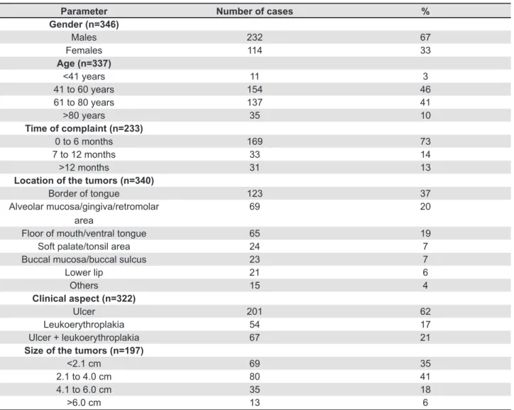
diagnosed in the laboratory on the selected period. Males represented two thirds of the affected
patients (232 cases, 67%) with a male:female ratio
of 2:1. Mean age of all patients was 62.3 years (standard deviation - SD±13.2, ranging from 30 to 102 years); mean age of the males (59.9 years, SD±10.9, ranging from 30 to 87 years) was almost a decade lower than mean age of females (67 years, SD±15.9, ranging from 32 to 102 years) (p<0.001).
More than 80% of the patients were diagnosed in their forties to seventies and 3% of the patients
were younger than 41 years (Table 1 - demographic and clinical features are included according to the number of cases for each parameter for which precise information was available).
Mean time of complaint with the lesions reported by the patients before diagnosis was 10 months
(SD±20.8, ranging from 1 to 123 months) and most patients reported to have noticed the lesions
up to 6 months before diagnosis (169 cases, 73%).
Mean time of complaint was longer for females (14.9 months, SD±26.5, ranging from 1 to 120 months) than for males (7.6 months, SD±16.9, ranging from 1 to 123 months) (p=0.03). Site distribution showed that the most common location
of the tumors was the border of the tongue (37%),
followed by the alveolar mucosa and gingiva
(20%) and loor of the mouth and ventral tongue (19%). Clinical aspect of the tumors revealed that
ulcers, ulcers associated to leukoerythroplakias and leukoerythroplakias represented, respectively,
62%, 21% and 17% of the sample. Mean size of
the tumors showed that most OSCC were diagnosed
with up to 4 cm in their greater diameter (76%),
showing a mean of 3.4 cm (SD±1.9, ranging from 0.2 to 12 cm). There was no difference on the mean size of the tumors when comparing affected males (3.6 cm, SD±1.9, ranging from 0.2 to 12 months) and females (3.1 cm, SD±1.9, ranging from 0.3 to 10 months) (p=0.091) (Table 1). Information about tobacco use was available for 281 patients
and showed that 225 patients (80%) were present
or past tobacco users. For alcohol use, information was available for 208 patients and showed that
146 patients (70%) were present or past alcohol
users (Table 3).
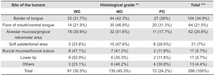
Histological diagnosis and grade of the tumors rendered after analysis of the He-stained slides revealed that, from 303 cases of conventional
invasive OSCC, 93 cases (30.7%) were classiied as WD OSCC, 138 cases (45.5%) as MD tumors and 72 (23.8%) as PD tumors. Although there were some
differences on the histological grade of the tumors when comparing each location, these results were
not statistically signiicant (p=0.381) (Table 2).
From the remaining 43 OSCC samples, 26 cases
Site of the tumors Histological grade ** Total ***
WD MD PD
Border of tongue 33 (31.7%) 44 (42.3%) 27 (26%) 104 (34.9%)
Floor of mouth/ventral tongue 14 (21.9%) 30 (46.9%) 20 (31.3%) 64 (21.5%)
Alveolar mucosa/gingiva/ retromolar area
19 (30.6%) 32 (51.6%) 11 (17.7%) 62 (20.8%)
Soft palate/tonsil area 5 (23.8%) 10 (47.6%) 6 (28.6%) 21 (7%)
Buccal mucosa/buccal sulcus 8 (47.1%) 7 (41.2%) 2 (11.8%) 17 (5.7%)
Lower lip 9 (52.9%) 6 (35.3%) 2 (11.8%) 17 (5.7%)
Others 3 (23.1%) 6 (46.2%) 4 (30.8%) 13 (4.4%)
Total 91 (30.5%) 135 (45.3%) 72 (24.2%) 298 (100%)
Table 2- Distribution of the histological grade of the tumors according to the site of the lesions*
(7.5%) were diagnosed as microinvasive OSCC and
the remaining 17 cases were diagnosed as OSCC
variants, including 9 verrucous carcinomas (2.6%), 5 spindle cell carcinomas (1.4%), 2 basaloid OSCC (0.6%) and 1 papillary OSCC (0.3%).
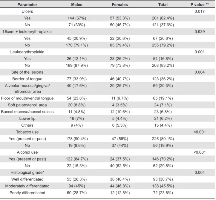
Clinical aspect of the tumors from the whole sample showed that the presence of ulcers was more common in males (p=0.017) while leukoerythroplakias were more frequently found in females (p=0.001). Distribution of the site of the tumors revealed that OSCC in males mostly
affected the border of tongue, loor of the mouth/
ventral tongue and alveolar mucosa/gingiva, while they were more frequent on the border of tongue, alveolar mucosa/gingiva and buccal mucosa/buccal sulcus in females (p=0.004). Past
and present tobacco and alcohol use were more common in males than in females (p<0.0001, both). Distribution of the histological grade of the conventional invasive OSCC (n=303) was also different when comparing the gender of the patients, as males were predominantly affected by MD and PD tumors, while females presented mostly with MD and WD tumors (p=0.004) (Table 3).
Microinvasive carcinoma equally affected males and females with a mean age of 67.2 years (SD±13.05, ranging from 42 to 87 years). Mean time of complaint was 18.4 months (SD±22.7, ranging from 1 to 72 months) and most lesions
showed leukoerythroplakic areas (73.1%), while ulcerated areas were found in 50% of the cases.
The tumors affected mostly the border of tongue
Parameter Males Females Total P value **
Ulcers 0.017
Yes 144 (67%) 57 (53.3%) 201 (62.4%)
No 71 (33%) 50 (46.7%) 121 (37.6%)
Ulcers + leukoerythroplakia 0.939
Yes 45 (20.9%) 22 (20.6%) 67 (20.8%)
No 170 (79.1%) 85 (79.4%) 255 (79.2%)
Leukoerythroplakia 0.001
Yes 26 (12.1%) 28 (26.2%) 54 (16.8%)
No 189 (87.9%) 79 (73.8%) 268 (83.2%)
Site of the lesions 0.004
Border of tongue 77 (33.9%) 46 (40.7%) 123 (36.2%)
Alveolar mucosa/gingiva/ retromolar area
40 (17.6%) 29 (25.7%) 69 (20.3%)
Floor of mouth/ventral tongue 54 (23.8%) 11 (9.7%) 65 (19.1%)
Soft palate/tonsil area 20 (8.8%) 4 (3.5%) 24 (7.1%)
Buccal mucosa/buccal sulcus 11 (4.8%) 12 (10.6%) 23 (6.8%)
Lower lip 16 (7%) 5 (4.4%) 21 (6.2%)
Others 9 (4%) 6 (5.3%) 15 (4.4%)
Tobacco use <0.001
Yes (present or past) 178 (90.4%) 47 (56%) 225 (80.1%)
No 19 (9.6%) 37 (44%) 56 (19.9%)
Alcohol use <0.001
Yes (present or past) 122 (84.7%) 24 (37.5%) 146 (70.2%)
No 22 (15.3%) 40 (62.5%) 62 (29.8%)
Histological grade* 0.004
Well differentiated 55 (26.3%) 38 (40.4%) 93 (30.7%)
Moderately differentiated 94 (45%) 44 (46.8%) 138 (45.5%)
Poorly differentiated 60 (28.7%) 12 (12.8%) 72 (23.8%)
Table 3- Distribution of the clinical aspect of all OSCC, site of the lesions, history of tobacco and alcohol use and histological grade of the tumors according to the gender of the affected patients
(56%) and lower lip (16%). Size of the tumors
showed a mean of 1.9 cm (SD±1.6, ranging from 0.2 to 6 cm) and most patients reported no tobacco
(52.6%) and alcohol (53.3%) use. Verrucous
carcinoma mostly affected the elderly with a mean age of 73.2 years (SD±14.3, ranging from 48 to
95 years), with predilection for females (66.7%).
Lesions showed leukoerythroplakic and ulcerated
areas, respectively, in 78% and 56% of the cases,
and tumors were mostly located on the alveolar
mucosa/gingiva (44.4%) and buccal mucosa/ buccal sulcus (33.3%). Mean time of complaint
was 24 months (SD±41.9, ranging from 1 to 120 months). Size of the tumors showed a mean of 3.8 cm (SD±1.3, ranging from 2 to 5 cm), and
most patients reported tobacco use (60%) but not alcohol use (66.7%). Spindle cell carcinoma was
diagnosed in 5 male adults (mean age of 57 years, SD±10.4, ranging from 48 to 74 years). Mean time of complaint was 6 months (SD±5.3, ranging from 2 to 12 months) and ulcerated areas were found
in 80% of the cases while leukoerythroplakic areas were present in only 20%. The tumors affected the border of tongue (40%), alveolar mucosa/gingiva (20%), soft palate (20%) and other sites (20%).
Mean size of the tumors was 4.3 cm (SD±1.5, ranging from 3 to 6 cm) and the 5 patients reported both tobacco and alcohol use. Basaloid OSCC affected the buccal mucosa of a 62 year-old male, with 123 months of evolution and an ulcerated and leukoerythroplakic clinical aspect with 10 cm in size; and the border of tongue of a 63 year-old female, with 24 months of evolution and also with an ulcerated and leukoerythroplakic clinical aspect with 2.5 cm in size. One of the patients reported tobacco use. The papillary OSCC affected the buccal mucosa of a non-tobacco and non-alcohol user 64 year-old male as an ulcerated lesion measuring 5 cm.
DISCUSSION
Although many studies have emphasized that
OSCC represent 80 to 90% of all oral malignant
tumors11, the present study has shown that about one third of all oral malignant tumors diagnosed in the Oral Pathology Laboratory, State University of Rio de Janeiro, were not OSCC. As this service is a local reference center for Oral Pathology in a metropolitan area, it is supposed that the additional referral of more complex and challenging malignant cases can be responsible for this bias in OSCC frequency.
OSCC predominantly affects males with variable male:female ratios ranging in recent studies from 6:1 to 2:11,8,9,16, in accordance with the present results. Several other recent studies have shown an increase in the number of affected females,
with a mean male:female ratio lower than 2:1, probably due to changes in social and daily activities
associated to modern women social proile and way
of living, leading to higher exposure to carcinogenic agents, such as tobacco and alcohol consumption and exposure to biological agents, such as high-risk HPV subtypes3,6,7,10,11,13,17,25.
Most studies have reinforced that OSCC is mostly
diagnosed in adults with mean ages in their ifties
to seventies1,3,8,9,11,16,17, a inding also corroborated
by the present results. Only 3% of the patients
reported in the present study were under 41 years of age, similarly to the results presented by Jainkittivong, et al.10 (2009) (4,7%) and the means reported by the literature, usually ranging from 4
to 6%11. It seems that there are geographical and populational differences in the mean age of the
affected patients, as demonstrated by Efiom, et
al.6 (2008) in Nigeria, which revealed a mean age
of 45.3 years-old and 40% of the patients with
ages below 40 years-old. Similarly to the results of the present study, other authors have also demonstrated that the mean age of males affected by OSCC is lower than the mean age for females6,13.
Present and past tobacco and alcohol consumption are considered the most important risk factors for OSCC11. The present results showed that both deleterious habits were frequently reported by OSCC-affected patients and that both were more common in males than in females, similarly to other studies2,13,19. This pattern can be responsible for the differences in the male:female ratio of OSCC-affected patients. Younger patients OSCC-affected by OSCC sometimes do not report tobacco and alcohol use as possible risk factors or the interval of use is
not long enough to support a deinite carcinogenic
effect. Other possible risk factors have been
suggested in this speciic group, such as dietary/
nutritional factors and genetic predisposition, and high-risk HPV types (especially HPV type 16) have been demonstrated in OSCC in youngsters more frequently than in control adult/elderly groups11,12,18.
Oral potentially malignant disorders (OPMD) are relatively common, showing a global prevalence
from 1 to 5% and a gender, age and site predilection
similar to OSCC11. Although the exact malignant transformation rate for OPMD is unknown, it is expectable that leukoerythroplakic areas can be encountered in association with OSCC. This pattern
was found in 35% of the patients included in the
importance of considering the possibility of OSCC when dealing with leukoplakias and erythroplakias, and the need of obtaining biopsy specimens from all lesions from this group.
OSCC can affect any site of the oral mucosa and large lesions can invade several continuous areas. The present results showed that the border
of tongue, gingiva/alveolar mucosa and loor of
mouth/ventral tongue were the most commonly affected locations. Although the border of tongue is considered the most common site for OSCC in America and europe14,17, the buccal mucosa is the most common site for OSCC in southeastern Asia, due to habits of areca nut- and tobacco-chewing11. Jainkittivong, et al.10 (2009) have additionally
reported that 50% of their OSCC affected the gingiva and alveolar ridge, which could be justiied by the
different etiological factors associated with the
development of OSCC in their speciic population. Similarly, Efiom, et al.6 (2008) have shown, in Nigeria, that the lower and upper gingiva were the most common affected sites in their sample, followed by the tongue. Most studies focusing on Brazilian and other occidental populations have
shown that the border of tongue and loor of mouth
are the most common OSCC-affected regions8,9,16. It is important to call attention to the fact that, when considered together with intraoral locations, the lower lip is the most common site for OSCC1. Andisheh-Tadbir, Mehrabani and Heydari3 (2008) have demonstrated that the tongue and the buccal mucosa were the two most common locations for OSCC in Iran, and Nemes, et al.19 (2008) have
shown that, in Hungary, the loor of mouth, lips and
the tongue were the most commonly affected areas. As demonstrated by the present results, location of the tumors can be different when comparing
males (border of tongue, loor of mouth/ventral
tongue and gingiva/alveolar mucosa) and females (border of tongue, gingiva/alveolar mucosa, buccal
mucosa/buccal sulcus). This gender-speciic pattern
was also reported by Kruse, Bredell and Grätz13 (2011), who have reported that females were more affected by OSCC located on the palate and alveolar mucosa. The exact reasons for this differential site predilection for OSCC in males and females are unknown.
Although delay in diagnosis is a major problem in early detection of OSCC, the exact reasons for
these dificulties, including social and health-related
behavior and tumor characteristics, are not well-understood27. Most tumors included in the present
series (76%) were diagnosed as cT1 or cT2 tumors,
but mean interval of complaints prior to professional assistance was 10 months. In contrast, Gervásio, et al.8 (2001), reviewing 740 OSCC patients in the state of Minas Gerais, also located in southeastern
Brazil, revealed that almost 50% of their patients
with OSCC were diagnosed as T4 tumors. Although this former study was published a decade ago and it seems that tumors are being diagnosed in earlier stages in recent years, some biases could justify these differences. The present study has presented data derived from an Oral Pathology public service responsible for diagnosis of OSCC mostly in patients of low socioeconomic status. After diagnosis, these patients are referred to public cancer treatment centers and some additional delay is expected,
both due to patient and institutional dificulties and
limitations, before starting treatment.
Most OSCC are histologically diagnosed as MD or WD tumors1,3,10,14,19,22,28, as was also shown by
the present results. In contrast, Efiom, et al.6
(2008) have shown that 47.6% of their cases were histologically classiied as PD tumors, while WD tumors represented 32.6% of their sample. When
analyzing the present results, it was also observed that males were predominantly affected by MD and PD OSCC, while females were mainly affected by WD and MD tumors. It was also shown that histological grade can also be possibly associated with the site of the tumors, as OSCC affecting the buccal mucosa/buccal sulcus and lower lip were predominantly WD, while tumors affecting the
border of tongue, loor of mouth/ventral tongue
and alveolar mucosa/gingiva were predominantly MD, although the differences were not statistically
signiicant.
Several OSCC variants have been reported in the literature and the establishment of the specific appropriate histological diagnosis is essential, as some histological subtypes and distinct clinicopathological entities are managed with different treatment protocols and present variable prognosis22,28. Microinvasive OSCC was
diagnosed in 7.5% of the present cases and, in this speciic pattern, careful evaluation of serial
sections and careful analysis of the basal lamina
were performed in all cases, in order to conirm
early invasion and restriction of the tumor cells to the papillary lamina propria. Verrucous carcinoma
was diagnosed in 9 cases (2.6%) in the present
sample and the mean time of complaint was much longer than the mean time of complaint of the
whole sample, which can be justiied by the indolent
and painless course of this subtype. Rekha and Angadi24 (2010) have recently reported a series of
133 verrucous carcinomas, representing 16% of all SCC diagnosed in their iles from India. Their cases have affected mostly males in their ifties
females were the most affected group and that the tumors showed a predilection for the alveolar mucosa/gingiva and buccal mucosa/buccal sulcus, but the few cases included in the present series avoid any conclusive distinct pattern of distribution of the disease in the studied population. Although it has been reported in a recent review22 that some studies have named verrucous carcinomas with minor foci of invasion as “hybrid verrucous carcinoma – WD OSCC”, we have considered these “hybrid” tumors as WD OSCC in the present series. Basaloid OSCC is a rare aggressive variant with predilection for male adults, usually tobacco and alcohol users, which is predominantly seen as an ulcerated exophytic mass22. Both cases diagnosed in the present series affected adults in their sixties and tumors were located on the tongue and buccal mucosa. Spindle cell carcinoma was diagnosed in 5 cases in the present series and these cases have been comparatively evaluated elsewhere26.
The most representative limitations on the methods and results from the present study are associated with the retrieval of clinical and histological information from, respectively, laboratory records and incisional biopsies. On
the other hand, the sample represents the proile
of OSCC submitted to diagnosis in the studied population, including both patients that are going to be submitted to curative and palliative OSCC treatment and patients who are not going to be submitted to any treatment due to advanced
disease. These data would properly relect the epidemiological proile of OSCC in a public Oral
Pathology diagnostic service in southeastern Brazil.
OSCC age and gender proile, as well as site
predilection, shows a heterogeneous pattern of distribution in different countries, in different regions from the same country and in different ethnic groups from the same region, which can be associated with both genetic factors and cultural habits/behavior15. Studies focusing on specific regions are welcome as they show the demographic
and clinical proile of OSCC in restricted geographic
locations, offering an enhanced comprehension of
these tumors and the possibility of planning speciic
strategies of prevention, diagnosis and treatment.
CONCLUSION
Demographic and clinical profile of OSCC-affected patients in the studied population revealed that females were affected about a decade older than males, that males reported both tobacco and alcohol consumption more frequently than females and that the tumors in males mostly affected the
border of tongue, loor of mouth/ventral tongue
and alveolar mucosa/gingiva, while they were more frequent on the border of tongue, alveolar
mucosa/gingiva and buccal mucosa/buccal sulcus in females. OSSC in males were predominantly poorly and moderately differentiated tumors while they were mostly well and moderately differentiated tumors in females. There were several differences when comparing the clinicopathological data from OSCC-affected male and female patients in the studied population and other studies focusing on different Brazilian and foreign populations are
encouraged to conirm these features.
ACKNOWLEDgEMENTS
This study was supported by FAPeRJ, Rio de Janeiro, Brazil.
REFERENCES
1- Al-Rawi NH, Talabani NG. Squamous cell carcinoma of the oral cavity: a case series analysis of clinical presentation and histological grading of 1425 cases from Iraq. Clin Oral Invest. 2008;12:15-8.
2- Albuquerque R, López-López J, Marí-Roig A, Jané-Salas e, Roselló-Llabrés X, Santos JR. Oral tongue squamous cell carcinoma (OTSCC): alcohol and tobacco consumption versus non-consumption. A study in a Portuguese population. Braz Dent J. 2011;22:517-21.
3- Andisheh-Tadbir A, Mehrabani D, Heydari ST. epidemiology of squamous cell carcinoma of the oral cavity in Iran. J Craniofac Surg. 2008;19:1699-702.
4- Barnes L, eveson JW, Reichart P, Sidransky D. World Health
Organization classiication of tumors - pathology & genetics - head
and neck tumors. Lyon: IARC Press; 2005.
5- Carvalho AL, Singh B, Spiro RH, Kowalski LP, Shah JP. Cancer of the oral cavity: a comparison between institutions in a developing and a developed nation. Head Neck. 2004;26:31-8.
6- Efiom OA, Adeyemo WL, Omitola OG, Ajayi OF, Emmanuel
MM, Gbotolorun OM. Oral squamous cell carcinoma: a clinical and pathologic review of 233 cases in Lagos, Nigeria. J Oral Maxillofac Surg. 2008;66:1595-9.
7- Gaitán-Cepeda LA, Peniche-Becerra AG, Quezada-Rivera DQ. Trends in frequency and prevalence of oral cancer and oral squamous cell carcinoma in Mexicans. A 20 years retrospective study. Med Oral Patol Oral Cir Bucal. 2011;16:e1-5.
8- Gervásio OL, Dutra RA, Tartaglia SM, Vasconcellos WA, Barbosa AA, Aguiar MC. Oral squamous cell carcinoma: a retrospective study of 740 cases in a Brazilian population. Braz Dent J. 2001;12:57-61.
9- Grimm M. Prognostic value of clinicopathological parameters and outcome in 484 patients with oral squamous cell carcinoma: microvascular invasion (V+) is an independent prognostic factor for OSCC. Clin Transl Oncol. 2012;14:870-80.
10- Jainkittivong A, Swasdison S, Thangpisityotin M, Langlais RP. Oral squamous cell carcinoma: a clinicopathological study of 342 Thai cases. J Contemp Dent Pract. 2009;10:e033-40.
11- Johnson NW, Jayasekara P, Amarasinghe AA. Squamous cell carcinoma and precursor lesions of the oral cavity: epidemiology and etiology. Periodontol 2000. 2011;57:19-37.
12- Kaminagakura e, Villa LL, Andreoli MA, Sobrinho JS, Vartanian JG, Soares FA, et al. High-risk human papillomavirus in oral squamous cell carcinoma on young patients. Int J Cancer. 2012;130:1726-32.
13- Kruse AL, Bredell M, Grätz KW. Oral cancer in men and women: are there differences? Oral Maxillofac Surg. 2011;15:51-5. 14- Larsen SR, Johansen J, Sørensen JA, Krogdahl A. The
prognostic signiicance of histological features in oral squamous
15- Liu L, Kumar SK, Sedghizadeh PP, Jayakar AN, Shuler CF. Oral squamous cell carcinoma incidence sectioned by sublocations among diverse racial and ethnic populations in California. Oral Surg Oral Med Oral Pathol Oral Radiol endod. 2008;105:470-80. 16- Losi-Guembarovski R, Menezes RP, Poliseli F, Chaves VN, Kuasne H, Leichsenring A, et al. Oral carcinoma epidemiology in Paraná State, Southern Brazil. Cad Saude Publica. 2009;25:393-400.
17- Marocchio LS, Lima J, Sperandio FF, Corrêa L, Sousa SO. Oral squamous cell carcinoma: an analysis of 1564 cases showing advances in early detection. J Oral Sci. 2010;52:267-73. 18- Marur S, D'Souza G, Westra WH, Forastiere AA. HPV-associated head and neck cancer: a virus-related cancer epidemic. Lancet Oncol. 2010;11:781-9.
19- Nemes JA, Redl P, Boda R, Kiss C, Márton IJ. Oral cancer report from Northeastern Hungary. Pathol Oncol Res. 2008;14:85-92. 20- Neville BW, Day TA. Oral cancer and precancerous lesions. CA Cancer J Clin. 2002;52:195-215.
21- Oliveira DT, Moraes RV, Fiamengui Filho JF, Fanton Neto J, Landman G, Kowalski LP. Oral verrucous carcinoma: a retrospective study in São Paulo Region, Brazil. Clin Oral Investig. 2006;10:205-9.
22- Pereira MC, Oliveira DT, Landman G, Kowalski LP. Histologic subtypes of oral squamous cell carcinoma: prognostic relevance. J Can Dent Assoc. 2007;73:339-44.
23- Rastogi T, Devesa S, Mangtani P, Mathew A, Cooper N, Kao R, et al. Cancer incidence rates among South Asians in four geographic regions: India, Singapore, UK and US. Int J epidemiol. 2008;37:147-60.
24- Rekha KP, Angadi PV. Verrucous carcinoma of the oral cavity: a clinical and pathologic appraisal of 133 cases in Indians. Oral Maxillofac Surg. 2010;14:211-8.
25- Rivero eR, Nunes FD. HPV in oral squamous cell carcinomas
of a Brazilian population: ampliication by PCR. Braz Oral Res.
2006;20:21-4.
26- Romañach MJ, Azevedo RS, Carlos R, Almeida OP, Pires FR. Clinicopathological and immunohistochemical features of oral spindle cell carcinoma. J Oral Pathol Med. 2010;39:335-41. 27- Scott Se, Grunfeld eA, McGurk M. Patient's delay in oral cancer: a systematic review. Community Dent Oral epidemiol. 2006;34:337-43.