I Guidelines For Perioperative Evaluation
OrganizatiOn
Bruno Caramelli, Cláudio Pinho, Daniela Calderaro
Danielle Menosi Gualandro, Pai Ching Yu
Writing COmmittee members
Danielle Menosi Gualandro, Claudio Pinho, Gilson Feitosa, Bruno Caramelli
task FOrCe members
Alina Coutinho Rodrigues Feitosa, Beatriz Ayub, Bruno Caramelli, Carisi A. Polanczyk, Carolina L.
Zilli Vieira, Claudio Pinho, Daniela Calderaro, Danielle Menosi Gualandro, Denise Iezzi, Dirk Schreen,
Dimas T. Ikeoka, Elbio Antonio D’Amico, Elcio Pfeferman, Emerson Quintino de Lima, Emmanuel de
Almeida Burdmann, Fábio Santana Machado, Filomena Regina Barbosa Gomes Galas, Gilson Soares
Feitosa-Filho, Heno Ferreira Lopes, Henrique Pachón , João César Nunes Sbano, José Augusto
Soares Barreto Filho, José L. Andrade, Roberto Henrique Heinisch, Luciana Moraes dos Santos,
Luciana S. Fornari, Ludhmila Abrahão Hajjar, Luis Eduardo P. Rohde, Luiz Francisco Cardoso,
Marcelo Luiz Campos Vieira, Maristela C. Monachini, Pai Ching Yu, Paula Ribeiro Villaça, Paulo
Grandini, Renato S. Bagnatori, Roseny dos Reis Rodrigues, Sandra F. Menosi Gualandro, Walkiria
Samuel Avila, Wilson Mathias Jr.
suppOrt
1. Definition of the Problem
2. General Assessment
A) History
B) Physical examination
C) Comorbid diseases I. Thyroid Diseases 1. Hypothyroidism 2. Hyperthyroidism II. Renal Impairment III. Blood Disorders 1. Sickle-cell Disease
2. Antiphospholipid Syndrome 3. Hereditary Thrombophilia 4. Hemophilia
5. Von Willebrand Disease IV. Adrenal Insufficiency 1. Symptoms and Signs
2. Identification of patients at risk of adrenal insufficiency
3. Corticosteroid supplementation doses V. Obesity
1. Obesity-associated surgical risk
2. Preoperative assessment according to BMI and surgery classification
3. Recommendations to reduce risk
D) Additional Tests I. ECG
II. Chest x-ray III. Full blood count
IV. Hemostasis / Coagulation tests V. Serum creatinine
E) Algorithms for Perioperative Evaluation I. Advantages of the ACP algorithm II. Disadvantages of the ACP algorithm III. Final considerations
3. Disease-specific approaches
A) Coronary artery disease (CAD) I. Patients with known CAD II. Patients with riks factors for CAD
B) Hypertension
C) Congestive heart failure (CHF)
D) Valvular Heart Disease
E) Cardiac arrhythmias and conduction disorders I. Cardiac arrhythmias
II. Atrioventricular and intraventricular conduction disorders
F) Implanted pacemakers and cardioverter-defibrillators I. Individuals with conventional single- or dual-chamber pacemakers
II. Individuals with multisite cardiac resynchronization devices
III. Individuals with Implantable Cardioverter-Defibrillators (CDI)
IV. Emergency cardioversion or defibrillation V. Recommendations
G) Transplants
H) Heart disease and Pregnancy
I) Dental procedures
I. Dental procedures in patients taking anticoagulants II. Dental procedures and prevention of infective endocarditis
4. Additional assessment
A) Shortcut for non-invasive test
B) Assessment of ventricular function at rest C) Exercise electrocardiogram
D) Stress myocardial perfusion scintigraphy E) Stress echocardiogram
F) Holter monitor G) Coronary angiography
5. Steps to reduce surgical risk
A) Medication therapy I. Beta-blockers II. Statins III. Alpha-agonists IV. Aspirin
B) Myocardial revascularization
D) Endocarditis prophylaxis
E) Perioperative anticoagulation
F) Glycemic control
G) Anesthetic and intraoperative considerations I. Choosing the anesthetic technique and agent II. Management of body temperature
III. Nitroglycerin IV. Catheters
V. Intra-aortic balloon pump
H) Monitoring
I. ST-segment monitoring II. Perioperative AMI
6. Emergency surgery
Guidelines for Perioperative Evaluation
1. Definition of the problem
A) Purpose of these guidelines:
Knowledge of surgical interventions is being transferred surprisingly fast to medical practice. New technological resources and the technical refinement of medical teams increase the confidence of physicians and patients. Consecutive successes associated with a lower rate of postoperative complications increase the number of surgery candidates. With less invasive, faster and more efficient technologies becoming available, cases that were once considered inoperable are now being operated. Consequently, a growing number of interventions are being performed in a population that is progressively older and of higher risk.
Now it is necessary to organize the knowledge of the phenomena that occur before, during and after a surgical intervention. This task demands a great effort when one considers the enormous variability of the characteristics of patients in these conditions and the difficulty to establish common criteria and references for observation and comparison, the basic methodology for the accumulation of scientific knowledge. For this area of knowledge, some authors proposed the name Perioperative Evaluation.
The goals of these guidelines are:
• Refine and unify the terminology used by the entire multidisciplinary team, including the patients and their family;
• Establish new routines, change indication for surgery according to the information obtained during the perioperative evaluation;
• Do not provide clearance for surgery but inform the patient of the possible risks. Based on these data, personal experience, knowledge of the other side of the story, the underlying disease, its risks and the risk attributable to the surgery itself, the surgeon can decide with the patient and their family if the risk/benefit ratio favors the intervention.
• Data or scientific evidences are not always available to allow all the different situations to be analyzed. As customary in medical practice, minute analysis of the patient and problem and the common sense of the team must prevail.
• The surgical intervention does not finish when the patient is bandaged or leaves the operating room. The concept of the word perioperative includes the need for a postoperative surveillance whose intensity is determined by the individual level of risk of the patient.
B) Methodology and Evidence:
The participants of these guidelines were chosen among health sciences specialists with hands on and academic experience, thus being characterized as clinical researchers. The basics of perioperative evaluation and the current recommendations were established in order to decrease perioperative complications. Unfortunately, we have not yet eliminated the stress caused by surgery or all its consequences, but the reader will notice that there is much that can be done for the surgery to run smoothly and have a successful outcome without hurting scientific truth. The adopted methodology and evidence levels were the same as those used in earlier documents by the Brazilian Society of Cardiology.
Recommendations:
• The guidelines must be based on evidences;
• Class division must be used when applicable;
• Degrees of recommendation must be used when applicable, according to the levels of evidence;
Degree or Class of Recommendation:
• Class I: Conditions for which there is evidence for and/or general agreement that the procedure/therapy is useful and effective.
• Class II: Conditions for which there is conflicting evidence and/or a divergence of opinion about the usefulness/ efficacy of performing the procedure/therapy.
Class IIa: Weight of evidence/opinion is in favor of usefulness/efficacy.
Class IIb: Usefulness/efficacy is less well established by evidence/opinion.
• Class III: Conditions for which there is evidence for and/or general agreement that the procedure/therapy is not useful/effective and in some cases may be harmful.
Levels of Evidence:
• A: Sufficient evidence from multiple randomized trials or meta-analyses;
non-randomized studies;
• C: Evidence only from case reports and series;
• D: Expert opinion or standard of care.
2. General Approach to the Patient
A) History
Medical history investigation by talking with the patient or family member is the first step in a perioperative evaluation. It should be done in a comfortable environment and at least one week before an elective surgery so that medications that decrease perioperative risk can be introduced or those that interfere with surgery can be discontinued, thus increasing surgical success. On the other hand, talking with the patient or family member right before emergency interventions can reveal important information that will help the surgeon decide if the patient should be sent to the ICU after surgery or if there are coexisting clinical conditions that can influence the early postoperative period.
Medical history investigation consists of the following essential actions:
• Minute investigation of the past surgical or anesthetic history of the patient that can lead to potentially preventable complications and allergies or the existence of comorbidities;
• Investigation of the clinical condition of the patient and the need to compensate for coexisting diseases;
• Determination of what medications are being taken and if they interfere with surgery;
• The surgeon’s opinion on the urgency and risk of the procedure;
• Degree of anxiety and doubts of the patient and their family regarding the procedure and its risks;
B) Physical examination
Physical examination is useful during the perioperative risk assessment process and it should not be limited to
the cardiovascular system. The objectives are: to identify preexisting or potential heart disease (risk factors), define the severity and stability of the heart disease and identify comorbidities, if any.
Patients with heart disease whose general condition is compromised by other conditions such as neurological diseases, renal failure, infections, liver abnormalities, malnourishment or pulmonary dysfunction are at higher risk of cardiac complications since these conditions exacerbate surgical stress.1
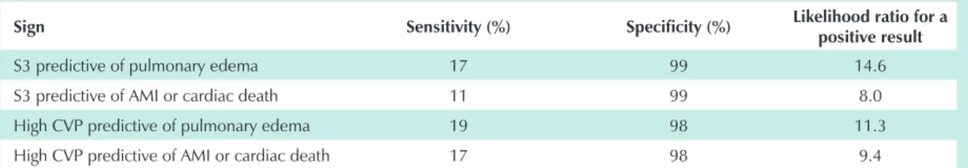
The incidence of ischemic heart disease in patients with peripheral arterial disease is high and it is a predictive factor for perioperative complication. Information obtained upon physical examination such as changes in pulse rate or carotid bruit should be investigated. On the other hand, jugular vein distension signaling high central venous pressure (CVP) indicates that the patient may develop pulmonary edema after surgery.2,3 Finding the third heart sound (S3) during
perioperative evaluation indicates a bad prognosis with increased risk of pulmonary edema, myocardial infarction or cardiac death.4
The presence of bilateral lower limb edema must be analyzed together with the presence of jugular vein distension. If the amplitude of the pulse wave of the internal jugular vein reveals high CVP, then heart disease and pulmonary hypertension are at least partially responsible for the patient’s edema. If CVP is not high, then the edema is probably caused by something else such as liver disease, nephrotic syndrome, chronic venous insufficiency or some medication. The presence of edema and unknown CVP is not a definite sign of heart disease.6 If heart murmurs are present, the physician
should be able to distinguish organic from functional murmurs, determine if they are significant or not and determine their origin. The origin will indicate if endocarditis prophylaxis or assessment of valvular lesion severity is necessary.
References:
1. Eagle KA, Berger PB, Calkins H, Chaitman BR, Ewy GA, Fleischmann KE, Fleisher LA, Froehlich JB, Gusberg
Table 1 - Physical examination and risk of perioperative complications
Sign Sensitivity (%) Specificity (%) Likelihood ratio for a
positive result
S3 predictive of pulmonary edema 17 99 14.6
S3 predictive of AMI or cardiac death 11 99 8.0
High CVP predictive of pulmonary edema 19 98 11.3
High CVP predictive of AMI or cardiac death 17 98 9.4
RJ, Leppo JA, Ryan T, Schlant RC,Winters WL Jr. ACC/ AHA guideline update for perioperative cardiovascular evaluation for noncardiac surgery: a report of the American College of Cardiology/ American Heart Association Task Force on Practice Guidelines (Committee to Update the 1996 Guidelines on Perioperative Cardiovascular Evaluation for Noncardiac Surgery) 2002. American College of Cardiology Web site. Available at: http:/www. acc.org/clinical/guidelines/perio/dirIndex.htm.
2. Goldman L, Caldera DL, Nussbaum SR, Southwick FS, Krogstad D, Murray B, Burke DS, O’ Malley TA, Goroll AH, Caplan CH, Nolan J, Carabello B, Slater EE. Multifactorial index of cardiac risk in noncardiac surgical procedures. N Engl J Med 1977; 297:845-850.
3. Goldman L, Caldera DL, Nussbaun SR, Southwick FS. Cardiac risk factors and complications in noncardiac surgery. Medicine 1978; 57:357-70.
4. McGee SR. Evidence-based physical diagnosis. Philadelphia: W.B. Saunders Company, 2001.
5. Jaeschke R, Guyatt G, Sackett DL for the Evidence-Based Medicine Working Group. Users’ guides to the medical literature, III: how to use an article about a diagnostic test, B: what are the results and will they help me in caring for my patients? JAMA 1994; 271: 703-707.
6. Butman SM, Ewy GA, Standen JR, Kern KB, Hahn E. Bedside cardiovascular examination in patients with severe chronic heart failure: importance of rest or inducible jugular distension. J Am Coll Cardiol 1993; 22: 968-974.
C) Comorbid Diseases
I. Thyroid diseases
Thyroid disease is a very common clinical condition. In endemic areas, goiter affects 15 to 30% of the adult population. In addition to the technical difficulties of managing the airways of the patient with goiter, the hormonal imbalance may be a considerable source of morbidity and mortality.1
Tetraiodothyronine (T4) represents 80% of thyroid hormone production and 40% of it is converted by peripheral organs to triiodothyronine (T3), which is five times more active. Another 50% of the T4 produced by the thyroid is converted to 3,5-triiodothyronine (rT3), which does not have biological activity. Only 0.2% and 0.3% of the circulating T3 and T4 respectively are in the free and biologically active form while the remaining is bound to plasma proteins (albumin, transthyretin and thyroid-binding globulin). T3 and rT3 are converted by the liver, kidneys and CNS into inactive compounds. Severe systemic diseases, trauma and drugs may block the peripheral conversion of T4 to T3 leading to the euthyroid sick syndrome in critically ill patients, a physiological mechanism that conserves energy in critical situations.
I. 1. Hypothyroidism
It is estimated that 5 out of every 1000 patients have hypothyroidism and the prevalence of subclinical hypothyroidism is three times greater. Hypothyroidism is 10 times more common in women than in men and may
be due to iatrogenic causes (radioactive iodine therapy or surgical resection) or autoimmune thyroiditis (Hashimoto´s), among others. In addition to the signs and symptoms (Table 2), TSH and free T4 and T3 must be determined to confirm the diagnosis.
Complications during the perioperative period are rare when hypothyroidism is subclinical, light or moderate. Special attention should be given to the severe cases since complications are more likely to occur.1-6
Table 2 - Relevant clinical manifestations of hypothyroidism during perioperative evaluation
Hypothermia
Myocardial depression
Slow respiratory rate and difficulty in weaning from mechanical ventilation
Slow heart rate
Altered baroreceptor response
Hypotension or hypertension
Angina, myocardial infarction
Hypovolemia
Anemia
Hypoglycemia
Hyponatremia (syndrome of inappropriate antidiuretic hormone secretion)
Abdominal distension
Decreased hepatic metabolism of drugs
Recommendations: 1-6
Class I, Level of evidence D.
• Assess all the risk factors of the patient;
• Do not worry about subclinical hypothyroidism when TSH < 10mU/dl;
• Elective surgery should only take place when the thyroid function of the patient is normal;
• Patients < 45 years should be given the full L-thyroxine dose which is usually from 1.6 to 2.2 mcg/kg or from 100 to 200 mcg per day. It takes from 4 to 6 weeks of treatment for TSH levels to normalize;
• Patients > 45 years should start with a 25-50 mcg/day dose to be increased at every two-week intervals;
• Coronary patients should start with a 15 mcg/day dose to be increased weekly until TSH normalizes;
• Do not postpone surgery in patients with light hypothyroidism but start oral hormone replacement therapy;
• Hypothermia prophylaxis, cardiovascular monitoring and hydrocortisone administration (100 mg at every 8 hours for 24 hours since adrenal insufficiency may occur) must be done in hypothyroid patients submitted to surgery;
Thus, a patient who is taking T4 does not need to take it on the day of surgery while the patient who is taking T3 does;
• Radiograph the cervical region to determine if goiter is going to interfere with tracheal intubation.
R e c o m m e n d a t i o n s f o r p a t i e n t s w i t h s e v e r e hypothyroidism or myxedema coma undergoing urgent surgeries:1-6
Class I, Level of evidence D.
• Administer 200-500 mcg of L-thyroxine or 40 mcg of intravenous T3 or 10-25 mcg of T3 at 8-hour intervals before surgery. This should correct hemodynamic and electrocardiographic changes. Divide the dose into 50% of T4 and 50% of T3 during the perioperative period;
• Maintenance dose should be 40 to 100 mcg of T4 or 10 to 20 mcg of T3 given intravenously at 24-hour intervals;
• Administer 100 mg of hydrocortisone at 6-hour intervals for a long period;
• Start hormone replacement therapy by digestive route using the doses listed above as soon as possible.
I. 2. Hyperthyroidism:
Thyrotoxicosis affects 2% of women and 0.2% of men. Clinical and subclinical hyperthyroidism prevalence in the USA is 0.2 and 1% respectively. The most common causes are: Graves´ disease, toxic nodular goiter, several types of thyroiditis and iatrogenic causes. Clinical manifestations with perioperative repercussions are listed in Table 3. Adrenergic effects are high risk factors for complications such as cardiac arrhythmias (10 to 15% being atrial fibrillation). They are associated with an increased number or sensitivity of beta-adrenoreceptors. Hyperthyroidism mortality is associated with cardiovascular events.1-6
Laboratory tests should be done to confirm diagnosis. TSH levels should be low and free T4 levels should be normal (subclinical hyperthyroidism) or high. Many conditions can raise total T4 because of increased thyroxine-binding globulin levels, yet they do not affect free T4 which is the biologically active substance: pregnancy, cirrhosis, acromegaly, Cushing’s syndrome, lithium, contraceptives, propranolol, amiodarone and iodinated contrasts. A patient must be treated for hyperthyroidism before being submitted to elective surgery.
Specific complications may occur in cases of thyroidectomy: patients with large goiters may present complications during intubation and extubation (up to 35% of these patients present some degree of tracheal obstruction), recurrent laryngeal nerve injury, tracheomalacia and edema of the glottis. Hypocalcemia may occur in 20% of the cases up to 36 hours after thyroidectomy and calcium must be replaced intravenously during this phase. Only 3% of the patients become permanently hypocalcemic and need to take oral calcium for life.
Recommendations:1-6
Class I, Level of evidence D.
• Antithyroid medications: the most common are propylthiouracil (PTU) and methimazole. They inhibit the synthesis of thyroid hormones by preventing iodide oxidation and organification. When used in high doses, PTU has the additional advantage of inhibiting the peripheral conversion of T4 to T3, therefore it is used more often during the perioperative period. The standard dose is 100 mg at 8-hour intervals and the maximum dose is 400mg at 8-hour intervals. Methimazole doses vary from 10 to 120 mg in a single daily dose. The dose should be reassessed at 4-6-week intervals. Adverse effects are rarely severe: skin rash, fever, itching and arthralgia, transient elevation of liver enzymes and leucopenia. More severe and less frequent complications that require discontinuation of medication are agranulocytosis (0.5%), severe hepatitis, lupus-like syndrome and thrombocytopenia;
• Beta-blockers:They are only used to control adrenergic signs and symptoms. They do not affect hormone levels. Propranolol is used most often at 10 to 80 mg at 6-8-hour intervals (1 mg intravenously during surgery). Esmolol can be given during surgery at a loading dose of 500 mcg/kg for 1 minute and a maintenance dose of 25-300mcg/kg/min.
Recommendations for urgent or emergency surgeries:1-6
Class I, Level of evidence D.
• Antithyroid medications – PTU in high doses is the medication of choice (1000 to 1200 mg divided in 3 doses per day);
• Beta-blockers – intravenous is the preferred route of administration;
• Iodine – can be used for a maximum of 10 days since the inhibition of iodine organification (Wolff-Chaikoff effect) is transient and after this time an escape phenomenon occurs and hyperthyroidism worsens. The most commonly used agent is Lugol´s iodine. It contains 5% iodine and 10% potassium Table 3 - Clinical manifestations of hyperthyroidism with perioperative
repercussions
Cardiovascular
Increased cardiac inotropism and chronotropism with decreased systemic vascular resistance
Left ventricular hypertrophy Higher incidence of angina, heart failure, arrhythmias and embolic events
Blood
Anemia, thrombocytopenia,
neutropenia, increased factor VIII levels Decreased levels of vitamin K-dependent coagulation factors, bleeding.
Gastrointestinal Inadequate medication absorption
Metabolic
Hypercalcemia, hypoalbuminemia, ketoacidosis
Increased medication clearance Glucose intolerance, weight loss and protein catabolism
Pulmonary Myopathy with respiratory failure
iodide. The dose varies from 0.1 to 0.3 ml at 8-hour intervals (3 to 5 drops);
• Iodinated contrasts –Sodium ipodate and iopanoic acid are used for compensation. They present two advantages: there is less escape and the peripheral conversion of T4 to T3 is inhibited. The dose is 500mg at 8-hour intervals;
• Corticosteroid – must be given when hyperthyroidism is not compensated during and after surgery because of elevated peripheral degradation of cortisol. The induction dose is 100mg followed by 100mg at 8-hour intervals for the first 24 hours;
• Anesthesia – pay special attention to increased metabolism of anesthetic agents and intubation difficulty due to goiter;1
• Thyroid storm – is associated with mortality rates varying from 20 to 30%. Given the sudden nature of the signs and symptoms, treatment should begin right away even if laboratory tests have not yet confirmed the condition. Table 4.
References:
1. Mostbeck A,Galvan G,Bauer P, et al.The incidence of hyperthyroidism in Austria from 1987 to 1995 before and after an increase in salt iodization in 1990. Eur J Nucl Med 1998;124:367.
2. Bennet-Guerrero E, et al. Effect of chronic and acute thyroid hormone reduction on perioperative outcome. Anesth Analg1997; 85:30.
3. Pronovost PH,et al. Perioperative management of thyroid disease: prevention of complications related to hyperthyroidism and hypothyroidism. Postgrad Med1995; 98:83.
4. Graham GW, et al. Perioperative management of selected endocrine disorders. IntAnaesthesiol Clin2000; 38:31. 5. Murkin JM. Anaesthesia and hypothyroidism: a review
of thyroxine, physiology, pharmacology and anaesthetic implication. Anaesth Analg 1982; 61:371.
6. Stehling LC. Anaesthetic management of the patient with hyperthyroidism. Anesthesiology 1974; 41:585.
II. Renal Impairment
Patients with renal failure are more prone to perioperative complications, increased hospital stay, greater costs during hospital stay and higher morbidity than those without renal failure.1,2 Preoperative creatinine > 2.0 mg/dL is among the
risk factors found in prognostic models for cardiovascular complications after non-cardiac surgeries. Even moderate chronic renal failure (creatinine between 1.5 and 3.0 mg/dL or glomerular filtration rate between 30 and 60 ml/min) is a risk factor for cardiac and non-cardiac complications after surgery, doubling the mortality rate of these patients in relation to patients with normal renal function.1,2 Patients on peritoneal
dialysis or hemodialysis must undergo dialysis before surgery to avoid hypervolemia, correct electrolyte and acid-base disturbances and reduce the likelihood of uremic bleeding. Immunosuppression in patients with kidney transplant must be carefully adjusted by the nephrologist before and after surgery to avoid an acute rejection episode and nephrotoxicity.
Acute renal failure (ARF) occurs in the postoperative period in 1-30% of the cases, depending on the type of surgery, with a mortality rate of roughly 50%. ARF prevention during the perioperative period depends on identifying risk factors for its onset (especially preoperative renal failure), avoiding nephrotoxic medications, keeping adequate hydration and avoiding hypotension. Attempts to prevent ARF with diuretics and vasoactive amines are not effective. “Renal dose” dopamine does not prevent renal failure and does not reduce the need for dialysis and ARF mortality rate. Potentially nephrotoxic medications should be avoided or used properly, that is, corrected for renal function. Aminoglycoside antibiotics, amphotericin B, radiological contrast agents and non-steroidal anti-inflammatory agents are examples of nephrotoxic substances commonly used in the perioperative period. The effects of anti-inflammatory agents with cyclooxygenase-II-selective inhibition properties in renal function are not different from the effects caused by the non-selective anti-inflammatory agents and their use should be avoided in patients at risk of anti-inflammatory-associated nephrotoxicity (old age, history of renal failure, heart failure, dehydration, concomitant use of angiotensin-converting enzyme inhibitor, diuretics or other nephrotoxic agents).3-6
The risks for postoperative complications are well defined in renal failure patients and all patients with preoperative creatinine greater than 1.5 mg/dL should be assessed by a nephrologist. Always bear in mind that creatinine is not a very sensitive marker for renal function. Therefore, creatinine below 1.5 mg/dL does not necessarily mean that renal function is normal, especially among the elderly and those with reduced muscle mass. Perioperative assessment is an opportunity to be in contact with the patient and the clinical and surgical teams and plan actions that will prevent the deterioration of renal function and later retard the progression of chronic renal failure.
Table 4 - Treating thyroid storm 6
Supportive care Specific treatment
Hydration Cooling
Respiratory support Metabolic control Inotropic agents
Loading PTU dose: 1000 mg by digestive route
Maintenance PTU dose: 200mg at 6-hour intervals by digestive route Loading hydrocortisone dose: 300mg intravenously Maintenance hydrocortisone: 100mg at 8-hour intervals. Lugol´s iodine given orally or intravenous iodine: 1g at 8-hour intervals
References:
1. O’Hare AM, Feinglass J, Sidawy AN, Bacchetti P, Rodriguez RA, Daley J, Khuri S, Henderson WG, Johansen KL. Impact of renal insufficiency on short-term morbidity and mortality after lower extremity revascularization: data from the Department of Veterans Affairs’ National Surgical Quality Improvement Program. J Am Soc Nephrol 2003; 14:1287-1295.
2. Chertow GM, Lazarus JM, Christiansen CL, Cook EF, Hammermeister KE, Grover F, Daley J. Preoperative renal risk stratification. Circulation 1997; 95:878-884.
3. Burdmann EA: Epidemiologia. In: Insuficiência Renal Aguda: Fisiopatologia, Clínica e Tratamento. Schor N, Santos O, Boim M, São Paulo, Editora Sarvier, 1997, pp 1-7.
4. Lima EQ, Castro I, Zanetta DMT, Yu L. Validation of predictive mortality models in acute renal failure. J Am Soc Nephrol 2002;
5. Kellum JA, Decker M: Use of dopamine in acute renal failure: a meta-analysis. Crit Care Med 2001; 29:1526-1531.
6. Ott E, Nussmeier NA, Duke PC, Feneck RO, Alston RP, Snabes MC, Hubbard RC, Hsu PH, Saidman LJ, Mangano DT. Efficacy and safety of the cyclooxygenase 2 inhibitors parecoxib and valdecoxib in patients undergoing coronary artery bypass surgery. J Thorac Cardiovasc Surg 2003; 125:1481-1492.
III. Blood disorders
Many blood disorders can increase the morbidity and mortality rates of individuals submitted to non-cardiac surgeries. Anemia is a condition that leads to an overload of the cardiovascular system, increasing cardiac output. Individuals with cardiovascular diseases are less tolerant of anemia as its presence can aggravate underlying myocardial ischemia and heart failure. Instructions regarding blood transfusions in the perioperative period are limited but its benefits and risks should nevertheless be questionned.1 In order to decide if the
patient needs to receive a blood transfusion, the physician must weigh the duration of anemia, intravascular volume, extent of surgery, likelihood of massive bleeding and presence of associated conditions, such as pulmonary diseases, heart failure, myocardial ischemia and peripheral or cerebral vascular disease. Always bear in mind that one unit of RBC (red blood cells) increases hemoglobin by approximately 1g/dL and hematocrit by 3%.1
Recommendations for RBC transfusions:2
• Symptomatic individuals should receive enough blood to minimize their symptoms; Class I, Level of evidence D;
• Hemoglobin below 7.0 g/dL in patients with acute anemia; Class I, Level of Evidence A.
• In cases of acute coronary disease, hemoglobin should be kept around 9.0 and 10.0 g/dL; Class I, Level of Evidence D.
There are many other blood conditions that should be taken into account during perioperative assessment for non-cardiac surgery and many of them require the presence of a hematologist in the multidisciplinary team that cares for the patient. Patients with platelet count equal to or above 50,000/mm3 usually tolerate surgeries well, do not bleed in
excess and do not require platelet transfusion.2
Recommendations for platelet transfusion: Class I, Level of Evidence B.
• Platelet count below 50,000/mm3;
• Platelet count below 100,000/mm3 if surgery is
neurological or ophthalmologic.
Recommended perioperative procedures for noncardiac surgeries in patients with other blood conditions
III. 1. Sickle-cell disease (SS/SC/ Sβthal) Class I, Level of Evidence C.
• Increase hemoglobin to 10 g/dL by RBC transfusion. If
hemoglobin ≥9 g/dL, ask a specialist;
• Monitor hematocrit, peripheral perfusion and oxygenation before surgery;
• Monitor blood pressure, heart rhythm and rate, oxygenation and body temperature during surgery, avoiding hypothermia;
• Monitor hydration, oxygenation and body temperature after surgery.3
III. 2. Primary antiphospholipid syndrome Class I, Level of Evidence C.
• Perioperative thrombosis prophylaxis in patients taking anticoagulant medications;
• Postoperative thrombosis prophylaxis in patients who are not taking anticoagulant medications;4
III. 3. Congenital thrombophilia Class I, Level of Evidence C.
• Perioperative thrombosis prophylaxis in patients taking anticoagulant medications;
• Postoperative thrombosis prophylaxis in patients who are not taking anticoagulant medications. Doses will vary according to the type of congenital thrombophilia.4
III. 4. Hemophilia
Class I, Level of Evidence B.
• Perform laboratory tests to determine if inhibitors are present;
• During surgery, correct the coagulation factor deficiency by infusion of the specific factor;
to type and classification of surgery (minor/major);
• Keep plasma levels of the lacking factor under strict control with the aid of laboratory tests.5
III. 5. Von Willebrand Disease Class I, Level of Evidence B.
• During surgery, correct the plasma level of the lacking factor with factor VIII/von Willebrand factor concentrates;
• After surgery, factor VIII and von Willebrand factor levels and activities will vary according to type and classification of surgery;
• Depending on the type of surgery and results of the DDAVP test, consider using this medication.6
References:
1. Armas-Loughran B, Kalra R, Carson JL. Evaluation and management of anemia and bleeding disorders in surgical patients. Med Clin North Am 2003; 87:229-242. 2. Madjdpour C, Spahn DR. Allogenic blood cell transfusions:
efficacy, risks, alternatives and indications. British Journal of Anaesthesia 2005; 95(1):33-42.
3. The management of sickle cell disease. National Institute of Health / National Heart, Lung, and Blood Institute / Division of Blood Diseases and Resources. 2002.
4. Middeldorp S, Büller HR, Prins MH, Hirsh J. Approach to the thrombophilic patient. In: Hemostasis and Thrombosis. Basic principles & Clinical practice. Colman RW, Hirsh J, Marder VJ, Clowes AW, George JN, eds. 4 ed, Lippincott Williams & Wilkins, Philadelphia 2001. P. 1085-100. 5. Arun B, Kessler CM. Clinical manifestations and therapy
of the hemophilias. In: Hemostasis and Thrombosis. Basic principles & Clinical practice. Colman RW, Hirsh J, Marder VJ, Clowes AW, George JN, eds. 4 ed, Lippincott Williams & Wilkins, Philadelphia 2001. P. 815-24.
6. Mannucci PM. Treatment of von Willebrand disease. Haemophilia 1998; 4:661-64.
IV. Adrenal Insufficiency
The increase in cortisol levels during acute stress is an important protective response. However, metabolic stress caused by surgery can precipitate acute adrenal insufficiency in individuals with clinical and subclinical disorders that affect the hypothalamic-pituitary-adrenal axis (HPA) and the results may be catastrophic, leading to multiple complications and even death.
Physical stress increases adrenocorticotropic hormone (ACTH) levels and cortisol secretion. Increased cortisol, noradrenalin and adrenalin levels characterize the hormonal changes induced by stress. These changes are minimal when surgical stress is low and rises progressively as surgical stress increases from moderate to severe, lasting no more than 24 hours in surgeries without complications. The HPA axis is most activated by surgery, recovery from anesthesia
and extubation, raising the cortisol levels which normalize within 24 to 48 hours.1 Increased corticosteroid requirement
may lead individuals with impaired adrenal activity and reserves to develop acute adrenal insufficiency (AAI). Therefore, it is essential to identify these individuals early and establish an adequate perioperative plan in order to avoid complications.
IV. 1. Signs and Symptoms of Adrenal Insufficiency (AI)
- Hypotension and hemodynamic shock (that may be resistant to vasopressors) with multiple organ failure;
- Hypoglycemia; - Tachycardia;
- Electrolyte disturbances: hyponatremia, hyperkalemia (in primary adrenal insufficiency), hypercalcemia, acidosis;
- Decreased cardiac contractility; - Anemia, eosinophilia and neutropenia;
- Nausea, emesis, weakness, orthostatic hypotension, dehydration, abdominal or flank pain (acute adrenal hemorrhage), fatigue, weight loss;
- Vitiligo, skin color changes, hypogonadism, hypothyroidism.
AI should be suspected when hypotension and unexplained, refractory or hypovolemic shock occur during or after surgery, or when there is discrepancy between the condition of the patient and severity of the disease, high fever without apparent cause (negative cultures) or refractory to antibiotic therapy, unexplained mental changes, apathy or depression without a specific psychiatric disorder. These cases should be regarded and treated as acute AI and confirmed later. Class I, Level of Evidence C.
IV. 2. Identification of patients at risk of AI • Patients with an already established AI diagnosis.2 • Patients at risk of AI3 and patients with relative AI (limited
adrenocortical reserve):
- Pituitary tumors (macroadenomas); - Radiotherapy in the pituitary region; - Previous pituitary surgery;
- Postoperative period of surgery for Cushing’s disease, bilateral adrenalectomy or unilateral adrenalectomy if the other adrenal is compromised;
- Chronic corticosteroid use (>7.5 mg of prednisone or equivalent for more than 30 days or > 20 mg for more than two weeks);
- Patients with type I diabetes mellitus or autoimmune diseases (Hashimoto’s disease, ovarian or primary testicular failure, hypoparathyroidism, vitiligo);
Recommendations:
• Confirm the diagnosis with tests that are appropriate for patients at risk of AI. It is advisable to have an endocrinologist involved; Class I, Level of Evidence B.
• If tests are necessary to confirm AI, use dexamethasone as it does not interfere with the tests; Class I, Level of Evidence C.
• If untreated hypothyroidism and AI coexist, replace AI hormones first; Class I, Level of Evidence C.
• Mineralocorticoid supplementation is not necessary since corticosteroid supplementation doses for surgical stress have mineralocorticoid activity Class I, Level of Evidence C.
• If it is impossible to confirm the diagnosis before surgery, the following corticosteroid supplementations are recommended; Class IIa, Level of Evidence D.
IV. 3. Corticosteroid supplementation doses:4-6
Recommendations:
• High corticosteroid supplementation doses are not necessary to prevent acute AI;
• High doses may increase the likelihood of complications such as hypertension and diabetic decompensation; Class IIa, Level of Evidence C.
A. Mild surgical stress:
• Double or triplicate corticosteroid dose in patients with established AI and chronic users. Bear in mind that adrenal suppression may occur rapidly with high doses or even after a long time of corticosteroid discontinuation (up to 48 months
Class IIa, Level of Evidence C.
• If the oral route in fasting subjects is not possible, administer 50mg of intramuscular or intravenous hydrocortisone right before surgery followed by 25mg of hydrocortisone twice daily or equivalent (dexamethasone 0.75mg twice daily). Reduce to the regular dose after 24 hours or as soon as stress is over;
Class IIa, Level of Evidence C.
• Patients who have not been diagnosed with AI but it is highly suspected, proceed with treatment for AI; Class IIb, Level of Evidence C.
B. Moderate surgical stress:
• Administer 25mg of intramuscular or intravenous hydrocortisone or equivalent at 8-hour intervals on the day of surgery. Reduce the dose daily by 50% until the regular dose is reached; Class IIa, Level of Evidence C.
C. High surgical stress:
• Administer 50mg of intramuscular or intravenous hydrocortisone or equivalent at 6-hour intervals on the day of surgery and maintain this dose until the metabolic stress is over. Metabolic stress usually lasts 48 hours following surgeries without complications (infections or other intercurrences). Then reduce the dose daily by 50% until the regular dose is reached; Class IIa, Level of Evidence C.
D. Cushing´s syndrome special situation:
• It is advisable for an endocrinologist to be involved;
• Start corticosteroid therapy as soon as the patient arrives at the ICU or on the day following surgery;
• In these cases, some groups will only replace hormones if there are signs or symptoms of acute AI or laboratory tests confirming the need for hormone replacement therapy;
References:
1. Udelsman R, Norton JA, Jelenich SE, Goldstein DS, Linehan WM, Loriaux DL, Chrousos GP. Responses of the hypothalamic-pituitary-adrenal and renin-angiotensin axes and the sympathetic system during controlled surgical and anesthetic stress. J Clin Endocrinol Metab 1987 May;64(5):986-94. 2. Oelkers W Adrenal Insufficiency. N Engl J Med 1996;
335:1206-1212.
3. Grinspoon SK, 1994, May ME, Vaughn ED, Carey RM. Adrenocortical insufficiency – clinical aspects. In: Vaughn ED Jr, Carey RM, eds. Adrenal Disorders. New York: Thieme Medical, 1989: 171-89.
4. Salem M, Tainsh RE Jr, Br J, Loriaux DL, Chernow B. Perioperative glucocorticoid coverage. A reassessment 42 years after emergence of a problem. Ann Surg 1994; Apr;219(4):416-25.
5. Cooper MS, Stewart PM. Corticosteroid insufficiency in acutely ill patients. N Engl J Med 2003; Feb 20;348(8):727-34.
6. Axelrod L. Perioperative management of patients treated with glucocorticoids. Endocrinol Metab Clin North Am 2003 Jun;32(2):367-83.
V. Obesity
Overweight and obesity prevalence is increasing worldwide at alarming rates. Prevalence increased by 50% from the 1980´s to today. It is estimated that roughly 40% of the adult Brazilian population presents excess weight (body mass index – BMI - > 25kg/m2) and that 8.9% of the men and 13.1% of the
women are obese. Prevalence tends to increase with age.
Severity of obesity can be characterized by degrees: Obesity grade 1: BMI 30-34.9 kg/m2
Obesity grade 2: BMI 35-39.9 kg/m2
Obesity grade 3: BMI ≥ 40 kg/m2
V. 1. Surgical risk associated with obesity:
• Surgical risk increases with severity of obesity, especially regarding the respiratory (airways and lungs) and cardiovascular systems;
pulmonary auscultation, abdominal palpation);
• Clinical history may result in underestimation of symptoms (great functional limitation) and surgical risk, especially among those with obesity grades 2 and 3;
• Risk scores do not contemplate obesity as an independent risk factor;
• Difficult intubation;
• Hypoxemia by hypoventilation, restrictive lung disease, postoperative atelectasis, central and obstructive sleep apnea, hypercapnia;
• Risk of aspiration of gastric content;
• Underdiagnosed decompensated congestive heart failure and precipitation of myocardial ischemia;
• Thromboembolic events;
• Difficulty to determine blood pressure and access veins;
• Sensitivity to opioids and sedative agents;
• Surgical wound infection;
• Rhabdomyolysis;
• The risks associated with comorbidities commonly found among the obese: hypertension, diabetes, cardiovascular disease, ventricular hypertrophy.
V. 2. Preoperative assessment according to BMI and surgery classification
A. Obesity of any grade and low-risk surgery:
• Same assessment as that for nonobese individuals; Class IIa, Level of Evidence D.
B. Obesity grades 1 and 2 and intermediate or high-risk surgery:
• Complete medical history and physical examination;
• Clinical assessment of obstructive sleep apnea; Class I, Level of Evidence B.
• ECG; Class IIa, Level of Evidence B.
• Fasting glucose; Class IIa, Level of Evidence B.
• Creatinine determination if the patient is diabetic, hypertensive or has a history of renal disease; Class IIb, Level of Evidence C.
• Polysomnography in selected patients; Class IIb, Level of Evidence C.
• Resting and overnight non-invasive oximetry if apnea score is intermediate or high in the clinical scoring system or if diagnosis of sleep apnea has been confirmed by polysomnography; Class IIb, Level of Evidence D.
• Echocardiographic assessment of diastolic function if there are signs or symptoms that suggest CHF; Class IIb, Level of Evidence D.
C. Obesity grade 3 and intermediate or high-risk surgery:
• Electrocardiogram; Class IIa, Level of Evidence B.
• Fasting glucose; Class IIa, Level of Evidence B.
• Creatinine determination if the patient has diabetes, hypertension or a history of renal disease; Class IIa, Level of Evidence C.
• Echocardiographic assessment of diastolic function; Class IIa, Level of Evidence D.
• Resting and overnight oximetry; Class IIb, Level of Evidence D.
Observations:
• Additional tests such as coagulation studies, noninvasive tests for cardiac ischemia, chest x-ray and pulmonary function tests are not mandatory and should not be done routinely during the preoperative assessment of obese individuals. Additional tests are selected according to medical history.
Class IIa, Level of Evidence B.
• Restrictive and mixed bariatric surgeries are considered intermediate-risk surgeries.
V. 3. Recommendations to reduce risk:
• Smoking cessation 6 weeks before surgery;1Class I, Level
of Evidence B.
• Respiratory physiotherapy; Class IIa, level of Evidence D.
• If patient has sleep apnea confirmed by polysomnography or if apnea risk score is high, consider using a CPAP during the preoperative period in patients who do not use it and do not discontinue its use in patients who do; Degree of
Recommendation IIa, Level of EvidenceB.
A. Intraoperative recommendations:
• Monitor blood pressure with an inflatable cuff that is appropriate for obese individuals or in a different location (forearm) adjusted for obese patients;2 Class I, Level of
Evidence B.
• Induce anesthesia with the patient in the reverse Trendelenburg position; Class IIa, Level of Evidence B.
• The use of sevoflurane general anesthesia results in faster extubation and a better initial recovery period Class IIa, Level of Evidence B.
• Perform pre-oxygenation in the sitting or elevated head position; Class IIa, Level of Evidence B.
• Use the rapid-sequence induction of anesthesia with application of cricoid pressure during intubation; Class IIa, Level of Evidence B.
• Make sure that the stretcher that can accommodate obese patients and watch out for pressure sores; Class IIa, Level of Evidence D.
• Invasive pressure monitoring should be done whenever necessary; Class IIb, Level of Evidence D.
B. Postoperative recommendations:
• CPAP in cases of confirmed sleep apnea;3 Class I, Level
of Evidence B.
system diseases (sleep apnea, alveolar hypoventilation); Class IIa, Level of Evidence B.
• High risk patients with comorbidities, patients who had failure on postoperative airway extubation program, patients who suffered complications during surgery or super obese patients (BMI>70kg/m2) should remain in the ICU after
surgery;4 Class IIa, Level of Evidence C.
• Maintain normal blood volume; Class IIa, Level of Evidence D.
• Perform continuous oximetry during recovery from anesthesia (Class IIb, Level of Evidence C), determine oxygen saturation after recovery from anesthesia (if it is normal it does not need to be measured again) and perform overnight oximetry (in cases of intermediate or high-risk surgeries); Class IIb, Level of Evidence D.
• All patients submitted to intermediate or high-risk surgeries are to undergo respiratory physiotherapy; Class IIa, Level of Evidence D.
• Deep vein thrombosis (DVT) prophylaxis:
- Encourage ambulation and use prophylatic heparins; Class I, Level of Evidence B.
- When indicated, both the low-molecular-weight heparin and unfractionated herapin can be used in regular regimens.5
If patient’s weight > 100kg, consider monitoring factor Xa activity; Class IIa, Level of Evidence B.
- Higher doses (40mg of enoxaparin at 12-hour intervals) result in less thromboembolic events and can be useful; 6Class
IIa, Level of Evidence B.
References:
1. Moller AM, Villebro N, Pedersen T, Tonnesen H. Effect of preoperative smoking intervention on postoperative complications: a randomised clinical trial. Lancet 2002; Jan 12;359(9301):114-7.
2. Pickering TG, Hall JE, Appel LJ, Falkner BE, Graves J, Hill MN, Jones DW, Kurtz T, Sheps SG, Roccella EJ; Subcommittee of Professional and Public Education of the American Heart Association Council on High Blood Pressure Research. Recommendations for blood pressure measurement in humans and experimental animals: Part 1: blood pressure measurement in humans: a statement for professionals from the Subcommittee of Professional and Public Education of the American Heart Association Council on High Blood Pressure Research. Hypertension 2005 Jan;45(1):142-61.
3. Lojander J, Maasilta P, Partinen M, Brander PE, Salmi T, Lehtonen H. Nasal-CPAP, surgery, and conservative management for treatment of obstructive sleep apnea syndrome. A randomized study. Chest 1996 Jul;110(1):114-9.
4. Helling TS. Operative experience and follow-up in a cohort of patients with a BMI > or =70 kg/m2. Obes Surg 2005 Apr;15(4):482-5.
5. Hamad GG, Choban PS. Enoxaparin for thromboprophylaxis
in morbidly obese patients undergoing bariatric surgery: findings of the prophylaxis against VTE outcomes in bariatric surgery patients receiving enoxaparin (PROBE) study. Obes Surg 2005 Nov-Dec;15(10):1368-74.
6. Scholten DJ, Hoedema RM, Scholten SE. A comparison of two different prophylactic dose regimens of low molecular weight heparin in bariatric surgery. Obes Surg 2002 Feb;12(1):19-24.
D) Additional tests
The request for additional tests in medicine is controversial. Technological advances increase the types of tests available exponentially. Physicians need to be familiar with the attributes of a test, its indications, advantages, disadvantages, cost, availability and risks before requesting it. However, one must always remember that medical history and physical examination continue to be essential instruments in clinical diagnosis.
I. Electrocardiogram (ECG):
The ECG can detect arrhythmias, conduction defects, ischemia or myocardial necrosis, overloaded chambers, digitalis intoxication or suggest electrolyte disturbances. It is also important to have a baseline ECG to assess changes that occur during the perioperative period.1
High risk electrocardiographic changes include severe arrhythmias (third-degree atrioventricular block, symptomatic ventricular arrhythmias with underlying heart disease, supraventricular tachycardia with high heart rate), medium risk ECG changes include the presence of pathological Q waves and low risk ECG changes include ventricular hypertrophy, left bundle-branch block, ST segment abnormalities and T wave abnormalities.2
On the other hand, routinely using a test with a limited specificity for some diseases may lead to false-positive results in patients who do not present heart diseases. For example, ST segment and T wave abnormalities may be found both in normal individuals as in patients with coronary artery diseases.3
Electrocardiographic changes usually worry the surgical team and prompts a visit to the specialist. Surgeries of individuals with varying electrocardiographic changes are cancelled more often than those of individuals with normal ECGs.4
Recommendations for requesting an ECG:5,6
Class I:
• All patients older than 40 years or all patients with a history of and/or physical examination abnormalities that suggest cardiovascular disease regardless of age;
• Patients with a recent episode of ischemic chest pain or considered to be at high risk after algorithmic assessment;
• Patients with diabetes;
Class IIa:
• Asymptomatic obese patients;
• Routinely request an ECG for asymptomatic individuals who will be submitted to low-risk surgeries.
II. Recommendations for requesting a chest x-ray: 5,6 • Request a chest x-ray for patients with a history of chest-related abnormalities or those with chest-chest-related abnormalities detected during the physical examination; Class I, Level of Evidence D.
III. Recommendations for requesting full blood count (FBC):
Class I, Level of Evidence D.
• Request a FBC for all patients older than 65 years;
• Request a FBC when anemia is suspected during physical examination or when chronic diseases associated with anemia are present;
• Request a FBC for all patients who will be submitted to moderate/high-risk surgeries if a need for transfusion is anticipated;
IV. Recommendations for requesting hemostasis / coagulation tests:
Class I, Level of Evidence D.
• Request test for all patients on anticoagulation therapy;
• Request test for all patients with liver failure;
• Request test for all patients with coagulation disorders;
• Request for all patients who will be submitted to intermediate or high-risk surgeries;
V. Determination of serum creatinine: Class I, Level of Evidence D.
• Determine serum creatinine in all patients older than 40 years;
• Determine serum creatinine in all patients with kidney disease, diabetes mellitus, hypertension, liver failure and/or heart failure and whose serum creatinine has not been determined in the last 12 months.
• Determine serum creatinine for all patients who will be submitted to intermediate or high-risk surgeries;
References:
1. Ikeoka DT, Caramelli B. Aplicações clínicas do eletrocardiograma na avaliação perioperatória de cirurgia não-cardíaca. Rev Soc Cardiol Estado de São Paulo 1999, 9: 424-7.
2. Eagle KA, Brundage BH, Chaitman BR et al. Guidelines for preoperative cardiovascular evaluation for non-cardiac surgery. Report of the American College of Cardiology / American Heart Association task force on guidelines (Committee on perioperative cardiovascular evaluation for non-cardiac surgery). Circulation 1996; 93:1280-1316. 3. Goldenberger AL, O’Konski M. Utility of the routine
eletrocardiogram before surgery and on general admission: critical review and new guidelines. Ann Intern Med 1996; 105: 552-7.
4. Heinisch RH, Nunes Fo. JR, Heinisch LMM. OO
eletrocardiograma na avaliação de risco cirúrgico para cirurgia não cardíaca. Arq Bras Cardiol 2003; 81: 124. 5. Eagle KA, Berger PB, Calkins H, et al. ACC/AHA GuidelinesACC/AHA Guidelines
update for perioperative cardiovascular evaluation for noncardiac surgery – executive summary. J Am Coll Cardiol 2002; 39:542-53.
6. Garcia-Miguel FJ, Serrano-Aguilar PG, López-Bastida J. Preoperative assessment. Lancet 2003, 362: 1749-57.
E) Perioperative evaluation algorithms
An algorithm for perioperative evaluation of cardiovascular risk of noncardiac surgeries should cover the following stages in sequence:1
• The clinical conditions of the patient;
• Cardiovascular functional capacity;
• The intrinsic risk associated with surgery;
• The need to use invasive or non-invasive cardiovascular diagnostic methods;
• Alternative strategies if risk is high;
• Optimization of pharmacological treatment;
• The need for additional therapeutic measures that reduce cardiovascular morbidity and mortality;
• The need for global cardiac monitoring during the perioperative period;
• The need for instructions and follow-ups after the perioperative period;
There are many perioperative evaluation algorithms in literature that can be used and some cover the items above only partially. All of them have positive and questionable points. In order to illustrate, we will discuss one of the algorithms, the one from the American College of Physicians (ACP), its advantages and disadvantages:2
I. Advantages of the ACP algorithm:
• Most of the variables used are well associated with perioperative cardiac events in many studies;
• It has been validated in Brazil by a study performed in the Clinical Hospital of University of São Paulo Medical School. In this study, the probability of cardiac events is 61.1%, 11.6% and 2.2% for Class II-III, Class I (intermediate risk) and Class I (low risk) respectively. This study shows that this algorithm presents a better post-test probability when compared with other indices.3
• It uses many clinical variables allowing for a better stratification;
• It stratifies stable heart disease (intermediate risk) and unstable heart disease (high risk);
• I t c o v e r s t h e i n f o r m a t i o n o b t a i n e d i n a n electrocardiogram;
• It covers some noncardiac clinical variables and emergency surgeries;
• It covers the type of surgery and there is a grading system for vascular and nonvascular surgeries;
predictive value like most algorithms. Therefore, patients are better selected.
II. Disadvantages of the ACP algorithm
• Low risk and intermediated risk surgeries are not differentiated;
• It does not cover functional capacity;
III. Final considerations
• Algorithms are not to be used in patients who need emergency surgery intending to postpone surgery but only to quantify risk and suggest strategies that minimize it;
• The algorithm that this guideline recommends is the ACP´s algorithm;2
• Some cases are far beyond the scope of an algorithm so other data must be used to assess the surgical risk/benefit
ratio;
• The algorithm is a guideline for perioperative evaluation and it does not replace the attending physician´s opinion. When the physician does not agree with the algorithm´s results, this fact must be stated in the perioperative evaluation. For instance, patients with reduced functional capacity who will be submitted to non-vascular high risk surgeries, may have their risk understimeted by the ACP´s algorithm.
Perioperative evaluation algorithm from the American College of Physicians
• Total score:
- Class I = 0-15 points - Class II = 20-30 points - Class III = > 30 points
AMI<6m (10 points) or AMI>6m (5 points) Class III angina (10 points) or Classe IV angina (20 points) APE in the last week (10 points) or Previous history of APE (5 points)
Suspected critical aortic stenosis (20 points)
Non-sinus rhythm or SR w/ SVES in ECG (5 points) or >5 VES in ECG (5 points) PO2<60, pCO2>50, K<3, U>50, C>2.3 or bedridden (5 points)
Age > 70 years (5 points) Emergency surgery (10 points)
1º step: attribute points according to the following indings:
AMI - acute myocardial infarction; SVES - supraventricular extrasystoles; VES - ventricular extrasystole; LVH - left ventricular hypertrophy; AHA - American Heart Association; APE - Acute pulmonary edema; MR - myocardial revascularization; CE - cardiac events; NIT - non-invasive test; CHF - congestive heart failure.
References:
1. Pinho,C e Caramelli,B. A consulta clínica pré-operatória In: Tratado de Cardiologia-SOCESP.1a Ed, São Paulo. Editora
Manole, 2006, pág:1545-52.
2. Palda, AV, Detsky, .AS.. Guidelines for Assessing and Managing the Perioperative Risk from Coronary Artery Disease Associates with Major Noncardiac Surgery. Report of the American College of Physicians. Ann Intern Med 1997; 127:309-328.
3. Machado FS. Determinantes Clínicos das Complicações Cardíacas Pós-operatórias e de Mortalidade Geral em até 30 dias após Cirurgia Não Cardíaca. Tese (Doutorado) – Faculdade de Medicina da Universidade de São Paulo. USP/FM/SBD-054/2001.
4. Pinho C, Grandini PC, Gualandro DM, Calderaro D, Monachini MC, Caramelli B. Multicenter study of perioperative evaluation for noncardiac surgeries in Brazil (EMAPO). Clinics 2007; 62(1):17-22.
3. Specific assessment
A) Coronary Artery Disease (CAD)
I. Patients with known CAD
It is essential to clearly distinguish the surgical risk of each specific CAD condition in order to prevent and reduce the morbidity associated with perioperative events. Around four decades ago, perioperative risk assessment of coronary disease patients consisted exclusively of determining the time elapsed between an ischemic cardiovascular event and surgery date.1,2 Today we weigh not only the time elapsed but also all
the factors that are known to be relevant in the prognosis of patients with CAD, regardless of perioperative context, such as symptoms of angina or heart failure, electrocardiographic signs of ischemia, degree of ischemia, ischemic threshold and coronary anatomy in pertinent cases.3,4
There are no proven benefits of routinely and indiscriminately requesting additional tests, especially functional tests and coronary cineangiography for patients with diagnosed CAD. A careful investigation of the medical history of a patient associated with diagnostic tests that focus on the circulatory system, such as resting electrocardiogram and chest x-ray, is often enough to determine the surgical risk of CAD patients.
II. Patients with risk factors for CAD
There are no evidences to recommend functional tests for all patients with risk factors for CAD. However, it is essential to assess this population with a higher degree of clinical suspicion and vigilance, analyze the electrocardiogram carefully, attempt to estimate the functional capacity of these individuals (to avoid trusting perceived symptoms in individuals that can be very debilitated by other diseases) and weigh the invasiveness of surgery. It is equally important to assess the nature of the surgery proposed, especially in cases of vascular procedures,
since the disease that lead to surgery not only shares the same physiopathology as CAD (atherosclerosis) but also indicates the severity of the disease. If many risk factors for CAD are present, a functional test can be requested to asses myocardial ischemia as suggested by some algorithms from the American Heart Association, American College of Cardiology1 and
American College of Physicians.2
References:
1. ACC/AHA Guideline Update on Perioperative Cardiovascular Evaluation for Noncardiac Surgery. J Am Coll Cardiol 2002; 39: 542-553.
2. American College of Physicians. Clinical Guideline, Part I. Guidelines for Assessing and Managing the Perioperative Risk from Coronary Artery Disease Associated with Major Noncardiac Surgery. Ann Intern Med. 1997; 127: 309-312.
3. Tarhan S., Moffitt E.A., Taylor W.F., Giuliani E.R. Myocardial Infarction After General Anesthesia. JAMA 1972; 220: 1451-1454.
4. Goldman L., Caldera D., Nussbaum S.R., Southwick F.S., et al. Multifactorial Index of Cardiac Risk in Noncardiac Surgical Procedures. N Engl J Med 1977; 297:845-850.
B) Hypertension
Stage 3 hypertension (systolic blood pressure > 180 mmHg and diastolic blood pressure > 110 mmHg) must be controlled before surgery but when stages 1 and 2 hypertension without metabolic or cardiovascular changes are found, there is no benefit of postponing surgery.1
Patients with some degree of autonomic disorder, including hypertension, are more susceptible to hypotension during induction of anesthesia and surgery than patients with normal blood pressure. This is particularly true for patients who take angiotensin-converting enzyme (ACE) inhibitors before surgery. In most of the patients, this may be associated with reduced intravascular volume. Thus, it is crucial to avoid hypovolemia in the perioperative period. The withdraw of these medications before surgery may cause an hypertensive response or heart failure. Impaired autoregulation of brain blood flow in hypertensive individuals make their brains more vulnerable to blood pressure variations.
In individuals with known CAD, blood pressure changes during surgery have already been associated with ischemic ECG changes. Many studies show that the introduction of beta-blockers during the preoperative period allows for a better control of major blood pressure oscillations and ischemic episodes during the perioperative period. Furthermore, beta-blockers reduce in-hospital mortality rates and incidence of cardiovascular complications in patients with CAD or at risk of CAD submitted to surgery.3,4 During surgery, it is critical to
monitor the hemodynamics of hypertensive patients in order to detect blood pressure changes and signs of ischemia as soon as possible. Hypertension is not only a risk factor for CAD but it is also associated with ventricular hypertrophy, a known independent cardiovascular risk factor. This aspect must the taken into account when doing the perioperative management of the blood volume of hypertensive patients with changes in ventricular geometry and artery elasticity, especially elderly patients.5
Recommendations:
• If blood pressure is high and there is time enough time before surgery to reduce it with proper medications, do so;
Class I, Level of Evidence D.
• If blood pressure is high and there is not enough time before surgery to reduce it with proper medications, administer a cardioselective beta-1 receptor blocker with rapid onset (esmolol) to keep the blood pressure from rising during intubation. Clonidine can be used when esmolol is contraindicated; Class I, Level of Evidence C.
• The antihypertensive medication (including ACE inhibitors) must be continued during the perioperative period, including on the day of the procedure. Class I, Level of Evidence D.
• If the patient’s potassium level is low, intravenous administration of potassium is recommended; Class I, Level of Evidence D.
• The reintroduction of antihypertensive medication, preferably the one that the patient was using before surgery, should be done as soon as possible. Class I, Level of Evidence D.
• Volume manegment should be rigorous during the perioperative period. Class I, Level of Evidence C.
References:
1. Eagle KA, Brundage BH, Chaitman BR, et al. GuidelinesGuidelines for perioperative cardiovascular evaluation for noncardiac surgery. Report of the American College of Cardiology/ American Heart Association Task Force on Practice Guidelines. Committee on Perioperative Cardiovascular Evaluation for Noncardiac Surgery. Circulation 1996; 93:1278-317.
2. Ghignone M, Calvillo O, Quintin L. Anesthesia and hypertension: the effect of clonidine on perioperative hemodynamics and isoflurane requirements. Anesthesiology 1987; 67:3-10.
3. Mangano DT, Layug EL, Wallace A, Tateo I. Effect of atenolol on mortality and cardiovascular morbidity after noncardiac surgery. Multicenter Study of Perioperative Ischemia Research Group. N Engl J Med 1996; 335:1713-20. 4. Poldermans D, Boersma E, Bax JJ, et al. The effect of
bisoprolol on perioperative mortality and myocardial infarction in high-risk patients undergoing vascular surgery. Dutch Echocardiographic Cardiac Risk Evaluation Applying Stress Echocardiography Study Group. N Engl J Med 1999; 341:1789-94.
5. Deague JA, Wilson CM, Grigg LE, Harrap SB. Physiological relationships between central vascular haemodynamics and left ventricular structure. Clin Sci (Lond) 2001; 101:79-85.
C) Congestive Heart Failure
Patients with signs and symptoms of decompensated heart failure must be considered at high risk of perioperative cardiovascular complications.1-3 Such patients need careful
treatment in order to optimize their hemodynamic balance and ensure a safer surgery. The use of pharmacological and dietary resources to improve the clinical status of the patient is very valuable but does not cancel the pathophysiological effects of the underlying disease. Fluid administration must be done with caution during and after surgery. Regarding anesthetic agents, prefer those that cause less myocardial depression.
When clearly symptomatic patients (NYHA functional classes III and IV) are submitted to urgent surgeries, they must be monitored closely during the postoperative period, preferably in the ICU. The use of flow-directed pulmonary artery catheter is indicated for this group to monitor hemodynamic parameters during and after surgery. Although there are no evidences that this practice improves survival rate, it allows for a better management of fluids and vasoactive agents in these circumstances.4
Recommendations: Class I, Level of Evidence D
• Assessment of patients with CHF symptoms must focus on determining its etiology and the patients functional class (NYHA);
• Treatment must be optimized before surgery and patient must continue to take medications during the entire perioperative period (including the day of surgery);
• Anesthetic agents that depress myocardial contractility must be avoided in patients with CHF;
• Volume management must be rigorous. Invasive monitoring can be useful during the intraoperative and early postoperative periods of patients with severely depressed cardiac function.
• Careful evaluation of the fluid and electrolyte balance must be done.