Comparative morphometric study of the
sigmoid sinus sulcus and the jugular
foramen
Estudo morfométrico comparativo do sulco do seio sigmóideo e do forame jugular
Silvio Pereira Ramos-Junior1,2, Sebastião Natanael da Silva Gusmão3, Jair Leopoldo Raso1,4, Arthur Adolfo Nicolato1, Marcileia Santos1, Isabela Maciel Caetano1
ABSTRACT
Objective:To compare the right and left sides of the same skulls as far as the described landmarks are concerned, and establish the craniometric differences between them.Method:We carried out measurements in 50 adult dry human skulls comparing both sides.
Results:The sigmoid sinus width at the sinodural angle level was larger on the right side in 78% of the cases and at the level of the digastric notch in 72%. The jugular foramen width was also larger on the right side in 84% of the cases. The sigmoid sinus distance at the level of the digastric notch was larger on the right side in 64% of the cases, and the sigmoid sinus distance at the level of the digastric notch to the jugular foramen was larger on the right side in 70% of the cases.Conclusion:Significant craniometric differences were found between both sides of the same skulls.
Keywords:skull base, posterior fossa, jugular foramen, craniometric points, sigmoid sinus.
RESUMO
Objetivo:Comparar os lados direito e esquerdo no mesmo crânio nos pontos referenciais descritos e definir as diferenças craniométricas entre ambos.Método: Realizamos mensurações em 50 crânios secos de humanos adultos comparando os lados direito e esquerdo.
Resultados:Como resultado, obtivemos as medidas da largura do seio sigmóideo na altura do ângulo sinodural maiores no lado direito em 78% dos casos e na altura do ponto digástrico em 72%. A largura do forame jugular foi também maior no lado direito em 84% dos casos. A distância do seio sigmóideo na altura do ângulo sinodural até a altura do ponto digástrico foi maior do lado direito em 64% dos casos, e a distância do seio sigmóideo na altura do ponto digástrico até o forame jugular foi maior do lado direito em 70% dos casos.Conclusão:
Diferenças craniométricas significativas foram encontradas entre os dois lados do crânio.
Palavras-chave:base do crânio, fossa posterior, forame jugular, pontos craniométricos, seio sigmóideo.
The jugular foramen, with its internal and neighboring vital structures, represents a major challenging the treat-ment of lesions in that region. Knowledge about the ana-tomy and the microsurgical techniques, associated with knowing the landmarks on the internal and external sur-faces of this region mitigate morbidity and improve the pro-gnoses of surgical interventions, preserving nervous and vascular structures.
In this paper we report a comparative study of the sig-moid sinus and the jugular foramen using the following structures as key landmarks: the sigmoid sinus sulcus the
transition point between the sigmoid sinus sulcus and the transverse sinus (sigmoid/transverse sinus) and the digastric notch in both sides of the same skull. With such measures we tried to create reference points to help surgeons approach the sigmoid sinus region.
To establish reference values between the right and left sides for the sigmoid sinus sulcus and the jugular foramen using the transverse/sigmoid sinuses junction point and the digastric notch as landmarks.
Assess whether or not there are differences in the values measured between the left and right sides of the same skull.
1Universidade Federal de Minas Gerais, Belo Horizonte MG, Brazil;
2Departamento de Neurocirurgia, Santa Casa de Caridade de Diamantina, Diamantina MG, Brazil; 3Hospital das Clínicas, Universidade Federal de Minas Gerais, Belo Horizonte MG, Brazil; 4Instituto Mineiro de Neurocirurgia, Belo Horizonte MG, Brazil.
Correspondence:Isabela Maciel Caetano; Alameda das Amendoeiras, 235 Ouro Velho; 34000-000 Nova Lima MG, Brasil; E-mail: [email protected]
Conflict of interest:There is no conflict of interest to declare.
Received 13 February 2014; Received in final form 07 June 2014; Accepted 27 June 2014.
DOI:10.1590/0004-282X20140117
METHOD
We assessed 50 dry human skulls belonging to the Anatomy Center of the Federal University of the Jequitinhonha and Mucuri River Valleys (11 skulls), from the Surgical Anatomy Laboratory of the Medical School of the Federal University of Minas Gerais (7 skulls) and from the Department of Morphology of the Institute of Biological Sciences of the Federal University of Minas Gerais (32 skulls).
On the outer surface of each side of the skulls we iden-tified the digastric notch, the asterion, the occipitomastoid suture, the jugular notch, the stylomastoid foramen and the jugular foramen.
On the skull’s inner surface, we identified the sigmoid sinus sulcus, the sinodural angle, the digastric notch and the jugular foramen.
In each skull side we measured the sigmoid sinus sulcus width and length, and the jugular foramen width on the fol-lowing sites:
N
Sigmoid sinus sulcus width at the sinodural angle;N
Sigmoid sinus sulcus distance from the sinodural angleall the way to its anterior curvature at the digastric notch level;
N
Sigmoid sinus sulcus width near the digastric notch;N
Sigmoid sinus distance from the digastric notch level allthe way to the jugular foramen;
N
Jugular foramen width, measured on the infero-externalskull surface (Table 1).
The distances and widths were all measured bilaterally in 50 dry adult human skulls, without gender separation. The measures were carried out using a digital pachymeter. We did three measurements for each distance and width, and the mean value was considered the checked value for statist-ical analysis purposes.
In order to define the digastric notch counterpart on the internal skull surface we used skull transillumination with LASER. We used the LASER source mounted on a pen/ pointer. The pen was placed on the digastric notch and,
by means of transillumination with LASER, the correspond-ing point on the inner skull surface was established and marked with a pencil (Figure).
When the skull thickness did not allow for LASER trans-illumination (8 skulls), the correspondent to the digastric notch on the skull’s inner surface was defined using the cali-per, with its tips making a 90 degree angle-one of the tips was placed on the digastric notch and the other was on the correspondent point on the inner surface, we marked this point with a pencil so as to serve as a correspondent for the proposed measurements.
Sample calculation
The present study sample size was defined assuming a normal distribution for each one of the quantita-tive variables.
In order to study two dependent groups, right and left sides of the same skull, we chose the paired t-test for ana-lysis. The sample calculation depended on the mean and standard deviation, since we still did not have these figures, we used figures established in the literature such as“small”
(0.3), “medium”(0.7) or“large”(1.1).
Considering the mean effect and the minimum power above 90%, we decided for 50 pairs of observations on the right and left sides of the skull.
We made an exploratory analysis of the total sample, encompassing 50 dry adult skulls. We used descriptive mea-sures (mean, standard deviation, minimum and maximum) for the variables. The descriptive analysis aimed at establish-ing general reference values by side: right and left, for the distances and angles measured. We tested the normality of the data in order to decide on which test should be employed. We found a normal distribution in all the mea-sures, except for the FJLD. For the measures with normal distribution we utilized a parametric test. For the FJLD measurement, we used a non-parametric test. In order to assess the differences between the mean values from the
Table 1.List of Acronyms used for the variables.
Full name of the measure Acronym
Left side width - sinodural angle ASDLE Right side width - sinodural angle ASDLD Left side distance - sinodural angle-digastric notch ASDDE Right side distance - sinudural angle-digastric notch ASDDD Left side width - digastric notch PDLE Right side width - digastric notch PDLD Left side distance - digastric notch jugular foramen PDFJDE Right side distance - digastric notch jugular foramen PDFJDD Left side widht - jugular foramen FJLE Right side width - jugular foramen FJLD
right and left sides we used the paired t-test. We studied the two sides of the same skull. When the distribution was not normal, we used the Wilcoxon test. In all the statistical tests used, we considered 5% as the level of significance. Thus, we considered statistically significant associations those which p value was lower than 0.05. The analyses were carried out using the SPSS statistical software package, version 8.0. Because we made three measurements for each data, we utilized the intraclass correlation coefficient to test the intraexaminer reliability. The closer it was from 1, the more accurate the measurement. We found coefficients very close to 1, showing that the measurements were compliant.
The hypothesis test was as follows: Ho : md=me versus
H1:md?me.
RESULTS
We used the mean values of three measurements for the following comparisons (Table 2).
The results depicted on Table 2 show that in our sample the right-side measures were larger than those made on the left side. We carried out a study to check and see whether such difference was statistically significant.
For a Gaussian distribution of the meanmand the stand-ard deviation s, intervals (m-s; m+s), (m-2s; m+2s) and (m-3s;m+3s) represent 68.3%, 95.4% and 99.7% of the distri-bution, respectively. For samples larger than 30 it is expected thatmwould be closer to the sample mean, andswould be closer to the sample’s standard deviation.
We noticed an indication that the variables do have a Gaussian distribution, except for the FJLD variable, which pre-sented a significant difference in the first percentage value.
We also noticed that the mean values of all the variables were higher on the right side. We ran the hypothesis test to check whether or not this difference was statistically signific-ant. We used the t-test for two paired samples of the vari-ables with a Gaussian distribution; for the FJLD variable we used the Wilcoxon non-parametric test for paired data. We considered 5% as significant.
The results from the tests had the p values presented on Table 3:
Considering the p value (below 0.05 for all the left-right pairs), we must reject the null hypothesis of equality of the
mean values. Thus, we conclude that the mean right-left measures’differences were significant for all the variables in the study.
DISCUSSION
The identification of superficial anatomical landmarks, known as craniometric points, which represent projections of vascular and neural structures on the skull enables us to define landmarks for cranial accesses. Detailed knowledge about the anatomical relations of craniometric points is paramount for neurosurgeons, especially when approaching the skull base. Moving from one landmark to another, the neurosurgeon may broaden craniotomies to better expose the area to be operated, identifying and preserving vascular and neural structures, reducing the morbidity directly asso-ciated with the surgical approach.
With today’s technological evolution, many instruments have been added to neurosurgery, such as neuronavigation and intraoperative monitoring of cranial nerve function. The correct use and interpretation of the information sup-plied by these instruments is only possible when the surgeon has full anatomical knowledge.
There are many craniometric points described for tem-poral bone approaches, most of them associated with the cranial sutures, which represent natural landmarks.
The asterion has been studied by different authors as the landmark used for a precise finding of the transverse sinus and the transverse/sigmoid sinus transition. These studies were carried out with the aim of guiding surgeons as to the site for trephinations in lateral suboccipital approaches1,2,3,4,5,6,7. The asterion was not very useful as a
landmark because of its anatomical variability and it is not always promptly identifiable8.
This variation in the preference for a trephination site in this small anatomical area involves two aspects: a poten-tial risk of damaging the venous sinuses and the limited bone opening.
Few studies instruct the surgeon on doing the trephina-tions using the lower limits of the sigmoid sinus as land-marks. One of them used the upper nuchal line as a landmark, making the trephination immediately below it and posterior to the axis established by the mastoid tip
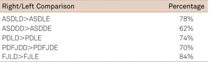
Table 2.Comparing the Right/Left Sample Ratio.
Right/Left Comparison Percentage
ASDLD.ASDLE 78%
ASDDD.ASDDE 62% PDLD.PDLE 74%
PDFJDD.PDFJDE 70% FJLD.FJLE 84%
Table 3.p value of the tests.
Compared pairs p value
and the junction of the squamous and parietomastoid sutures7. Other study used the occipitomastoid suture at
the mastoid incisure level, considering that a proper place to make the trephination and expose the posterior and inferior margins of the sigmoid sinus2.
In anatomical dissections aiming at illustrating the trans-petrosal approach with partial labyrinthectomy, was noticed that the sigmoid sinus curved anteriorly towards the jugular bulb9. This curvature was located near the top of the
mas-toid incisure. Although easily identifiable in dry skulls, the cranial sutures may be difficult to pinpoint during surgical procedures and in anatomical dissections. In a study were dissected heads and properly pinpointed the sutures in about 60% of the cases investigated8. In that same study,
they did not find the occipitomastoid suture in 14 of the 24 sides studied.
A study described a new craniometric point, associating it with the anterior curve of the sigmoid sinus10. This
land-mark - called digastric notch, was defined as the point located immediately posterior to the mastoid incisure in dry skulls, or the notch immediately superior to the posterior belly of the digastric muscle in dissections and surgical pro-cedures. The digastric notch is projected on the sigmoid sinus sulcus on the internal skull surface in 49.6% of the cases on the right side and in 29.9% on the left. When not coinciding with the sigmoid sinus sulcus, this notch was located at 3.10 mm higher than average (SD 3.11 mm), being shorter on the right side, thus showing a lack of symmetry between skull sides.
This notch was used as a landmark, making the trephina-tion to gain access to the posterior fossa at 1cm medial to it, to avoid exposing the sigmoid sinus8. This study was carried
out in 10 dry skulls and in 12 heads injected with colored silicone in the arteries and veins, they described that the mastoid incisure completely covered the sigmoid sinus in 85% of the specimens.
Very few papers assessed skull differences reflecting size differences in vascular structures such as the sigmoid sinus. Based on one of them we may conclude that the sigmoid sinus is larger on the right side10. In a study involving 263
digital brain angiographies with bone subtraction, was found symmetric sigmoid sinuses in 49% of the cases11
. In about 36% of the cases, the right-side sigmoid sinus was dominant, or the only one present.
It was to be expected that anatomical studies would find differences in the distance measures involving anatomical landmarks, such as the sigmoid sinus sulcus, between both sides of the skull. However, in two studies there was no dif-ference between skull sides vis-à-vis the measures carried out2,3. It was not compared the sides in their craniometric
measurements8. Considering measurements between the
mastoid tip and the lateral margin of the jugular foramen, the lateral margin of the stylomastoid foramen and the lat-eral margin of the occipital condyle, there were no sidewise differences among the 25 skulls investigated12.
When the number of skulls studied is higher, one usually finds differences between the two sides. In two studies invol-ving 100 dry skulls6
and 84 skulls7
, were found differences between the right and left sides vis-à-vis the asterion and the transverse sinus.
A group studied the jugular foramen microanatomy in ten cadavers13. The right-side jugular foramen was wider
than the left one in 70% of the cases. In this paper, with 5 times more specimens, the difference increased to 84%.
In the present study we investigated the entire extension of the sigmoid sinus and jugular foramen, and we measured the sulcus width near the point corresponding to the sinodural angle, which marks the beginning of the sigmoid sinus, near the digastric notch, corresponding to its anterior curvature and jugular foramen width. We also measured the sigmoid sinus sulcus distance from the sinodural angle all the way to its anterior curvature at the level of the digastric notch, and from this point all the way to the jugular foramen.
The sigmoid sinus width values at the level of the sino-dural angle are larger on the right side in 78% of the cases and at the digastric notch level in 72%. The jugular foramen width was also higher on the right side in 84% of the cases. The sigmoid sinus distance at the level of the sinodural angle all the way to the digastric notch was higher in the right side in 64% of the cases, and the sigmoid sinus distance from the digastric notch to the jugular foramen was higher in the right side in 70% of the cases.
Our findings confirm the asymmetry between skull sides. The comparative measures vis-à-vis the jugular foramen width and the sigmoid sinus sulcus, in two reference points show that it is higher on the right side, thus contributing with one more piece of information for the surgical planning of lesions involving this region of difficult approach and elo-quent structures.
In conclusion the craniometric measurements carried out in this study and their statistical analyses allow us to reach the following conclusions:
1) In most of the cases, the sigmoid sinus sulcus width at the transverse/sigmoid sinuses transition point, the sigmoid sinus sulcus distance from the transverse/sigmoid sinuses transition level all the way to its anterior curvature at the level of the digastric notch, the sigmoid sinus sulcus width near the digastric notch, the sigmoid sinus sulcus distance from the digastric notch all the way to the jugular foramen are larger on the right side of the same skull;
References
1. Ribas GC. Estudo das relações topográficas das suturas lambdóide, de occipitomastóidea e parietomastóidea com os seios transverso e sigmoide etrepanações da região. São Paulo: Tese de Doutoramento, Faculdade de Medicina da Universidade de São Paulo, 1991.
2. Ribas GC, Rhoton AL, Cruz OR, Peace D. Temporo-parieto-occipital burr hole sites study and systematized approaches proposal. In: Samii M, (edit.) Skull Base Surgery: First Int Skull Base Congr. Basel, Karger,1994:723-730.
3. Ribas GC, Rhoton AL, Cruz OR, Peace D. Suboccipital burr holes and craniectomies. Neurosurg Focus 2005;19:E1.
4. Gusmão S, Silveira, RL, Arantes, A. Pontos referenciais nos acessos cranianos. ArqNeuropsiquiatr. 2003;61:303-308.
5. Day JD, Kellogg JX, Tschabitscher M, Fukushima T. Surface and superficial surgical anatomy of the posterolateral cranial base: significance for surgical planning and approach. Neurosurgery 1996;38:1079-1083.
6. Day JD, Tschabitscher M. Anatomic position of the Asterion. Neurosurgery 1998;42:198-199.
7. Bozbuga M, Boran BO, Sahinoglu K. Surface anatomy of the posterolateral cranium regarding the localization of the initial burr-hole for a retrosigmoid approach. Neurosurgery 2006;29:61-63.
8. Avci E, Kocaohullar Y, Fossett D, Caputy A. Lateral posterior fossa venous sinus relationships to surfacelandmarks. Surg Neurol 2003;59:392-397.
9. Sekhar LN, Schessel DA, Bucur, SD, Raso JL, Wright DC. Partial Labyrinthectomy petrous apicectomy approach to neoplastic and vascular lesions of the petroclival area. Neurosurgery. 1999;44:537-552.
10. Raso J, Gusmão S, A new landmark for finding the sigmoid sinus in suboccipital craniotomies. Neurosurgery 2011;68(Suppl):1-6.
11. Shima T, Okita S, Okada Y, Nishida M, Yamane K, Hatayama T. Anatomical dominance of venous sinuses and jugular vein examined by intravenous digital subtraction angiography. In: Hakuba A (Ed). Surgery of the Intracranial Venous System. 1996.
12. Tedeschi H, Rhoton Jr, AL. Lateral approaches to the petroclival region. SurgNeurol 1994;41:180-216.