Transcranial Doppler in sickle cell anaemia
Evaluation of brain blood low parameters in
children of Aracaju, Northeast - Brazil
Hyder Aragão de Melo
1, José Augusto S. Barreto-Filho
2,
Roberto César P. do Prado
2, Rosana Cipolotti
2Abstract – Background: Environmental factors interfere on sickle cell anaemia (SCA). Transcanial Doppler (TCD) is important to evaluate cerebrovascular disease. Objective: To evaluate brain haemodynamic profile of children with SCA in Sergipe. Methods: Cross sectional study (group1: SCA patients aged 3-18; group2: age and sex matched healthy individuals). Baseline brain flow was evaluated. Results: Group1=34 patients; group 2=81 controls. SCA patients had mean velocity (MV)=125.69 cm/s±23.40; pulsatility index (PI)=0.66±0.10; middle cerebral artery ratio (MCAr)=14.53±15.23; right anterior cerebral artery/right middle cerebral artery=0.77±0.20; left anterior cerebral artery/left middle cerebral artery=0.78±0.20. Controls had MV=79.44±15.54; PI=0.82±0.11; MCAr=13.19±13.77; right anterior cerebral artery/right middle cerebral artery=0.80±0.16; left anterior cerebral artery/left middle cerebral artery=0.84±0.18. MV and PI differences were statistically significant between groups. MV was related to age but not to gender. Conclusion: MV evaluation using TCD was similar to international standards and possible to be used in our setting.
KEy WorDS: transcranial Doppler, sickle cell anaemia, cerebral blood flow, pulsatility, children.
Doppler transcraniano em portadores de anemia falciforme: estudo dos parâmetros de fluxo sangüíneo cerebral em crianças de aracaju, sergipe.
Resumo – Introdução: Aspectos ambientais interferem na apresentação da anemia falciforme (AF). Doppler transcraniano (DTC) é útil na avaliação do risco para doença cerebrovascular em pacientes com AF. Objetivo:
Avaliar o perfil hemodinâmico cerebral de crianças com AF em Sergipe. Método: Estudo transversal (grupo1: portadores de AF 3-18 anos; grupo2: indivíduos saudáveis, pareados por idade e gênero). Foram avaliadas medidas de fluxo sangüíneo cerebral basal. Resultados: Grupo1 (n=34): velocidade média (Vm)=125,69 cm/s
±23,40; índice de pulsatilidade (Ip)=0,66±0,10; relação entre artéria cerebral média (ACMs)=14,53±15,23; artéria cerebral anterior (ACA)/ACM direita=0,77±0,20; ACA/ACM esquerda=0,78±0,20. Grupo 2 (n=81): Vm=79,44 cm/s
±15,54; Ip=0,82±0,11, relação entre ACMs=13,19±13,77, ACA/ACM direita=0,80±0,16; ACA/ACM esquerda=0,84±
0,18. Vm foi maior e Ip menor no grupo1. Vm se correlacionou com idade mas não com gênero. Conclusão: o perfil hemodinâmico cerebral de crianças com AF em Sergipe assemelha-se às referências internacionais. Não se observou interferência de fatores ambientais sobre os resultados.
PALAVrAS-CHAVE: Doppler transcraniano, anemia falciforme, fluxo sanguíneo cerebral, pulsatilidade, crianças.
Department of Medicine and University Hospital, Federal University of Sergipe, Aracaju SE, Brazil: 1MD, MSc; 2MD, MSc, PhD.
received 18 July 2007, received in inal form 27 February 2008. Accepted 20 March 2008.
Dr. Hyder Aragão de Melo – Rua Dom José Thomaz 473 - 49015-090 Aracaju SE - Brasil. E-mail: [email protected]
It is well established that environmental conditions may interfere on the presentation and severity of sickle cell disease (SCD). The levels of fetal haemoglobin, con-comitant occurrence with alfa-thalassemia and the dif-ferent haplotypes related to hemoglobin S (HbS) may act as genetic modulators and be inluenced by the en-vironment, socioeconomic status (SES), nutrition, pre-ventive measures and access to health services1-3.
chance of occurrence of such event was of 11% up to the second decade of life, but increasing to 40% in the next 3 years for a second ictus6,7. Transcranial Doppler (TCD) is an ultrasonographic method that uses 1 to 2 MHz puls-es which can penetrate skull. It was irst used clinically by Aaslid in 19828. It is considered of low cost and free of side effects. Its use in clinical research for SCD is justiied by the disease’s physiopathology, which involves great ce-rebral vessels, the same very well accessed by TCD6. The hemodynamic parameters of blood low in children with SCD without antecedent of stroke can be seen on Chart. Aracaju is the capital and largest city in Sergipe state. Located in the North-eastern costal area of Brazil, it has a tropical climate and rain fall index of 1000 mm to 2000 mm. The municipality population is 520,2079 reaching 900,000 population in its metropolitan area. The Paedi-atric Haematology clinic at University Hospital - Feder-al University of Sergipe is the reference service for the entire state and neighbour locations (Bahia and Alagoas). Three hundred SCD patients from 0 to 26 years of age are followed free of charge in this clinic. In general, patients are of low socioeconomic status and literacy, but they get partial support from 2 NGos which provide assistance during their stay in Aracaju and to get medicines.
We report, using TCD, the blood low proile of great cerebral vessels of SCD patients free of cerobrovascular disease in Sergipe.
meTHoD
This was a controlled cross sectional study. The sample eval-uated constitutes 10% of the entire population and was enrolled sequentially during their routine appointments. We selected ho-mozygous HbS patients after 3 years of age (as it is necessary the patient collaboration). Patients with antecedent of cerebrovas-cular disease, using hydroxiurea, who had been submitted to blood transfusion or had any clinical event in the 30 previous days, were excluded. Controls (2 per case) were school children without any chronic disease, paired by age and gender. Anemic children (Hb<11g/dL) were excluded.
TCD evaluations were performed using the same device (WAKI-I, Átis Medical®, monochannel, probe of 2 MHz), by the same examiner and technique: patients were positioned dorsally and oriented about the procedures; they were asked to not laugh, speak, hold breath or sleep. The middle cerebral artery (MCA), anterior cerebral artery (ACA), posterior cerebral artery (PCA), vertebral (AV) and basilar were evaluated. Each vessel was exam-ined through its proper bone window (MCA, ACA, PCA via tem-poral window e VA e basilar via occipital windows) according to Aaslid technique10. Ultrasound signal was deepened by 2
millime-tres each time and the highest speed was chosen, in accordance with SToP study6,11. After TCD a blood sample was collected.
results were analysed using SPSS-15.0 software. For each
pa-tient it was considered one record of medium velocity (mV) and of pulsatility index (Pi), which corresponds to the arithmetic av-erage of mV of the MCA and respective Pi of both sides. Normal-ity of data was evaluated using Shapiro-Wilk test. Continuous variables were compared using t test for independent samples, and other continuous variables using Mann-Whitney test. Cat-egorical variables were compared using the Chi Square test for the 2 groups. Signiicance level was established as 5% (p<0.05).
The project was approved by the Federal University of Ser-gipe Ethics Committee.
resUlTs
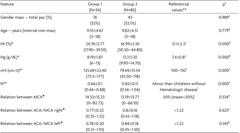
Thirty four SCD patients (Group 1) and 80 controls (Group 2) were studied. Age varied from 3 to 18 years and gender had similar distribution. Hemoglobin and hemato-crit values were signiicantly lower in SCD group (Table 1). This group presented higher mV and lower Pi than group 2. relation between CMAs, and ACA/MCA were not sig-niicant in both sides (Table 1).
Medium velocity was similar for paired vessels when considered each group. Speed values for each vessel were statistically higher in group 1, when compared to the cor-respondent in group 2 (Table 2). There were not VM dif-ferences between genders except for PCA in group 2. In this group women had higher mV.
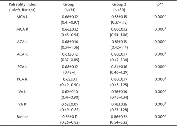
Group 1 had signiicantly lower Pi than control group in all vessels. otherwise, there was similarity between paired vessels for each group (Table 3). Correlation co-eficient was signiicant for age in both groups (group1: r=0.495; p=0.03 and group 2: r=0.430; p=0.000). Hemato-crit levels were related to mV in group 2 (r=0.229; p=0.04) but not in SCD group (r=0.158; p=0.37).
DiscUssion
Table 1. Characteristics studies between groups
Feature Group 1
(N=34)
Group 2 (N=80)
referencial values**
p#
Gender masc – total pac (%) 18
(52%)
43 (53.1%)
0.989A
Age – years (interval min-max) 9.55±4.67
(3–18)
9.82±4.51 (3–18)
0.779B
Ht (%)* 24.74±5.77
(17.90–39.50)
36.99±3.30 (30.50–44.80)
21.1±2.32 0.000B
Hg (g/dL)* 8.19±1.87
(6–13)
12.1±1.10 (9.90–14.70)
7.4±0.82 0.000B
mV (cm/s)*• 125.69±23.40
(75.5–177)
79.44±15.54 (43.50–118)
100–1301 0.000C
Pi*• 0.66±0.1
(0.46–0.88)
0.82±0.11 (0.56–1.04)
Minor than childrens without Hematologic disease1
0.000C
relation between MCA◆ 14.53±15.23
(0–82.72)
13.19±13.77 (0–66.10)
50% (mean<20%)1 0.534B
relation between ACA/MCA right◆ 0.77±0.22
(0.35–1.32)
0.8±0.16 (0.43–1.18)
<1.22 0.623C
relation between ACA/MCA left◆ 0.78±0.20
(0.31–1.10)
0.84±0.18 (0.45–1.50)
<1.22 0.341B
*Presentation mean±standard deviation; •Average velocitys at middle cerebral artery (average between MCA right and left); **Data are of the sickel cell anemia childrens; #Difference between Group 1 and Group 2; 1Babikian, 19998; 2Adams, 19986; AChi-square; BMann-Whitney; Ht, hematocrit; Hg,
hemoglobin; mV, mean velocity of middle cerebelar arteries; Pi, pulsatility index; MCA, middle cerebral artery; ACA, anterior cerebral artery.
Table 2. Mean velocity in vases. Average speed
(L=left; r right)
Group 1 (N=34)
Group 2 (N= 80)
referencial values*
p**
MCA L (cm/s) 126.24±27.33
(78–180)
78.16±18.48 (40–124)
110–130 0.000A
MCA r (cm/s) 125.15±24.78
(73–178)
80.72±16.55 (44–124)
110–130 0.000A
ACA L (cm/s) 97.12±27.81
(40–150)
64.35±14.00 (35–109)
85–1101 0.000A
ACA r (cm/s) 94.44±23.85
(51–133)
63.54±15.52 (34–118)
85–1101 0.000B
PCA L (cm/s) 65±21.43
(33–118)
46.62±10.19 (26–67)
65–851 0.000A
PCA r (cm/s) 73.53±20.62
(30–110)
49.43±9.94 (32–84)
65–851 0.000B
AV L (cm/s) 82.76±19.96
(36–120)
52.52±14.09 (21–110)
– 0.000B
AV r (cm/s) 84.91±20.11
(39–122)
54.33±13.01 (26–103)
– 0.000B
Basilar (cm/s) 93.79±21.10
(39–137)
59.67±13.97 (16–95)
– 0.000A
*Presentation mean±standard deviation (min-max); *This dados are of the sickel cell anemia children; **Difference between Group 1 and Group 21 (Adams, 19986); At student test; BMann-Whitney; MCA, middle cerebral artery; ACA,
As high MV indicates high blood flow, in this case caused by chronic anaemia, low Pis were expected be-cause a lower resistance is required for the arteries to re-ceive increased low. otherwise for both groups relations between mV and age and mV and hematocrit are inversely proportional. Both indings are in agreement with Babiki-an’s8 in a study with similar group of patients and controls.
In our study there was no gender difference in low MV, except for PCA in control group. This inding differs from a previous study12, but in that study participants were adults (predominantly women) without chronic disease. In our data
there is not difference in cerebral arteries mV between gen-ders in SCD patients, similarly to previous study in children8.
Clinical presentations of SCD may be altered by genet-ic and environmental factors2,13,14. Genetic factors include proportion of fetal hemoglobin, association with alfa thalassemia and with G6PD deiciency, esferocitosis and anti-oxidants enzymes, as well as with the haplotypes15. Environmental factors include physical environment, nu-trition, and access to medical, social and psychological support2. The hot weather in Brazilian Northeast and low SES and schooling level are factors that may worsen our parameters. These particularities have been shown in pre-vious studies for both weight and height impairment4 and for insidious cerebrovascular disease. But when cerebral blood low in children with SCD is considered, our indings are similar to the ones in different places, which makes DTC a good tool to routinely follow SCD patients, using the references from international studies.
In conclusion, cerebral blood low proile in children with SCD showed higher cerebral speed low and lower pulsatility index than in health children. relation between MCAs and ACAs/MCAs were not different when both groups were compared showing that the hiperdynamic effect of chronic anaemia is homogenous for these arter-ies. Cerebral blood low previously described is adequate
Chart. Reference values for sickle cell disease patients.
Feature referencial values
MCA 100–130 cm/s
ACA 85–110 cm/s
PCA 65–85 cm/s
Pulsatility index Minum than normal childrens
relation between MCAs <50%
relation ACA/MCA <1.2
relation velocity/age Not relation
relation velocity/hematocrit Negative
MCA, middle cerebral artery; ACA, anterior cerebral artery; PCA, posterior cerebral artery. Babikian, 1999.
Table 3. Pulsatitlity index in vases. Pulsatility index
(L=left, r=right)
Group 1 (N=34)
Group 2 (N=80)
p**
MCA L 0.66±0.12
(0.41–0.97)
0.83±0.15 (0.37–1.15)
0.000A
MCA r 0.66±0.12
(0.45–0.94)
0.80±0.12 (0.54–1.06)
0.000A
ACA L 0.68±0.16
(0.34–1.06)
0.81±0.15 (0.42–1.14)
0.000A
ACA r 0.63±0.12
(0.37–0.85)
0.80±0.17 (0.42–1.34)
0.000A
PCA L 0.68±0.12
(0.42–1)
0.84±0.16 (0.46–1.29)
0.000A
PCA r 0.65±0.1
(0.49–0.90)
0.80±0.17 (0.43–1.25)
0.000B
VA L 0.62±0.10
(0.41–0.90)
0.76±0.16 (0.45–1.34)
0.000B
VA r 0.62±0.09
(0.49–0.85)
0.78±0.16 (0.53–1.28)
0.000B
Basilar 0.56±0.11
(0.26–0.82)
0.86±0.36 (0.54–3.22)
0.000B
*Presentation mean±standard deviation (min-max); these data are of the sickle cell anemia children; referencial values: minor than childrens without hematologic disease1; **Difference between Group 1 and Group 2; 1Babikian, 19998; At
student test; BMann-Whitney; MCA, middle cerebral artery; ACA, anterior cerebral artery; PCA, posterior cerebral artery;
for our population. There were not any environmental indings that suggest interference on hemodynamic pa-rameters obtained with TCD.
reFerences
1. ANVISA, Manual de diagnóstico e tratamento de doenças falciformes, Brasil. Brasília 2002. Disponível em http://www.anvisa.gov.br/divulga/public/ sangue/hemoglobinopatia/diagnostico.pdf Acess 15 November 2005. 2. Naoum PC. Interferentes eritrocitários e ambientais na anemia
falci-forme. Rev Bras Hematol Hemoter 2000;22:5-22.
3. Romero WER, Renauld GFS. Haplótipos de la hemoglobina S: impor-tancia epidemiológica, antropológica y clínica, Rev Panam Salud Pu-blica 1998;3:1-8.
4. Cipolotti R, Caskey MFB, Franco RP, et al. Childhood and adolescent growth of patientes with sickle cell disease in Aracaju, Sergipe, north-east Brazil. Ann Trop Paediatr 2000;20:109-113.
5. Manci EA ,Culberson DE,Yang YM, et al. Causes of death in sickle cell disease: an autopsy study. Br J Haematol 2003;123:359-365.
6. Adams RJ, McKie VC, Hsu L, et al. Prevention of a irst stroke by trans -fusions in children with sickle cell anemia and abnormal results on transcranial doppler ultrasonogrphy, N Engl J Med 1998;339:5-11.
7. Ohene-Frempong K, Weiner SJ, Sleeper LA, et al. Cerebrovascular acci-dents in sickle cell disease: rates and risk factors. Blood 1998;91:288-294. 8. Babikian VL, Wechsler LR, Toole JF. Transcranial doppler
ultrasonog-raphy, 2nd ed. USA: Butterworth-Heinemann, 1999.
9. IBGE. Censo demográico, http://www.ibge.gov.br/home/estatistica/
populacao/contagem2007/popmunic2007layoutTCU14112007.pdf. Acess 04 December 2007.
10. Newell DW, Aaslid R. Transcranial Doppler. New York: Raven Press, 1992. 11. Nicchols FT, Jones AM, Adams RJ. Stroke prevention in sickle cell dis-ease (STOP) study guidelines for transcranial Doppler testing. J Neu-roimaging 2001;11:354-362.
12. Barbosa MF, Nitamar Abdala N, Carrete H Jr. Doppler transcraniano convencional em voluntários assintomáticos variabilidade e valores de referência para parâmetros de luxo sanguíneo. Arq Neuropsiquiatr 2006;64:829-838
13. Marouf R, Grupta R, Haider MZ, Adekile AD. Silent brain infarcts in adult Kuwaiti sickle cell disease patients, Am J Hematol 2003;73:240-243. 14. Pegelow CH, Wang W, Granger S, et al. Silent infarcts in children with
sickle cell anemia and abnormal cerebral artery velocity. Arch Neurol 2001;58:2017-2021.