DE MORSIER SYNDROME ASSOCIATED WITH
PERIVENTRICULAR NODULAR HETEROTOPIA
Case report
Mônica Jaques Spinosa
1, Paulo Breno Noronha Liberalesso
2,
Simone Carreiro Vieira
1, Alfredo Löhr Júnior
2ABSTRACT - Introduction: Septo-optic dysplasia (De Morsier syndrome) is defined as the association between optic nerve hypoplasia, midline central nervous system malformations and pituitary dysfunction. Case re-port: Third child born to nonconsanguineous parents, female, adequate pre-natal medical care, cesarean term delivery due to breech presentation, Apgar score 3 at the first minute and 8 at 5 minutes, symptomatic hypoglycemia at 18 hours. Neurological follow-up identified a delay in acquisition of motor and language developmental milestones. Epileptic generalized seizures began at 12 months and were controlled with phenobarbital. EEG was normal. MRI revealed agenesis of the pituitary stalk, hypoplasia of the optic chiasm and periventricular nodular heterotopia. Ophthalmologic evaluation showed bilateral optic disk hypopla-sia. Endocrine function laboratory tests revealed primary hypothyroidism and hyperprolactinemia. Con-clusion: The relevance of this case report relies on its uniqueness, since periventricular heterotopia had not been described in association with septo-optic dysplasia until 2006.
KEY WORDS: De Morsier syndrome, septo-optic dysplasia, periventricular nodular heterotopia, primary hy-pothyroidism.
Síndrome de De Morsier associada a heterotopia nodular periventricular: relato de caso
RESUMO - Introdução: Displasia septo-óptica (síndrome de De Morsier) é definida como a associação en-tre hipoplasia do nervo óptico, malformações de linha média do sistema nervoso central e disfunção pituitária. Relato de caso: Terceiro filho, pais não consangüíneos, sexo feminino, pré-natal adequado, parto cesário a termo por apresentação pélvica, Apgar 3 no primeiro minuto e 8 no quinto minuto, hipo-glicemia sintomática com 18 horas de vida. Durante o acompanhamento neurológico identificou-se atra-so na aquisição dos marcos de desenvolvimento motor e linguagem. Crises epilépticas generalizadas ini-ciaram com 12 meses de vida sendo controladas com fenobarbital. EEG era normal. Ressonância magnéti-ca revelou agenesia de haste pituitária, hipoplasia de quiasma óptico e heterotopia nodular periventricu-lar. Avaliação oftalmológica demonstrou hipoplasia bilateral de disco óptico. Investigação da função en-dócrina revelou hipotireoidismo primário e hiperprolactinemia. Conclusão: A relevância deste relato re-side em seu ineditismo, já que heterotopia periventricular não havia sido descrita em associação com dis-plasia septo-óptica até 2006.
PALAVRAS-CHAVE: síndrome de De Morsier, displasia septo-óptica, heterotopia nodular periventricular, hipotireoidismo primário.
Unidade de Neurologia Infantil Pequeno Príncipe, Curitiba PR, Brasil (UNIPP): 1Residente de Neuropediatria; 2Médico da Unidade
de Neurologia Infantil.
Received 8 November 2006, received in final form 9 February 2007. Accepted 2 April 2007.
Dra. Mônica Jaques Spinosa - Rua Johann Sebastian Bach 71 - 80820-140 Curitiba PR - Brasil. E-mail: [email protected]ônica Jaques Spinosa - Rua Johann Sebastian Bach 71 - 80820-140 Curitiba PR - Brasil. E-mail: [email protected] Arq Neuropsiquiatr 2007;65(3-A):693-696
In 1941, Reeves1
first described a patient with sep-tum pellucidum agenesis and optic nerve abnormal-ities. De Morsier2
, in 1956, associated optic nerve hy-poplasia to septum pellucidum agenesis and coined the term “septo-optic dysplasia” (SOD), later to be recognized as De Morsier syndrome. The following descriptions of patients with optic nerve hypopla-sia and septum pellucidum agenesis included endo-crinological abnormalities related to
malfunction-ing of the hypothalamic-pituitary axis, with predom-inance of growth hormone (GH) deficiency resulting in growth failure3-5
. Billson and Hopkins6
, in 1972, indicated that the absence of septum pellucidum could represent an inconstant feature. Patel et al.7
confirmed this finding in 1975, reporting 4 cases of SOD, 2 of which had normal septum pellucidum. In 1990, Brenner et al.8 reported midline abnormalities
sib-694 Arq Neuropsiquiatr 2007;65(3-A)
lings. Recent studies conclude that SOD has variable phenotypic expression that may include optic nerve hypoplasia, midline central nervous system malfor-mations and pituitary dysfunction.
This case report was approved by the ethics com-mittee of Pequeno Príncipe Hospital and parental written informed consent was obtained for publi-cation.
CASE
Third child born to nonconsanguineous parents, female, adequate pre-natal medical care, cesarean term delivery due to breech presentation, Apgar score 3 at the first min-ute and 8 at 5 minmin-utes, birth weight 3150g, symptomatic hypoglycemia at 18 hours. Neurological follow-up identi-fied a delay in acquisition of motor and language devel-opmental milestones. Electroencephalogram (EEG) was normal at 9 months. Magnetic resonance imaging (MRI) at 10 months revealed agenesis of the pituitary stalk and hypoplasia of the optic chiasm suggestive of SOD, normal septum pellucidum, multiple foci of periventricular nodu-lar heterotopia at left frontal and bilateral parieto-occipi-tal regions, focal thickening of the quadrigeminal plaque causing relative stenosis of the distal half of the cerebral aqueduct without any evidence of obstruction of the su-pratentorial ventricular system (Figs 1 and 2). At 12 months, was admitted at the pediatric neurology unit of Peque-no Príncipe Hospital, Curitiba, Brazil, for epileptic seizures. The patient presented with 7 episodes of epileptic seizures at the same day, described by the mother as a scream fol-lowed by hyperextension of all 4 limbs and cervical hyper-extension, evolving to a generalized clonic phase. All ep-isodes lasted less than 1 minute and some occurred while the patient was febrile. Epileptic crisis were controlled with phenobarbital 5 mg/kg/day given bid. Clinical and neuro-logical examination identified hypertelorism, midline fron-tal bulging, neurodevelopment delay with milestones
com-patible with a 5-month-old child and axial and apendicular hypotonia. EEG was repeated, again yielding normal re-sults. Ophthalmologic evaluation showed bilateral optic disk hypoplasia. Echocardiogram was normal. Endocrine function was assessed by measurement of TSH, T3, T4, GH, FSH, LH, ACTH, prolactin and cortisol levels. Results showed primary hypothyroidism (low T3 and T4 with high TSH lev-els) and high prolactin levels. Treatment with levothyroxine was instituted. The patient was not tested for HESX1 muta-tions due to the unavailability of this genetic test.
DISCUSSION
Concerning phenotypic variability, Thomas et al.9,
in 2001, emphasized that only 30% of the patients
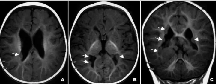
Fig 1. MRI showing multiple foci of periventricular nodular heterotopia at left frontal and bilateral parieto-occipital regions, on T1-weighted axial (A,B) and coronal (C) cuts.
Arq Neuropsiquiatr 2007;65(3-A) 695
present with the complete triad of optic nerve hy-poplasia, midline central nervous system malforma-tions and pituitary dysfunction. Birkebaek et al.10,
reported a series of 55 patients with optic nerve hy-poplasia, 49% with agenesis of septum pellucidum and 64% with hypothalamic-pituitary axis neurora-diologic abnormalities on MRI. Forty-nine percent of the patients had endocrine dysfunction, 85% of which also presented with abnormal neuroimage of the hypothalamic-pituitary axis. Agenesis of septum pellucidum was found by Birkebaek et al.10 to be
as-sociated to endocrinopathy, since the frequency of hormonal disturbances was higher in the group with abnormal septum pellucidum (56%) than in patients with no abnormalities in this structure (39%). How-ever, this difference was statistically significant only with respect to ADH deficiency (p=0.04). Our patient had a normal septum pellucidum and displayed no clinical or laboratorial signs of hypothalamic-pituitary axis dysfunction. Hyperprolactinemia was attributed to primary hypothyroidism, a well known association accredited to the stimulatory effect of TRH in the se-cretion of prolactin. There have been no reports of the association between SOD and primary hypothy-roidism and hyperprolactinemia until 2006. The as-sociation between SOD and hyperprolactinemia was described by Izenberg et al.11 in 2 of the 4 patients
in their sample. Birkebaek et al.10 reported that 35%
of patients with normal septum pellucidum and ab-normal hypothalamic-pituitary axis on neuroimage, such as our patient, showed no endocrine dysfunc-tion of central cause.
The genetic basis for SOD was suggested by Wales et al.12
in 1996, with the report of two siblings with SOD born of consanguineous parents. In 1998, Dat-tani et al.13 performed the genetic study of these
pa-tients, along with 18 other cases of sporadic SOD, searching for mutation in the HESX1 gene ( homeo-box gene expressed in embryonic stem cells), locus 3p21.2-p21.1. The choice of this candidate gene was based on the finding that mice with a null Hesx1 gene exhibited malformations of optic nerves and midline structures that were analog to SOD in hu-mans13
. The siblings were found to be carriers of a homozygous missense mutation in the HESX1 gene. Nine family members, including their parents, were asymptomatic carriers of the mutation in heterozy-gosis, suggesting an autosomal recessive mode of in-heritance. In this study, no mutations were found on patients with sporadic SOD. Mutations in the HESX1 gene were confirmed by other studies. Thomas et al.9 analyzed a sample of 228 patients with
congeni-tal pituitary defects, 105 with SOD. Three different heterozygous missense mutations were identified in 4 patients of 3 different pedigrees. Phenotype var-ied widely between isolated GH deficiency and SOD. Family members were scanned for HESX1 mutations. In all cases an asymptomatic parent was found to harbor the mutation, along with an asymptomatic sibling of one affected individual. This study showed that even though most sporadic cases cannot be at-tributed to HESX1 gene mutations, a pattern of het-erozygous autosomal dominant inheritance with in-complete penetration exists. This is consistent with a similar finding in mice, where heterozygous Hesx1 mutations were expressed as low penetrance mild phenotype13
. Tajima et al.14
described a Japanese pa-tient with sporadic SOD and a heterozygous frame-shift mutation of the HESX1 gene. Neither parent harbored the mutation, indicating a de novo occur-rence. Mutations in the HESX1 gene have been found in 5/93 patients with undescendent or ectopic poste-rior pituitary by Brickman et al.15
, indicating that this gene is implicated as a genetic basis for a variety of midline central nervous system defects.
The association between SOD and periventricular nodular heterotopia has not been reported in medical literature to this date. However, in 2002, Mitchell et al.16
described 20 children with ectopic posterior pituitary, four of which had associated periventricular hetero-topia. None of them displayed optic nerve or septum pellucidum abnormality. Two patients were screened for HESX1 mutations and in 1 of them a heterozygous mutation was found. In all 4 patients, similarly to our case, periventricular heterotopia was characterized by few subependymal nodules, ranging from 1 to 7. The authors concluded that their finding supports a role for the HESX1 gene in the genesis of both ectopic poste-rior pituitary and periventricular heterotopia and their place in the wide spectrum of SOD.
Schizencephaly has been reported by some au-thors in 50% of the patients with SOD17
. Barkovich et al.17
neuroradiologically classified SOD in two sub-types, according to the presence of schizencephaly or lack thereof. Sener18 described a patient with SOD
and bilateral rolandic cortical dysplasia, suggesting the term “cortico-septo-optic dysplasia”. Miller et al.19
named the association of SOD and disorders of neuronal organization “SOD-Plus”.
696 Arq Neuropsiquiatr 2007;65(3-A)
such as the septum pellucidum and the corpus cal-losum. A genetic basis is clearly implicated in some cases and the fact that the same gene has been shown to de defective in patients with different phe-notypes further strengthens the link between these disorders.
In conclusion, the relevance of this case report re-lies on its uniqueness, since periventricular heteroto-pia had not been described in association with SOD until 2006. However, periventricular heterotopia has been connected to ectopic posterior pituitary and HESX1 mutation, signaling that this neuronal organi-zation disorder may indeed be part of this spectrum, as other cortical development abnormalities seem to be. A thorough understanding of these complex as-sociations seems to lie in the comprehension of the intricate course of forebrain and cortical develop-ment and the role of HESX1 gene in this process.
REFERENCES
1. Reeves DL. Congenital absence of septum pellucidum. Johns Hopkins Med J 1941;69:61-71.
2. De Morsier G. Etudes sur les dysraphies cranioencéphaliques: III. Agénésie du septum pelucidum avec malformation du tractus optique. La dysplasie septoöptique. Schweiz Arch Neurol Psychiatr 1956;77:267-292.
3. Harris RJ, Haas L. Septo-optic dysplasia with growth hormone defi-ciency (De Morsier syndrome). Arch Dis Child 1972;47:973-976. 4. Hoyt WF, Kaplan SL, Grumbach MM, Glaser JS. Septo-optic dysplasia
with pituitary dwarfism. Lancet 1970;1:893-894.
5. Brook CG, Sanders MD, Hoare RD. Septo-optic dysplasia. Br Med J 1972;3:811-813.
6. Billson F, Hopkins IJ. Optic hypoplasia and hypopituitarism. Lancet 1972;1:905.
7. Patel H, Tze WJ, Crichton JU, McCormick AQ, Robinson GC, Dolman CL. Optic nerve hypoplasia with hypopituitarism: septo-optic dyspla-sia with hypopituitarism. Am J Dis Child 1975;129:175-180. 8. Benner JD, Preslan MW, Gratz E, Joslyn J, Schwartz M, Kelman S.
Sep-to-optic dysplasia in two siblings. Am J Ophthalmol 1990;109:632-637. 9. Thomas PQ, Dattani MT, Brickman JM, et al. Heterozygous HESX1 mu-tations associated with isolated congenital pituitary hypoplasia and septo-optic dysplasia. Hum Mol Genet 2001;10:39-45.
10. Birkebaek NH, Patel L, Wright NB, et al. Endocrine status in patients with optic nerve hypoplasia: relationship to midline central nervous system abnormalities and appearance of the hypothalamic-pituitary axis on magnetic resonance imaging. J Clin Endocrinol Metab 2003; 88:5281-5286.
11. Izenberg N, Rosenblum M, Parks JS. The endocrine spectrum of septo-optic dysplasia. Clin Pediatr (Phila) 1984;23:632-636.
12. Wales JK, Quarrell OW. Evidence for possible Mendelian inheritance of septo-optic dysplasia. Acta Paediatr 1996;85:391-392.
13. Dattani MT, Martinez-Barbera JP, Thomas PQ, et al. Mutations in the homeobox gene HESX1/Hesx1 associated with septo-optic dysplasia in human and mouse. Nature Genet 1998;19:125-133.
14. Tajima T, Hattorri T, Nakajima T, et al. Sporadic heterozygous frame-shift mutation of HESX1 causing pituitary and optic nerve hypopla-sia and combined pituitary hormone deficiency in a Japanese patient. J Clin Endocrinol Metab 2003;88:45-50.
15. Brickman JM, Clements M, Tyrell R, et al. Molecular effects of novel mutations in Hesx1/HESX1 associated with human pituitary disor-ders. Development 2001;128:5189-5199.
16. Mitchell LA, Thomas PQ, Zacharin MR, Scheffer IE. Ectopic posterior pituitary lobe and periventricular heterotopia: cerebral malformations with the same underlying mechanism? Am J Neuroradiol 2002; 23:1475-1481.
17. Barkovich AJ, Fram EK, Norman D. Septo-optic dysplasia: MR imag-ing. Radiology 1989;171:189-192.
18. Sener RN. Septo-optic dysplasia associated with cerebral cortical dys-plasia (cortico-septo-optic dysdys-plasia). J Neuroradiol 1996;23:245-247. 19. Miller SP, Shevell MI, Patenaude Y, Poulin C, O’Gorman AM.