Original Article
Heart Rate Variability in Patients with Iron Deficiency Anemia
Mustafa Tuncer
1, Yilmaz Gunes
1, Unal Guntekin
1, Hasan Ali Gumrukcuoglu
1, Beyhan Eryonucu
1, Niyazi Guler
1, Imdat
Dilek
2, Cengiz Demir
2Yuzunci Yil University, Faculty of Medicine, Cardiology Department1, Hematology Department2, Van - Turkey
Summary
Background: Heart rate variability (HRV) is associated with increased cardiac risk factor in several conditions. The iron status of an individual may play an important role in cardiovascular health.
Objective: To evaluate heart rate variability in patients with iron deficiency anemia.
Methods: Twenty-three patients with iron deficiency anemia (mean hemoglobin (Hb) 8.6±2.2 g/dL) and 10 healthy people (mean Hb 13.9±1.2 g/dL) were assessed with 24-hour ambulatory Holter recordings during in hospital course having limited physical activity.
Results: Although mean heart rate was significantly higher in patients with anemia, there was no significant difference regarding HRV parameters compared to the healthy group.
Conclusion: There was no significant difference in HRV parameters between patients with iron deficiency anemia with limited physical activity and healthy ambulatory people. (Arq Bras Cardiol 2009;92(5):368-371)
Key words: Heart rate; anemia, iron-deficiency.
Mailing address: Mustafa Tuncer •
Kazim Karabekir Street, Yuzuncu Yil University Research Hospital, Cardiology Department 9065100 TURKEY-Van
E-mail: [email protected]
Manuscript received November 19, 2007; revised manuscript received March 28, 2008; accepted April 07, 2008.
Introduction
Anemia is an independent risk factor for adverse cardiovascular outcomes in patients with kidney disease1 and possibly in the general population2. Decreased heart rate variability (HRV) is associated with increasedmortality and morbidity in various types of heart diseaseincluding myocardial infarction, cardiomyopathy, congestiveheart failure, and chronic mitral regurgitation3-7.
The iron status of an individual may play an important role in cardiovascular health, as either an excess of iron or iron deficiency may lead to significant problems8. It was previously suggested that anemia was associated with low HRV in ambulatory patients with stable coronary heart disease and that low HRV could potentially mediate the association of anemia with increased cardiac risk9. Anemia and HRV relation has been searched in several types of anemia; thalassemia10, vitamin B
12 deficiency and megaloblastic anemia11, sickle cell trait12.
In this study, we aimed at studying the association between HRV parameters and iron deficiency anemia.
Methods
Complete blood count, peripheral blood smear, serum ferritin, serum iron, iron binding capacity and transferrin saturations were examined. The diagnosis of iron deficiency anemia was established as hemoglobin <12 g/dl in women and <13 g/dl in men and serum ferritin <12 ng/ml, mean corpuscular volume (MCV) and mean corpuscular hemoglobin concentration (MCHC) values below the normal range (83 to 97 fl and 32 to 36 g/dl, respectively)13.
With an alpha level of 0.05 and a test power of 0.80, a sample size of 32 participants was considered adequate to define a 15 msec difference in heart rate variability parameters between groups. A total of 23 hospitalized patients diagnosed as having iron deficiency anemia (mean hemoglobin (Hb 8.6±2.2 g/dl) and 10 healthy people (mean Hb 13.9±1.2 g/dl) were enrolled in the study.
All patients underwent transthoracic echocardiographic examination. Patients with acute bleeding, malignancy, known to have chronic bleeding, thalassemia, sickle cell disease, structural heart disease, diabetes, hypertension, renal failure, hypo- or hyperthyroidism or any other systemic disease were excluded from study. All patients gave written informed consent to the study, and the study was approved by the hospital ethical committee.
Heart rate variability analysis
Twenty-four-hour ambulatory electrocardiographic Holter monitoring was performed using a Del Mar Avionics483
Original Article
Arq Bras Cardiol 2009;92(5):368-371
Tuncer et al HRV in iron deficiency anemia
channel recorder (Del Mar Medical Systems; Irvine, CA). The recordings were analyzed with a specialcomputer software (Del Mar Holter Analysis System; Del Mar MedicalSystems). All recordings were visually examined and manuallyoverread to verify beat classification by an experienced cardiologist. Abnormal beats and areasof artifact were automatically and manually identified and excludedfrom the analysis. Moreover, the longest and shortest true normal-to-normal intervals were identified for each recording in order to exclude all beats outside this range from the HRV analysis. Intervals within this range were carefully edited.
The following parameters were used in evaluation of HRV in timedomain:
1) Standard deviation (SD) of all normal-to-normal intervals (SDNN [milliseconds]);
2) Standard deviations of mean of all normal-to-normal intervals in all consecutive5-min segments of the entire recording (SDANN [milliseconds]);
3) the mean square root of differences between adjacent normal-to-normalintervals (MSRSD [milliseconds]);
4) mean of the SD in all5-min intervals (HRVM [milliseconds]); and
5) Standard deviations in all 5-minintervals (SDHRV [milliseconds]).
Statistics
Results were expressed as means ± SD.Using an SPSS package version 10.0, the data from both groups were compared with Student’s t-test for continuous variables and Chi-square test for dichotomous variables, and Mann-Whitney’s U-test for variables without normal distribution. A two-tailed p value < 0.05 was consideredsignificant.
Results
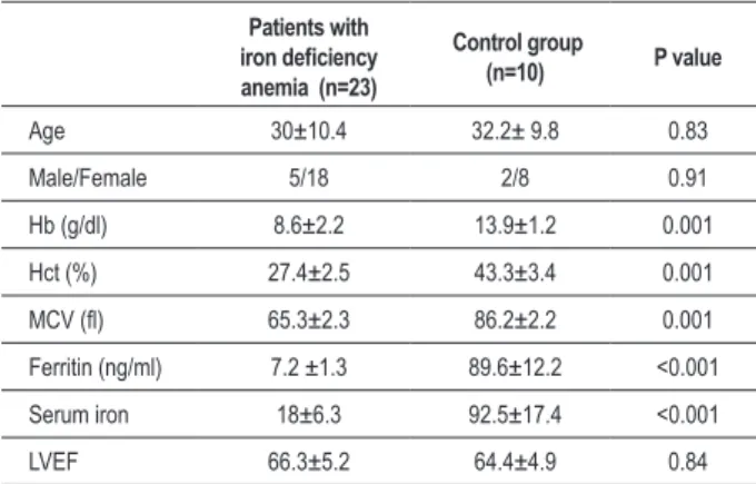
Clinical characteristics and Holter results of the study population are summarized in Table 1 and Table 2. All patients were in sinus rhythm throughoutthe recordings. No ST-segment changes have been recorded in the overall population. There were no pathologies at the echocardiographic examinations, except for slight valvular regurgitations within the normal limits. The mean heart rate was significantly higher among anemic patients compared to the control group (84.4±22.4 bpm vs. 72.6±13.2 bpm; p=0.005). There was no significant difference between the healthy group and iron-deficient patients in regard to HRV parameters.
Discussion
There is increasing evidence that anemia contributes to cardiac disease and death14,15. In patients with chronic kidney disease, for instance, anemia is an independent risk factor for the development of cardiovascular disease1. In patients with heart failure, anemia is associated with increased morbidity7.
Holter recordings play a pivotal role in identifying arrhythmias; its widely available association with heart rate variability (HRV) analysis provides information about
Table 1 - Baseline characteristics of patients with iron deiciency anemia and healthy group
Patients with
iron deiciency
anemia (n=23)
Control group
(n=10) P value
Age 30±10.4 32.2± 9.8 0.83
Male/Female 5/18 2/8 0.91
Hb (g/dl) 8.6±2.2 13.9±1.2 0.001
Hct (%) 27.4±2.5 43.3±3.4 0.001
MCV (l) 65.3±2.3 86.2±2.2 0.001
Ferritin (ng/ml) 7.2 ±1.3 89.6±12.2 <0.001
Serum iron 18±6.3 92.5±17.4 <0.001
LVEF 66.3±5.2 64.4±4.9 0.84
Hb - hemoglobin; Htc: hematocrit; MCV - mean corpuscular volume, LVEF
- left ventricular ejection fraction.
Table 2 - Heart rate variability parameters in patients with iron deiciency anemia and healthy group
Patients with
iron deiciency
anemia (n=23)
Control group
(n=10) P value
SDNN 128.3±43.7 136.5±37.1 0.62
MSRSD 33.3±19.5 27.3±11.3 0.73
HRVM 61.1±35.6 61.9±15.9 0.94
SDHRV 24.5±12.6 23.6±4.8 0.84
SDANN 111.6±40.2 120.9±36.5 0.55
Mean heart rate bpm 84.4±22.4 72.6±13.2 0.05
SDNN (milliseconds) -Standard deviation (SD) of all normal-to-normal intervals; MSRSD (milliseconds) - the mean square root of differences
between adjacent normal-to-normalintervals; HRVM (milliseconds) -
mean of the SD in allmin intervals; SDHRV (milliseconds) - SD in all
5-minintervals; SDANN (milliseconds) - SD of mean of all normal-to-normal
intervals in all consecutive5-min segments of the entire recording; bpm
- beats per minute.
the influences of the autonomic nervous system and sympathovagal balance. Heart rate variability is a method that determines the effects of the autonomic nervous system on the heart and spontaneous changes in heart rate. A decrease in heart rate variability reflects an autonomic dysfunction. Heart rate variability decreases after MI, as well as in diabetic neuropathy and heart failure. Currently, HRV is considered a predictor of sudden cardiac arrest and arrhythmias3-6.
Iron deficiency is the most common nutritional deficiency in developed and developing regions of the world. The physiologic response to anemia is a compensatory increase in cardiac output through increases in blood volume, preload, heart rate, and stroke volume, along with a decrease in afterload. Therefore, the increased sympathetic activity, evidenced as palpitation and tachycardia, is frequent in patients with anemia13.
Original Article
Arq Bras Cardiol 2009;92(5):368-371
Tuncer et al
HRV in iron deficiency anemia
References
1. Sarnak MJ, Levey AS, Schoolwerth AC, Coresh J, Culleton B, Hamm LL, et al. Kidney disease as a risk factor for development of cardiovascular disease: a statement from the American Heart Association Councils on Kidney in Cardiovascular Disease, High Blood Pressure Research, Clinical Cardiology, and Epidemiology and Prevention. Circulation. 2003; 108: 2154-69.
2. Sarnak MJ, Tighiouart H, Manjunath G, MacLeod B, Griffith J, Salem D, et al. Anemia as a risk factor for cardiovascular disease in the Atherosclerosis Risk in Communities (ARIC) study. J Am Coll Cardiol. 2002; 40: 27-33.
3. Task Force of European Society of Cardiology and the North American Society of Pacing and Electrophysiology. Heart rate variability, standards of measurement, physiological interpretation and clinical use. Circulation. 1996; 93: 1043-65.
4. Stein PK, Domitrovich PP, Kleiger RE, CASR investigators. Including patients with diabetes mellitus or coronary artery bypass grafting decreases the association
between heart rate variability and mortality after myocardial infarction. Am Heart J. 2004; 147: 309-16.
5. Karcz M, Chojnowska L, Zareba W, Ruzyllo W. Prognostic significance of heart rate variability in dilated cardiomyopathy. Int J Cardiol. 2003; 87: 75-81.
6. Stein KM, Borer JS, Hochreiter C, Okin PM, Herrold EM, Devereux RB, et al. Prognostic value and physiological correlates of heart rate variability in chronic severe mitral regurgitation. Circulation. 1993; 88: 127-35.
7. Anand IS, Kuskowski MA, Rector TS, Florea VG, Glazer RD, Hester A, et al. Anemia and change in hemoglobin over time related to mortality and morbidity in patients with chronic heart failure: results from Val-HeFT. Circulation. 2005; 112: 1121-7.
8. Turner LR, Premo DA, Gibbs BJ, Hearthway ML, Motsko M, Sappington A, et al. Adaptations to iron deficiency: cardiac functional responsiveness to An extreme result of iron deficiency is cardiomyopathy. The
pathogenesis of cardiomyopathy associated with anemia has not been ascertained. Several hypotheses have been advanced to explain how iron-deficiency anemia causes cardiomyopathy. It has been suggested that, similarly to other types of heart failure, high-output heart failure is driven by ongoing increased sympathetic nervous activity. In order to test this hypothesis, one study used β-blockade in an attempt to blunt the deleterious effect of the elevated sympathetic tone on the hearts of iron-deficient laboratory rats. β-Blockers, however, failed to prevent cardiac decompensation in those animals8.
The analysis of heart rate variability (HRV) is a reliable and reproducible technique for assessing autonomic activity in patients with cardiovascular disease. However, its use in patients with iron deficiency anemia has not been investigated thoroughly.
Anemia was associated with low HRV in ambulatory patients with stable coronary heart disease9. De Chiara et al10 reported that abnormal HRV might represent the early features of cardiac disease in thalassemic patients with no evidence of ventricular dysfunction at routine evaluation. Aytemir et al11 reported that HRV parameters were associated with vitamin B12 deficiency and parameters were improved with vitamin B12 replacement in patients with vitamin B12 deficiency and megaloblastic anemia. Connes et al12 showed that global variability (SDNN, HRVM) and parasympathetic (PNN50, MSRSD, high frequency domain (HF)) HRV indices were significantly lower in the sickle cell trait carriers and tried to explain this fact as a physiological adaptation to increase heart beat frequency, providing adequate O2 supplies to tissues despite the hemorheological alterations such as red blood cell deformability and increased blood viscosity.
In a recent study Yokusoglu et al16 reported an impairment in global (SDNN, SDANN) HRV indices that might be caused by increased sympathetic or decreased parasympathetic activity and decreased PNN50 (a parasympathetic activity index) in iron deficiency anemia. They suggested that low oxygen tension in tissues with resultant increased sympathetic activity may be responsible for altered autonomic function in iron deficiency anemia. In contrast to previous studies, however, no statistically significant differences were observed between iron-deficient anemia patients and the healthy control group, except for mean
heart rate. However, in our study, Holter monitoring was carried out during an in-hospital evaluation. Therefore, physical activity of the patients were limited, reflected by relatively lower mean heart rate compared to the study patients of Yokusoglu et al16 (84.4±22.4 bpm vs. 117±19 bpm). Therefore, combining our results with previous studies, it can be concluded that the increase in physical activity with consequent increase in tissue O2 demand may result in increased sympathetic and decreased parasympathetic activity.
Limitations
The sample size of the present study is relatively small to generalize our results. We were not able to use frequency domain parameters of HRV due to technical reasons. A follow-up would have been more informative. Furthermore, the reevaluation of HRV parameters after treatment of iron deficiency might have been meaningful in case of improvement of the HRV parameters.
Conclusion
In conclusion, in patients with iron deficiency anemia with restricted physical activity, heart rate variability does not change according to our results. Further and larger studies including follow-up data are needed to better understand the impact of iron deficiency on the autonomic system and the clinical implications of altered autonomic balance.
Potential Conflict of Interest
No potential conflict of interest relevant to this article was reported.
Sources of Funding
There were no external funding sources for this study.
Study Association
This study is not associated with any post-graduation program.
Original Article
Arq Bras Cardiol 2009;92(5):368-371
Tuncer et al HRV in iron deficiency anemia
norepinephrine, arterial remodeling, and the effect of beta-blockade on cardiac hypertrophy. BMC Physiol. 2002; 2: 1.
9. Gehi A, Ix J, Shlipak M, Pipkin SS, Whooley MA. Relation of anemia to low heart rate variability in patients with coronary heart disease (from the Heart and Soul study). Am J Cardiol. 2005; 95: 1474-7.
10. De Chiara B, Crivellaro W, Sara R, Ruffini L, Parolini M, Fesslova V, et al. Early detection of cardiac dysfunction in thalassemic patients by radionuclide angiography and heart rate variability analysis. Eur J Haematol. 2005; 74: 517-22.
11. Aytemir K, Aksoyek S, Buyukasık Y, Haznedaroglu I, Atalar E, Ozer N. Assessment
of autonomic nervous system functions in patients with vitamin B12 deficiency by power spectral analysis of heart rate variability. Pacing Clin Electrophysiol. 2000; 23: 975-8.
12. Connes P, Martin C, Barthelemy JC, Monchanin G, Atchou G, Forsuh A, et al.
Nocturnal autonomic nervous system activity impairment in sickle cell trait carriers. Clin Physiol Funct Imaging. 2006; 26: 87-91.
13. Lee GR. Iron deficiency and iron-deficiency anemia. In: Lee GR, Bithell TC, Foerster J, Athens JW, Lukens JN. (eds.). Wintrobe’s clinical hematology. 9th ed. Philadelphia: Lea & Fabiger; 1993. p. 808-10.
14. Hegde N, Rich MW, Gayomali C. The cardiomyopathy of iron deficiency. Tex Heart Inst J. 2006; 33: 340-4.
15. Petering DH, Stemmer KL, Lyman S, Krezoski S, Petering HG. Iron deficiency in growing male rats: a cause of development of cardiomyopathy. Ann Nutr Metab. 1990; 34: 232-43.
16. Yokusoglu M, Nevruz O, Baysan O, Demirkol S, Uzun M, Avcu F, et al. The altered autonomic nervous system activity in iron deficiency anemia. Tohoku J Exp Med. 2007; 212: 397-402.