Instituto do Coração do Hospital das Clínicas - FMUSP
Mailing address: Fernando Bacal – InCor – Av. Dr. Enéas C. Aguiar, 44 – 05403-000 – São Paulo, SP, Brazil
English Version by Stela Maris C. Gandour
Objective – To study the influence of immune and no-nimmune risk factors on the development of allograft vas-culopathy after cardiac transplantation.
Methods – We studied 39 patients with a mean age of 46±12 years. The following variables were analyzed: weight (kg), body mass index (kg/m2), donor’s age and sex,
rejection episodes in the first and second years after trans-plantation, systolic and diastolic blood pressures (mmHg), total cholesterol and fractions (mg/dL), triglycerides (mg/ dL), diabetes, and cytomegalovirus infection. The presence of allograft vasculopathy was established through coronary angiography.
Results - Allograft vasculopathy was observed in 15 (38%) patients. No statistically significant difference was observed between the two groups in regard to hypertension, cytomegalovirus infection, diabetes, donor’s sex and age, rejection episodes in the first and second years after trans-plantation, and cholesterol levels. We observed a tendency toward higher levels of triglycerides in the group with disease. Univariate and multivariate analyses showed statistically significant differences between the two groups when we analyzed the body mass index (24.53±4.3 versus 28.11±4.6; p=0.019).
Conclusion – Body mass index was an important marker of allograft vasculopathy in the population stu-died.
Key words: cardiac transplantation, allograft vasculopa-thy, risk factors, body mass index
Arq Bras Cardiol, volume 75 (nº 5), 421-428, 2000
Fernando Bacal, Viviane Cordeiro Veiga, Alfredo Inácio Fiorelli, Giovanni Bellotti, Edimar Alcides Bocchi, N oedir Antonio Groppo Stolf, José Antonio Franchini Ramires
São Paulo, SP - Brazil
Analysis of the Risk Factors for Allograft Vasculopathy in
Asymptomatic Patients after Cardiac Transplantation
Allograft vasculopathy has remained the most impor-tant late complication in the follow-up of patients undergoing cardiac transplantation, despite the great advances obtained in controlling rejection and infection episodes, with a conse-quent reduction in total mortality. Interest in understanding and controlling allograft vasculopathy had already been evi-dent since the beginning of transplantation programs, even in the experimental phase of technique development.
Lower et al 1 were the first to report the appearance of coronary atherosclerosis in a dog undergoing cardiac trans-plantation, showing that this complication could affect the late posttransplantation evolution. In 1969, however, Thomp-son et al 2 first reported coronary artery disease affecting human grafts one year after the first experimental report.
The term allograft vasculopathy has been adopted to represent the disease instead of posttransplantation acce-lerated coronary artery disease or atherosclerosis, because this disease is also found in renal, hepatic, and lung trans-plantations, contributing to the functional deterioration of the respective organs in late follow-up. This is the major cause of indication for retransplantations 3. Allograft vas-culopathy has remained the major cause of death in late posttransplantation follow-up, and its incidence has rea-ched levels of approximately 10% per year of the postope-rative period, reaching 40% to 50% in recipients who have completed the fifth year of follow-up.
Some immune and nonimmune risk factors seem to contribute to the development of the disease; this issue, ho-wever, remains controversial. The immune risk factors in-volved are HLA histocompatibility, rejection episodes, and donor’s sex. The nonimmune factors are duration of ische-mia, diagnosis of pretransplantation ischemic heart disease, cytomegalovirus infection, systemic hypertension, dyslipi-demia, obesity, diabetes, sedentary lifestyle, smoking, and the immunosuppression regimen used.
The endothelial lesion associated with cofactors, su-ch as cytokines and growth factors, could predispose a patient to proliferation of smooth muscle cells and macro-phages, which are typical changes of allograft vasculopa-thy. Understanding the pathophysiological mechanism of the disease has motivated several groups to discover thera-peutical alternatives in the attempt to reduce the complica-tions resulting from this disease.
The objective of this study was to assess the influen-ce of immune and nonimmune risk factors in the develop-ment of allograft vasculopathy in patients undergoing car-diac transplantation in our institution.
Methods
We studied 39 patients who had undergone orthotopic cardiac transplantation at the Instituto do Coração of the Hospital das Clínicas of FMUSP. Their mean age was 46±12 years, and the mean follow-up time was 86±31 months.
We chose the patients with a postoperative follow-up period longer than 2 years, who were clinically stable, asymptomatic, and who agreed to undergo the study proto-col, according to the Ethics Committee of the Hospital das Clínicas of FMUSP, through the signature of a formal con-sent form by the patient or guardian.
On periodical out patient medical visits, the patients without symptoms of angina or heart failure underwent echocardiography for assessing ventricular function. The patients with normal ventricular function and no segmentary hypocontractility were included in the study. The patients also underwent endomyocardial biopsy of the right ventricle when they had not undergone scintigraphy with gallium-67 in the last month, to exclude the diagnosis of acute rejection that could interfere with data analysis. The medications used for controlling postoperative arterial hypertension and dys-lipidemia were maintained during the period of examination. Clinical and laboratory data collection was carried out in a single week of the study. These data were correlated with information obtained from coronary angiography to analyze the influence of risk factors in the development of allograft vasculopathy.
The exclusion criteria were as follows: postoperative evolution shorter than 2 years, anginal or heart failure symp-toms, global or segmentary ventricular dysfunction evident on echocardiography at rest, acute rejection <3A, incomple-te adherence to the protocol.
Variables studied – We considered the results obtai-ned in the week of study, and occasional variations
between the preoperative and postoperative values were not considered. The variables studied were the following: 1) laboratory assessment: total cholesterol (mg/dL), HDL (mg/ dL), LDL (mg/dL), triglycerides (mg/dL); 2) constitutional characteristics of the recipients: age (years), weight (kg), height (m), and body mass index (BMI). The BMI was calcu-lated according to the formula: BMI = weight (kg)/height2 (m2) and the values were analyzed as follows: BMI <18.5, diagnosis: malnutrition; BMI >18.5, diagnosis: eutrophia; BMI >25.0, diagnosis: overweight or obesity; BMI >30.0, diagnosis: grade II obesity; BMI >40.0, diagnosis: grade III obesity; 3) postoperative time (months); 4) systolic and di-astolic blood pressure (mmHg) – we considered the measu-rements obtained on an out patient basis during the week of data collection; 5) number of rejection episodes during the first and second postoperative years – this was a retros-pective analysis. We studied the endomyocardial biopsy of patients undergoing cardiac transplantation available at the Incor-HCFMUSP department of pathology. Rejection episodes <3A were considered relevant, according to the criteria of the International Society of Heart-Lung Trans-plantation (ISHLT) 13; 6) donor’s age and sex; 7) we conside-red as cytomegalovirus infection those cases with sugges-tive clinical findings, such as fever, leukopenia, hepatitis or pneumonia, or both, confirmed by serologic examination or antigenemia higher than 10 cells, but we did not consider those cases of infection in which serology was positive (IgG+) but no clinical manifestation was found.
Coronary angiography – We adopted the technique of Sones et al 14. We used the Phillips device, Optimus 1050 model, with an image intensifier from 6.5 to 9 inches coupled with an Arritecno recording camera, with 30 frames per se-cond. The films were separately analyzed by two specialists from the hemodynamics service at Incor, who did not have previous knowledge of the clinical and laboratory data of the patients. We analyzed each artery to establish the degree of luminal reduction in at least two views: right ante-rior oblique and left anteante-rior oblique views. This method was considered the gold standard for definition of the presence or absence of allograft vasculopathy. The test was conside-red positive for allograft vasculopathy when luminal obs-truction was >50% 15-17, and we used the designations: di-sease YES or NO. Lesions < 50% were considered as pa-rietal irregularities and were not evaluated.
Statistical analysis – At first we analyzed all 39 pa-tients and summarized the quantitative data by the minimum and maximum values, median, mean, and standard devia-tion, and the qualitative data by the tables of absolute and relative frequencies.
The Student’s t test and the Wilcoxon test were used to compare the quantitative data between the patients developing the disease and those not developing the disease. With the first method, the means were compared; however, when the necessary suppositions for the use of the Student’s t test were not met, we used the Wilcoxon test, where the medians were compared.
To estimate the probability of developing coronary artery disease based on the variables selected in the univa-riate analysis, through a multivauniva-riate model, we used the lo-gistic regression technique 18. To select the most important variables, we used the procedure of selecting stepwise va-riables, assuming a significance level of 0.1 or 10% in the univariate analysis.
We established a significance level of 5% for analysis, and the entire calculation was performed with the SAS (Sta-tistical Analysis System) 19.
Results
Of the 39 patients studied, 15 (38.4%) had allograft vasculopathy on coronary angiography, characterized by an obstructive coronary lesion = 50% in at least one vessel. We studied 15 patients in the period from the 2nd to the 5th postoperative years, 17 patients from the 6th to the 9th posto-perative years, and 7 patients from the 10th postoperative year onwards.
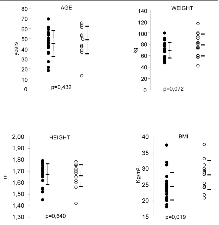
Table I shows the statistical data regarding age, hei-ght, weihei-ght, and body mass index. The group with disease had a tendency toward greater weight but with no
statisti-cal significance (68.83±13.54 kg versus 78.53±19.17 kg; p=0.072).
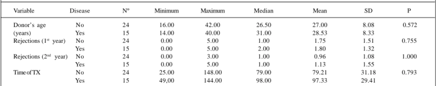
Body mass index was an independent risk factor for developing allograft vasculopathy, and the patients with di-sease showed higher values than the patients in the other group (28.11±4.60 kg/m2 versus 24.53±4.36 kg/m2; p=0.019). Other variables, such as donor’s age and number of re-jection episodes during the first and second postoperative years showed no statistically significant differences between the two groups (Table II). In regard to time of trans-plantation, the values obtained were 79.21±31.18 months for the group without disease versus 97.33±29.41 months for the group with disease (p=0.793).
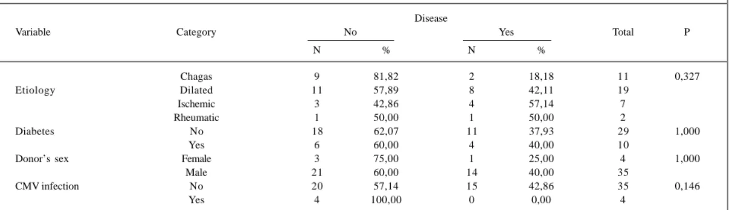
We observed a tendency toward higher levels of tri-glycerides in the group with allograft vasculopathy as com-pared with the patients with normal coronary angiography (p=0.097). The values of systolic and diastolic blood pressu-res, total cholesterol, HDL, and LDL are shown in table III, and no statistically significant differences were observed. When we analyzed the cause of pretransplantation basic cardiomyopathy in regard to the occurrence of allo-graft vasculopathy in individuals undergoing cardiac trans-plantation, we observed a predominance of patients with idiopathic dilated cardiomyopathy (8 patients), followed by ischemic patients (4), chagasic patients (2), and rheumatic patient (1). The prevalence of the disease was lower in cha-gasic patients; when comparing, however, the chacha-gasic group with the nonchagasic group in regard to develop-ment of allograft vasculopathy, we found no statistical sig-nificance (p=0.150). The presence of diabetes, the influence
Table I – Descriptive measures of the possible predictive factors according to the presence or absence of disease (allograft vasculopathy)
Variable Disease Nº Minimum Maximum Median Mean SD P
Age (anos) No 24 18.00 69.00 45.50 44.96 12.87 0.432
Yes 15 13.00 65.00 52.00 48.40 13.62
Height (m) No 24 1.45 1.79 1.68 1.67 0.09 0.640
Yes 15 1.42 1.78 1.68 1.66 0.10
Weight (kg) No 24 47.00 99.00 69.00 68.83 13.54 0.072
Yes 15 42.00 115.00 80.00 78.53 19.17
BMI (kg/m2) No 24 18.04 37.46 24.28 24.53 4.36 0.019
Yes 15 20.83 37.55 28.72 28.11 4.60
N – number of patients; BMI – body mass index; SD – standard deviation.
Table II – Descriptive measures of the possible predictive factors according to the presence or absence of disease (allograft vasculopathy)
Variable Disease Nº Minimum Maximum Median Mean SD P
Donor’s age No 24 16.00 42.00 26.50 27.00 8.08 0.572
(years) Yes 15 14.00 40.00 31.00 28.53 8.33
Rejections (1st year) No 24 0.00 5.00 1.00 1.75 1.51 0.755
Yes 15 0.00 5.00 2.00 1.80 1.32
Rejections (2nd year) No 24 0.00 3.00 1.00 0.96 1.08 1.000
Yes 15 0.00 5.00 1.00 1.13 1.55
Time of TX No 24 25.00 148.00 79.00 79.21 31.18 0.793
Yes 15 49,00 144.00 98.00 97.33 29.41
N – number of patients; SD – standard deviation; rejections (1st year) - episodes of rejection in the 1st year; rejections (2nd year) - episodes of rejection in the 2nd year;
of the donor’s sex, and posttransplantation cytomegalo-virus infection showed no influence on the development of allograft vasculopathy in either group (Table IV).
From the variables with a statistical trend or signifi-cance in the univariate analysis, we selected the following for the multivariate analysis: weight, body mass index, and triglycerides. The body mass index was the only predictive factor for developing allograft vasculopathy.
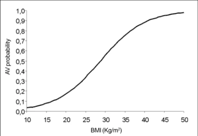
The logistic regression graph shows the probability of developing allograft vasculopathy as the body mass in-dex increases. At each unit increase in body mass inin-dex, the chance of developing disease increases 1.197 times. With an increase of 5 units in body mass index, the chance of de-veloping the disease increases 2.459 times (fig. 2).
Discussion
The real role of cellular rejection in the development of allograft vasculopathy still remains controversial. Helper T lymphocytes seem to play an important role in this process, as they are activated by class II HLA antigens, which are expressed in vascular endothelial cells and smooth muscle cells. When activated, these lymphocytes may also
produce interferon gamma, which stimulates the release of adhesion molecules leading to activation of macrophages, resulting in an increase in growth factors, such as interleu-kin-6, interleukin-1, and platelet-derived growth factors. These, when released, cause proliferation of smooth muscle cells, leading to a progressive luminal obstruction, characte-ristic of allograft vasculopathy. Another point not yet clari-fied is the influence of the number of rejection episodes and their consequent participation in the development of allograft vasculopathy. Uretsky et al 15 have found a relation between allograft vasculopathy and the occurrence of two or more rejection episodes. These data were corroborated by Radovancevic et al 20 when they analyzed the experience of the Texas Heart Institute; these results, however, were not confirmed by data of other study groups such as the one by Gao et al 21 at Stanford University. In the present series, we have found no statistically significant difference between the group with allograft vasculopathy and the group with a normal coronary angiography in regard to the number of rejection episodes. We should stress, however, that this observation takes into consideration that the biopsy is limited, reflecting a focal situation and not often representing the myocardium as a whole. Another
impor-Table III - Descriptive measures of the possible predictive factors according to the presence or absence of disease (allograft vasculopathy)
Variable Disease N Minimum Maximum Median Mean SD P
SBP (mmHg) No 24 120.00 160.00 147.50 144.38 11.16 0.326
Yes 15 120.00 170.00 150.00 148.33 11.29
DBP (mmHg) No 24 80.00 115.00 95.00 92.71 9.78 0.262
Yes 15 80.00 115.00 95.00 97.33 9.23
Cholesterol (mg/dL) No 24 131.00 279.00 185.00 192.79 40.98 0.173
Yes 15 138.00 322.00 202.00 215.67 62.08
HDL (mg/dL) No 24 27.00 66.00 45.00 44.67 10.61 0.869
Yes 15 22.00 116.00 42.00 45.73 23.33
LDL (mg/dl) No 24 60.00 184.00 111.50 117.00 33.86 0.318
Yes 15 77.00 246.00 105.00 130.73 51.11
TG (mg/dl) No 24 65.00 450.00 149.00 173.29 90.35 0.097
Yes 15 65.00 440.00 203.00 225.40 97.26
SBP – systolic blood pressure; DBP – diastolic blood pressure; HDL - HDL fraction of cholesterol; LDL - LDL fraction of cholesterol; TG - triglycerides; N – number of patients; SD – standard deviation.
Table IV – Distribution of disease (allograft vasculopathy) occurrence according to possible predictive factors
Disease
Variable Category No Yes Total P
N % N %
Chagas 9 81,82 2 18,18 11 0,327
Etiology Dilated 11 57,89 8 42,11 19
Ischemic 3 42,86 4 57,14 7
Rheumatic 1 50,00 1 50,00 2
Diabetes No 18 62,07 11 37,93 29 1,000
Yes 6 60,00 4 40,00 10
Donor’s sex Female 3 75,00 1 25,00 4 1,000
Male 21 60,00 14 40,00 35
CMV infection No 20 57,14 15 42,86 35 0,146
Yes 4 100,00 0 0,00 4
tant point is that the assessment of rejection episodes in stable out patients was performed with scintigraphy with gallium-67 and, occasionally, one episode of moderate rejec-tion may not have been diagnosed because of limitarejec-tions of the method. Humoral rejection also seems to play a role in the development of allograft vasculopathy through endo-thelial lesion. This concept is based on the presence of cyto-toxic antibodies directed against the HLA-DR antigen of the vascular endothelium, the relation of the presence of class I and II anti-HLA cytotoxic antibodies in patients with post-transplantation allograft vasculopathy, and the detection through immunofluorescence of immunoglobulins and
complement in the vascular endothelium 22,23. However, it is worth stressing that this type of rejection usually manifests with hemodynamic impairment and high mortality and, when we select the clinically stable patients with normal ventricular function for study, we may not be including the real representation of this type of rejection in the analysis. Hypertension may perhaps be the most common complication associated with the use of cyclosporine in patients undergoing cardiac transplantation, and its inci-dence has ranged from 50 to 100% in several series 15,24,25. The mechanisms involved in this process have not been to-tally clarified; cyclosporine, however, is known to cause a Fig. 1 – Mean values of the constitutional characteristics of the patients. ! no allograft vasculopathy; " allograft vasculopathy; BMI – body mass index.
AGE
WEIGHT
HEIGHT
BMI
state of avidity for sodium with a consequent decrease in its fraction of excretion. The association with its renal vascular effects, such as a reduction in renal blood flow and an in-crease in intrarenal vascular resistance, contributes to the state of hypertension7. Posttransplantation heart denerva-tion also seems to be involved in developing hypertension, because it causes loss of normal baroreflexes. From the prac-tical point of view, controlling the pressure levels of pa-tients is very difficult, despite the use of a large range of an-tihypertensive medications, probably because of its multi-factorial character added to the direct influence of immuno-suppressive medications, such as cyclosporine and predni-sone. In the general population, the influence of arterial hy-pertension on coronary events is well known. However in the population of cardiac transplantation patients, this rela-tion has not yet been shown, which confirms our results. It is important to stress that in our study, the pressure levels obtained in the two groups may have been influenced by the use of antihypertensive medication, as most of the pa-tients were using this class of drug during the study.
Cytomegalovirus infection has also been studied to better understand its real role in the genesis of conventional atherosclerosis and, more specifically, of posttransplanta-tion allograft vasculopathy 26. This infectious agent, which belongs to the herpes virus group, has an incidence ranging from 30 to 100% in patients awaiting cardiac transplantation, depending mainly on cultural and social aspects of each country. In the United States, the incidence of positive sero-logy for cytomegalovirus is 60 to 70% of the population 27; in Brazil, it reaches more than 90% 28. The mechanism by which cytomegalovirus may lead to atherosclerosis has not been well established; however, it is believed to stimulate the proliferation of smooth muscle cells and to cause procoa-gulant changes in the endothelium, contributing to the formation of atherosclerotic plaque 29. The first report on the association of cytomegalovirus and allograft vasculo-pathy was by Grattan et al 30 at Stanford University. Later, other groups showed the same association and since then the pretransplantation use of ganciclovir has been
propo-sed for prophylaxis in patients with negative serology for cytomegalovirus 31,32. Clinical manifestation of cytomega-lovirus infection may be found both in seronegative pa-tients, the so-called primary infection, and seropositive patients, the so-called clinical reactivation. Reactivation is usually more benign and has a mild and self-limited fever; primary infection, however, may present as a systemic illness characterized by fever, leukopenia, gastrointestinal involvement, pneumonitis, and has high morbidity and mortality. The risk factors for severe infections by cytome-galovirus are believed to be the following: seronegative in-dividuals who receive an organ from a seropositive donor, and patients requiring high doses of immunosuppressants for treating refractory rejections or as an inducing therapy with lymphocytolytic agents. As almost our entire popula-tion of patients is seropositive for cytomegalovirus in the pretransplantation period, we have chosen to evaluate only those with suggestive clinical findings, followed by a sero-logic confirmation or by antigenemia values higher than 10 cells, which happened in 4 patients. These patients were then treated with ganciclovir and had a good clinical evolu-tion. Coronary angiography revealed that these patients did not have allograft vasculopathy, i.e., in our series we found no relation between this risk factor and the development of allograft vasculopathy. These results, however, await con-firmation with studies involving a greater number of pa-tients and comparison with a subpopulation of seronega-tive patients in the pretransplantation assessment.
Another very important point in the context of the risk factors for developing allograft vasculopathy is dyslipide-mia. Its influence in atherosclerosis and, more precisely, in conventional coronary artery disease is well established; therefore, we currently use all resources for the appropriate control of cholesterol and triglyceride levels, mainly in pa-tients at higher risk. In papa-tients undergoing cardiac trans-plantation, dyslipidemia also seems to exert an influence on the multifactorial context, which culminates in allograft vas-culopathy. The levels of LDL-cholesterol and triglycerides begin to increase in the second postoperative week and reach a peak around the 3rd to the 6th month. Immunosup-pressants, mainly cyclosporine and corticosteroids, seem to directly contribute to posttransplantation dyslipidemia 33-37. Prednisone works by increasing the hepatic production of apolipoprotein B, predisposing the patient to insulin resis-tance, reducing the activity of the lipoprotein lipase, and decisively contributing to the weight gain observed in pa-tients in the postoperative period 38. Cyclosporine inhibits the hepatic clearance of prednisone and HDL-cholesterol because of its hepatotoxic effect, which results from its inte-raction in the P-450 cytochrome system 39. Kobashigawa et al 40 have shown that the use of pravastatin, an HMG-CoA reductase inhibitor, affects cholesterol reduction and de-creases the incidence of allograft vasculopathy. Because of the beneficial evidence of treating posttransplantation dyslipidemia, the introduction of drugs from the HMG-CoA reductase inhibitor group has met the same criteria used for the general population, i.e., LDL-cholesterol levels higher Fig. 2 – The logistic regression graph shows the relation between body mass
index (BMI) and the probability to develop allograft vasculopathy (AV).
BMI (Kg/m2)
A
V
than 130mg/dL 41. In our case series, we have found no relation between the levels of total cholesterol, HDL-cho-lesterol, and LDL-cholesterol in the groups with allograft vasculopathy and normal angiography. However, we have observed a tendency toward higher levels of triglycerides in the first group. When analyzing these results, we should consider that the patients used medication for hypercholes-terolemia on a routine basis, especially HMG-CoA reduc-tase inhibitors, which were introduced according to labora-tory results to maintain total cholesterol levels lower than 200mg/dL and LDL-cholesterol levels lower than 130mg/dL; these drugs were not suspended during the study. These drugs have an effect mainly on cholesterol; their effect on triglycerides is poor, as shown in a study by Kobashigawa et al 42. This point and the greater weight gain in the group with allograft vasculopathy partially explains the results observed in the triglycerides. A further analysis, however, with a larger group of patients and with no influence from medications may help in the future to understand the real role of the lipids in the development of posttransplantation allograft vasculopathy.
In the present study, the risk factor that showed the greatest correlation with allograft vasculopathy, both in the univariate and multivariate analyses, was body mass index, which better reflected the actual nutritional status of the patient. In studies with critically ill patients in intensive care units when we considered the body mass index, we obser-ved that malnourished and cachectic patients had a higher mortality index as compared with patients with normal body mass index 43. On the other hand, high levels of body mass index also correlate with an increase in the risk of cardiovas-cular events 44. Winters et al 45 and Hauptman et al 46 have
found a direct relation between the elevated levels of body mass index (>29kg/m2) and allograft vasculopathy in pa-tients undergoing cardiac transplantation. In our series, we have found moderate levels of body mass index compatible with grade I obesity in patients with allograft vasculopathy. Some factors seem to contribute to obesity evident in patients after cardiac transplantation. The immuno-suppressive therapy, mainly corticosteroid, directly influ-ences weight gain, as does the physical inactivity observed, despite the significant improvement in clinical findings and in functional capacity after transplantation. The nutritional conditions also improve because of a better absorption of nutrients due to reversion in heart failure and an increase in appetite, due to the use of corticosteroids. The actual role of body mass index in the context of allograft vasculopathy still awaits further clarification that goes beyond a simple assessment of the nutritional status. This index may be a marker for metabolic changes that may be occurring in patients with a greater predisposition for developing al-lograft vasculopathy in the late posttransplantation follow-up. Therefore, while these questions await clarification, so-me instructions should be provided to patients aiming to m-inimize these complications. Encouraging the practice of physical exercise, alimentary control, and attempts at wi-thdrawal of the corticosteroid after the 6th postoperative month may be useful measures for attaining this aim. Because allograft vasculopathy is multifactorial, the strict control of risk factors is extremely important, so that in the future with a better understanding of the actual physiopathological mechanism of this complication we will be able to interfere with its incidence and evolution, and, consequently, provide the cardiac transplantation patient with a longer survival.
1. Lower RR, Kontos HA, Kosek JC, Sewell DH, Graham WH. Experiences in heart transplantation. Am J Cardiol 1968; 22: 766-71.
2. Thomson JG. Production of severe atheroma in a transplanted heart. Lancet 1969; 2: 1088-92.
3. Hosenpud J, Mauck K, Hogan K. Cardiac allograft vasculopathy: IgM antibody responses to donor-specific vascular endothelium. Transplantation 1997; 63: 1602-06.
4. Fiorelli A, Stolf N, Graziosi P, et al. Incidência de coronariopatia após o trans-plante cardíaco ortotópico. Rev Bras Cir Cardiovasc 1994; 9: 69-80. 5. Gao SZ, Alderman EL, Schroeder JS, et al. Clinical and laboratory correlates of
ac-celerated coronary artery disease in the cardiac transplant patient. Circulation 1987; 76: 56-61.
6. Grattan MT, Moreno Cabral CE, Starnes VA, Oyer PE, Stinson EB, Shumway NE. Eight year results of cyclosporine treated patients with cardiac transplants. J Thorac Cardiovasc Surg 1990; 99: 500-09.
7. Greenberg ML, Uretsky BF, Reddy PS, et al. Long term hemodynamic follow-up of cardiac transplant patients treated with cyclosporine and prednisone. Circu-lation 1985; 71: 487-94.
8. Bocchi E, Vilas-Boas F, Pedrosa AA, et al. Doença coronariana após transplante cardíaco ortotópico. Arq Bras Cardiol 1994; 62: 195-200.
9. Pethig K, Besser K, Heublein B, Wahlers T, Harringer W, Haverich A. Coronary vasculopathy after heart transplantation – effect os temporal onset, severity and pro–gression on long-term prognosis. Z Kardiol 1999; 88: 498-506. 10. Lindelow B, Bergh C, Lamm C, Andersson B, Waagstein F. Graft coronary artery
disease is strongly related to the aetiology of heart failure and cellular rejection. Eur Heart 1999; 20: 1326-34.
References
11. Halle AA, Disciascio G, Massin EK, et al. Coronary angioplasty, atherectomy and bypass surgery in cardiac transplant recipients. J Am Coll Cardiol 1995; 26: 120-8.
12. Davies H, Al-Tikriti S. Coronary arterial pathology in the transplanted human heart. Intern J Cardiol 1989; 25: 99-118.
13. Billingham ME, Cary NRB, Hammond ME, et al. A working formulation for the standartization of nomenclature in the diagnosis of heart and lung rejection: he-art rejection study group. J Hehe-art Transplant 1990; 9: 587-601.
14. Sones FM, Shirey EK, Prondfit WL, Westcott RN. Cine-coronary arteriography. Circulation 1959; 20: 773.
15. Uretsky BF, Murali S, Reddy S, et al. Development of coronary artery disease in cardiac transplant patients receiving immunosupressive therapy with cyclos-porine and prednisone. Circulation 1987; 76: 827-34.
16. Smart FW, Ballantyne CM, Cocanougher B, et al. Insensitivity of noninvasive tests to detect coronary artery vasculopathy after heart transplant. Am J Cardiol 1991; 67: 243-7.
17. Hardesty RL, Griffith BP, Debsky RF, et al. Experience with cyclosporine in cardiac transplantation. Transplant Proc 1983; 15(suppl 1): 2553-8. 18. Hosmer DW, Lemeshow S. Applied Logistic Regression. John Wiley & Sons,
1976: 75-118.
19. SAS Institute Inc., SAS/STATâ User’s Guide. Version 6, Fourth Edition, volume 1. Cary, NC: SAS Institute Inc., 1989.
21. Gao SZ, Schroeder JS, Hunt SA, Valantine HA, Hill IR, Stinson EB. Influence of graft rejection on incidence of accelerated graft coronary artery disease: a new ap-proach to analysis. J Heart Lung Transplant 1993; 12: 1029-35.
22. Costanzo-Nordin MR. Cardiac allograft vasculopathy: relationship with acute cellular rejection and histocompatibility. J Heart Lung Transplant 1992; 11: 90-103.
23. Rose EA, Pepino P, Barr ML, Smith CR, Ratner AJ HO E, Berger C. Relation of HLA antibodies and graft atherosclerosis in human cardiac allograft recipients. J Heart Lung Transplant 1992; 11: 120-3.
24. Thompson ME, Shapiro AP, Johnsen AM, et al. The contrasting effects of cy-closporin A and azathioprine on arterial blood pressure and renal function follo-wing cardiac transplantation. Int J Cardiol 1986; 11: 219-29.
25. Olivari MT, Antolick A, Ring WS. Arterial hypertension in heart transplant re-cipients treated with three-drug immunosupressive therapy. J Heart Transplant 1989; 8: 34-9.
26. Hosenpud JD. Coronary artery heart transplantation and its relation to cytome-galovirus. Am Heart J 1999; 138 (5 Pt 2): 469-72.
27. Melnick JL, Adam E, Debakey ME. Cytomegalovirus and atherosclerosis. Eur Heart J 1993; 14: 30-8.
28. Camargo LF. A importância da sorologia, antigenemia e reação em cadeia por po-limerase no diagnóstico da infecção pelo citomegalovírus após-transplante cardíaco. Tese de Doutorado) – Faculdade de Medicina da Universidade de São Paulo. São Paulo, 1998: 163p.
29. Smiley ML, Mar EC, Huang ES. Cytomegalovirus infection and viral-induced transformation of human endothelial cells. J Med Virol 1988; 25: 213-26. 30. Grattan MT, Moreno-Cabral CE, Starnes VA, Oyer P, Stinson EB, Shumway NE.
Cytomegalovirus infection is associated with cardiac allograft rejection and atherosclerosis. J Am Med Assn 1989; 261: 3561-6.
31. Wu TC, Hruban RH, Ambinder RF, et al. Demonstration of cytomegalovirus nu-cleic acids in the coronary arteries of transplanted hearts. Am J Pathol 1992; 140: 739-47.
32. Merigan TC, Renlund DC, Keay S, et al. A controlled trial of ganciclovir to pre-vent cytomegalovirus disease after heart transplantation. N Engl J Med 1992; 326: 1182-6.
33. Keogh A, Simons L, Spratt P, et al. Hyperlipidemia after heart transplantation. J Heart Transplant 1988; 7: 171-5.
34. Becker DM, Chamberlain B, Swank R, et al. Relationship between corticosteroid ex-posure and plasma lipid levels in heart transplant recipients. Am J Med 1988; 85: 632-8. 35. Chang G, Denofrio D, Desai S, et al. Lipoprotein(a) levels and heart
transplanta-tion atherosclerosis. Am Heart Journal 1998; 136(suppl 2): 329-34. 36. Maranhão R, Santos RD, Furlaneto C, et al. Lipoproteína (a), apolipoproteínas e
perfil lipídico em fase tardia após-transplante cardíaco Arq Bras Cardiol 1994; 63: 465-8.
37. Bellotti G, Bocchi E, Goiato MAC, et al. Alterações do perfil lipídico na evolu-ção tardia após transplante cardíaco. Arq Bras Cardiol 1996; 66: 263-6. 38. Renlund DG, Bristow MR, Crandall BG, et al. Hypercholesterolemia after heart
tranplantation: Amelioration by corticosteroid-free maintenance imunossupres-sion. J Heart Transplant 1989; 8: 214-20.
39. Ballantyne CM, Podet EJ, Patsch WP, et al. Effects of cyclosporine therapy on plasma lipoprotein levels. JAMA 1989; 262: 53-6.
40. Kobashigawa JA, Katznelson S, Laks H, et al. Effect of pravastatin on outcomes after cardiac transplantation. N Engl J Med 1995; 333: 621-7.
41. II Consenso Brasileiro sobre Dislipidemias. Arq Bras Cardiol 1996; 67: 1-16. 42. Kobashigawa JA, Murphy FL, Stevenson WL, et al. Low-dose lovastatin safely
lowers cholesterol after cardiac transplantation. Circulation 1990; 82: 281-3. 43. Galanos AN, Pieper CF, Kussin PS, et al. Relationship of body mass index to
sub-sequent mortality among seriously ill hospitalized patients. Crit Care Med 1997; 25: 1962-75.
44. Hubert HB, Feinleib M, McNamara PM, Castelli WP. Obesity as na independent risk factor for cardiovascular disease: a 26 year follow-up of participants in the Framingham Heart Study. Circulation 1983; 67: 968-77.
45. Winters GL, Kendall TJ, Stanley J, et al. Posttransplant obesity and hyperlipi-demia: Major predictors of severity of coronary arteriopathy in failed human heart allografts. J Heart Transplant 1990; 9: 364-71.