1 5 6
Croti et al
Coronary bilateral ostial enlargement
Arq Bras Cardiol volume 74, (nº 2), 2000
Instituto do Coração do Hospital das Clínicas – FMUSP
Mailing address: Ulisses A. Corti – Incor – Av. Dr. Enéas C. Aguiar, 44 – 2o- S/10
– 05403-900 – S. Paulo, SP – Brazil Received on 6/24/99
Accepted on 9/15/99
Ulisses A. Croti, Francisco Gregori Jr, Miguel B. Marcial, Luís A. Dallan, Thelma Elisa F. Gregori, Divina S. O liveira
São Paulo, SP - Brazil
Coronary Bilateral Ostial Enlargement Using the Saphenous
Vein in a Patient with Syphilitic Aortitis
Case Report
A patient with tertiary syphilis presenting with bilate-ral coronary ostial lesions and aortic regurgitation under-went surgical reconstruction of the coronary ostia by the an-terior approach with autogenous saphenous vein grafting and substitution of the aortic valve with a bovine bioprosthe-sis. The procedure was easily performed and had good out-comes both early and late. The rarity of the association of a lesion in both coronary ostia with aortic regurgitation in sy-philis and the surgical technique employed are discussed.
Syphilis has been succesfully controlled in recent ye-ars; involvement of the heart and the aorta have become rela-tively rare events. The incidence and degree of seriousness of syphilitic aortitis is greater in the initial portion of the as-cending aorta involving the coronary ostia and the valvulae of the aortic valve. Heggtveit 1, in a clinicopathological review
of syphilitic aortitis, noted uni or bilateral coronary ostial stenosis with aortic regurgitation in 14% of patients.
Several surgical tactics for the correction of coronary ostial stenosis have been reported: endarterectomy 2-5,
aorto-coronary bridging with either arterial 6,7 or venous 4
grafts or both , and ostial reconstruction via the anterior 3,8
or posterior 8,9 approaches.
We have performed surgical treatment of a patient pre-senting with bilateral coronary lesions and aortic regurgitati-on by ostial amplificatiregurgitati-on via the anterior approach. Alregurgitati-ong with the aortic valve substitution, we used grafting with auto-genous saphenous vein to reconstruct both coronary ostia.
Case Report
A forty-eight-year old male Caucasian patient related a history of two months of intense nonirradiating precordial pain unrelated to effort, dyspnea, throbbing and decreased visual accuity. In his past medical history, he reported seve-ral episodes of veneseve-ral disease but denied the existence of familial heart disease.
Upon examination, the patient was in good physical condition, eupneic, with arterial blood pressure of 16/40mm-Hg in the right upper limb and 70/40mm16/40mm-Hg in the left upper limb; his left radial and arm pulses were of lower amplitude relative to the right, which showed strong pounding featu-re. The first heart sound was normal, the second was of de-creased intensity, with a protomesodiastolic regurgitative murmur ++/++++ in aortic and accessory aortic areas.
The thoracic radiograph revealed aortic ectasia and a normal cardiac area. The electrocardiogram showed a sinu-sal rhythm, QRS axis AT + 30o, T wave negative at 4V,
flatte-ned at V5 and V6, Dl and the VL with left ventricular hyper-trophy. Dopplerechocardiography showed diffuse hypoki-nesia of the left ventricle and moderate aortic regurgitation with ectasia of the ascending aorta. A cinecoronariographic study showed 70% obstruction of the left coronary ostium, 60% of the right coronary ostium, serious aortic regurgita-tion and a critical lesion in the left subclavian artery that was treated by percutaneous transluminal coronary angioplasty at the time of diagnosis. Results of routine laboratory tests were normal with the exception of the Venereal Disease Re-search Laboratory test (VDRL), reactive at 1:64 dilution and positive fluorescent treponemal antibody-absorption test (FTA-ABS).
The surgical approach was performed by medial ster-notomy, aortic and right atrial cannulation, and extracorpo-real circulation under moderate hypothermia at 30oC.
Myo-cardial protection was afforded by St. Thomas crystalloid solution.
Arq Bras Cardiol volume 74, (nº2), 2000
Croti et al Coronary bilateral ostial enlargement
1 5 7 propitiating adequate amplification of the left coronary
ostium, which increased from one to 5mm.
Inspection of the right coronary ostium confirmed its stenosis (1.5 mm diameter). A T-shape incision was made starting at the lower border of the aortotomy, through the ostium, reaching near to the sinusal branch. From this point on, a graft of the autogenous saphenous vein was sutured until the transversal incision of the aorta was reached, per-mitting ostial amplification to approximately 4.5mm.
The aortic valve did not allow for repair surgery and was replaced with a bovine pericardial prosthesis fixed in glutaraldehyde and preserved in formaldehyde 23mm (Braile-Biomédica – São José do Rio Preto, SP - Brazil). The perfusion time was 85min; myocardial anoxia lasted 73min. During the first hours of the postoperative period, the patient’s electrocardiogram indicated a subepicardial lesion in the antero-septal wall, without hemodynamic alterations; cardiac catheterization was performed. Good functioning of the biological prosthesis and a significant amplification of the coronary ostia (figs. 1 and 2) with adequate flow to all coronary arteries was observed; the cause of the electrocar-diographic chnges was not identified.
Pathological analysis of fragments removed from the aorta showed atherosclerosis and intense lymphoblastic
infiltration in the medial and adventitial layers of the vessel (fig. 3) confirming a luetic cause.
The patient had no further intercurrent events, receiving his hospital discharge on the eighth day following the ope-ration, He was followed on an outpatient basis with antibiotic therapy adequate for the treatment of tertiary syphilis. After a three-year follow-up period, he does not have angina, and myocardial scintigraphy does not indicate signs of ischemia.
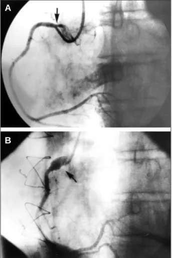
Fig. 1 – Right coronary – preoperative cinecoronariography: significant stenosis of the coronary ostium is observed; postoperative cinecoronario–graphy demonstra-tes adequate ostial amplification.
A
B
Fig. 2 – Left coronary – preoperative cinecoronariography: significant stenosis of the coronary ostium is observed; postoperative cinecoronariography demonstrates adequate ostial amplification.
A
B
1 5 8
Croti et al
Coronary bilateral ostial enlargement
Arq Bras Cardiol volume 74, (nº 2), 2000
Discussion
Syphilis affects the cardiovascular system in various ways.Aortitis is the most severe and frequent outcome, whi-ch leads to the formation of aneurysms, aortic regurgitation and coronary ostial stenosis. Heggtveit 1 in a study of 100
patients with aortic syphilis found 26 cases of ostial stenosis, 14 of which were associated with aortic regurgitation.
The first surgical disobstruction of coronary ostia due to syphilitic disease, was performed by endarterectomy in 1959 2. Conolly et al 5 in 1964 reported a further case of
endarterectomy of the right coronary ostium associated with valve repair due to aortic regurgitation.
Ostial lesions conventionally have been treated with myocardial revascularization techniques 4,6,7, including
ei-ther the left or the right internal thoracic artery or both of these, or their combination with saphenous vein grafting. This surgical option can be inconvenient because of coro-nary trunk occlusion, competitive flow, the corocoro-nary steal phenomenon, retrograde perfusion over an extensive myo-cardial area and the requirement for future surgical inter-ventions, especially in young patients.
Ostial reconstructive surgery was first reconsidered after 1983 when Hitchcock et al. 9 obtained favorable results by
approaching the coronary ostium and trunk, restoring phy-siological perfusion of the myocardium. The authors used the posterior approach to the aorta with an incision through its floor and ostial amplification with a segment of the autogenous saphenous vein. This technique was modified by others 3 who
aimed to control eventual bleeding in the suture area.
Sullivan and Murphy 3 advocated repair surgery by
the anterior approach with autogenic saphenous vein grafting rather than using the posterior approach, because it presents a more frontal vision and greater ease of execution. Dion et al 8 reported on a series of patients who
underwent ostial repair by the anterior and posterior approaches, using the saphenous vein and the autoge-nous pericardium as grafting materials. Bortolotti et al 10
described 4 patients with ostial coronary lesions treated by surgical repair; one of the patients had bilateral ostial lesions of syphilitic origin: amplification was made with the autologous pericardium.
In the present case, the surgical approach by the an-terior method using saphenous vein grafts for the re-construction of both coronary ostia, a rarely used proce-dure in luetic aortitis, was employed. This technique appeared to be the most adequate form of re-establishing coronary flow in this patient, because it permited the am-plification of stenosed ostia and avoided possible com-plications associated with classical myocardial revas-cularization.
We believe that our choice was adequate, yielding ex-cellent early and late post- operative results and adding a further surgical alternative to the treatment of ostial lesions of syphilitic origin.
Acknowlegment
We thank Dr. Danton R. Loures for bibliographic assistance.
1. Heggtveit HA. Syphilitic Aortitis. A Clinicopathologic Autopsy Study of 100 Cases, 1950 to 1960. Circulation 1964; 29: 346-55.
2. Dubost CH, Blondeau PH, Piwnica A, et al. Syphilitic coronary obstruction: correction under artificial heart-lung and profound hypothermia at 10ºC. Surge-ry 1960: 540-7.
3, Sullivan JA, Murphy DA. Surgical repair of stenotic ostial lesions of the left main coronary artery. J Thorac Cardiovasc Surg 1989; 98: 33-6.
4. Yamada T, Sakamoto T, Asano K. Surgical treatment of syphilitic coronary ostial stenosis with aortic regurgitation. J Cardiovasc Surg 1983; 24: 222-6. 5. Connolly JE, Eldrigde FL, Calvin JW, Stemmer EA. Proximal Coronary-Artery
Obstruction – Its Etiology and Treatment by Transaortic Endarterectomy. N Engl J Med 1964; 271: 213-9.
References
6. Vijayanagar R, Bognolo D, Eckstein P, et al. Safety and efficacy of internal mam-mary artery grafts for left main coronary artery disease. J Cardiovasc Surg 1987; 28: 576-80.
7. Ogino K, Tokuyasu Y, Motomiya T, Sugiura M, Endo M. Bilateral Coronary Os-tial Stenosis Associated with Aortitis Syndrome. Chest 1991; 99: 1286-7. 8. Dion R, Verhelst R, Matta A, Rosseau M, Goenen M, Chalant C. Surgical
angioplas-ty of the left main coronary artery. J Thorac Cardiovasc Surg 1990; 99: 241-50. 9. Hitchcock JF, Robles de Medina EOR, Jambroes G. Angioplasty of the left main
coronary artery for isolated left main coronary artery disease. J Thorac Cardiovasc Surg 1983; 85: 880-4.