1
Arquivos Brasileiros de Cardiologia - Volume 84, Nº 4, Abril 2005
Case Report
Transient Ventricular Dysfunction
(Takotsubo Cardiomyopathy)
José Marconi Almeida de Sousa, Marcos Knobel, Gustavo Buchelle,
José Augusto M. de Sousa, Cláudio H. Fisher, Daniel Born, Nelson Akamine, Elias Knobel
São Paulo, SP - Brazil
Hospital Israelita Albert Einstein
Mailing address: José Marconi Almeida de Sousa - Rua Vicente Felix, 60/121 - Cep 01410-020 - São Paulo, SP, Brazil
E-mail: [email protected], [email protected] Sent for publication: 05/28/2004
Accepted for publication: 09/29/2004 English version by Stela Maris Costalonga
The patient was a male with myasthenia gravis, hospitalized with acute respiratory failure due to decompensation of the underlying disease. He evolved with findings suggestive of acute myocardial infarction, with electrocardiographic and enzymatic alterations compatible with that diagnosis. The patient under-went emergency coronary angiography, which showed no severe coronary obstruction, although his left ventricle had significant systolic dysfunction with characteristic alterations, on ventricu-lography, of the syndrome described as transient ventricular dysfunction or Takotsubo syndrome. On evolution, complete re-covery of the electrocardiographic alterations and systolic ven-tricular function assessed on echocardiography occurred, con-firming the syndrome.
Left ventricular systolic dysfunction is the final result of any pathological process affecting the heart in its many structures. It often results from primary muscular impairment with consequent heart failure. In this situation, myocardial damage is usually irre-versible and prolonged. In some clinical situations, reirre-versible ven-tricular systolic dysfunction has been well described, its prototype being the myocardial dysfunction of sepsis 1. Recently, other
si-tuations have been reported evolving with myocardial dysfunction, also in a reversible form, but with peculiarities that distinguish them from septic dysfunction, mainly due to their characteristic clinical presentation, suggestive of acute myocardial infarction. Initially described in Japan 2, the Takotsubo (a bottle used for
trapping octopuses in Japan) syndrome or Takotsubo cardiomyopathy has been increasingly identified in daily clinical practice, including its important complications 3. We report a case of that syndrome
in a patient with respiratory failure of muscular etiology, which evolved with cardiogenic shock.
Case Report
The patient was a 64-year-old male with myasthenia gravis for 1 year, an ex-smoker, who had quit smoking 20 years earlier and had a familial history of coronary artery disease. The patient, who
regularly used pyridostigmine, was hospitalized due to worsening of his neurological disease, with intense and generalized muscular weakness and clinical and gasometrical signs of hypoventilation.
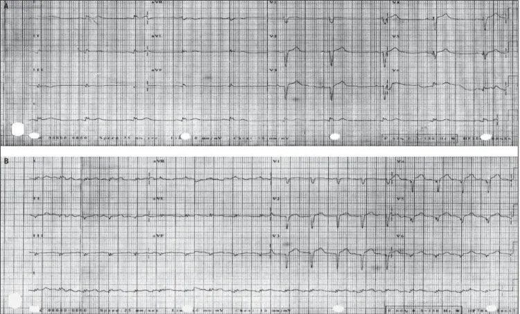
Initially, the patient underwent plasmapheresis sessions, with no significant improvement. At the ward, he experienced chest discomfort and worsening of the dyspnea, being transferred to the intensive care unit, where he underwent electrocardiography that showed an ST segment elevation of approximately 2 mm from V1 to V6 with amputated R from V1 to V6 (fig. 1A).
Blood gas analysis showed significant hypoventilation (PCO2= 105 mm Hg) with severe respiratory acidosis (PH=7.07; HCO3= 29.9 mEq/L). The electrolytes and renal function were normal; troponin I was 5.0 ng/dL (Nl=up to 1.0 ng/dL); and CKMb was 22 ng/dL (N=up to 3 ng/dL). The patient was intubated, put under mechanical ventilation, and underwent emergency cine coronary angiography. The examination showed that the left main coronary artery, the anterior descending artery, and the right coronary artery had no significant atherosclerotic lesions, and the left marginal branch of the circumflex artery had a 60% atherosclerotic lesion (fig. 2). The left ventricle had akinesia of the anteroapical wall and an ejection fraction of 33% (fig.3)
The patient evolved with arterial hypotension, which required 0.3µg/kg/min of noradrenaline. A catheter was passed through
the pulmonary artery, showing a cardiac index of 2.1 L/min and a pulmonary artery occlusion pressure of 12 mm Hg. On that occa-sion, his arterial lactate level was 31 mg/dL (normal, up to 22 mg/dL), the electrocardiogram showed an inactive zone from V1 to V6 (fig. 1B), and the echocardiogram showed apical dyskinesia, akinesia of the middle segment of all walls, and hypokinesia of the basal segment, mainly of the anterior and septal walls, with an ejection fraction of 20%. Dobutamine, at the dosage of 5 µg/
kg/min, was initiated.
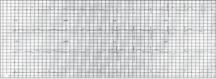
In the subsequent 24 hours, the patient evolved favorably. His blood pressure levels returned to normal with low doses of nora-drenaline, and his lactate level normalized. He had an enzymatic curve typical of acute myocardial infarction with maximum CKMB and troponin levels of 22.3 ng/dL and 9.6 ng/dL, respectively. A new echocardiogram showed an improvement in the ejection frac-tion to 37%. The electrocardiogram showed recovery of the R wave and an alteration in the ventricular repolarization of the anterior wall. After 48 hours, noradrenaline could be suspended, and, after 72 hours, dobutamine.
2
Arquivos Brasileiros de Cardiologia - Volume 84, Nº 4, Abril 2005
Transient Ventricular Dysfunction (Takotsubo Cardiomyopathy)
recovery of the electrocardiographic changes (fig. 4). After stabi-lization of the acute phase, the patient was transferred to another service, where, after one month, he died due to multisystem organ failure secondary to sepsis.
Discussion
The patient reported thoracic discomfort of acute onset, which evolved with electrocardiographic alterations compatible with acute myocardial infarction with an ST segment elevation, accompanied by severe left ventricular dysfunction. Complete spontaneous reversion of those alterations occurred after 10 days. This clinical cardiac syndrome was first described in 1990 by Satoh et al 2, and, in
1991, by Dote et al 4. On that occasion it was called
Takotsubo-type cardiomyopathy, due to the similarity, on ventriculography, to a device used for trapping octopuses in Japan. Most cases were described in that country, and this is the first case reported in Brazil under that name. In 2000, Franken et al 5 reported on a
similar case, which they called “myocardial pseudo-infarction during an episode of herpes zoster”; these authors, however, did not charac-terize their case as the one here reported.
The etiology and clinical characteristics of that syndrome have not been completely clarified. However, in a recent review, Tsu-chihashi et al 6 retrospectively studied 88 patients with that
diag-nosis. In that series, several conditions triggered the syndrome. Psychological factors were observed in approximately 20% of ca-ses, exacerbation of a systemic disease in 3%, neurogenic causes in 7%, pulmonary causes in 8%, gastrointestinal disorders in 7%, renal disease in 6%, and other unspecific factors in 23 (15%) patients. None of the patients had myasthenia gravis, and the number of women was 6 times greater than that of men. Systemic arterial hypertension was the most frequent (48%) coronary risk factor. The only risk factor of our patient was hypertension, although he was an ex-smoker. Initially, most patients (67%) had precordial discomfort, 56% had an enzymatic elevation, and 90% had an ST segment elevation in the acute phase. Persistent Q waves occurred in only 10%. Cardiogenic shock was reported in 15%, use of intra-aortic balloon in 8%, and dopamine or dobutamine, or both, in 19%. In-hospital mortality was 1%. Only one patient had heart failure on hospital discharge. According to the criteria of the MONICA project 7, 39% of the patients met 3 criteria for
acute myocardial infarction, and in 43% of patients that diagnosis
Fig. 2 - Left coronary artery in the cranial RAO and LAO views, showing coronary arteries with no severe obstructive lesions.
Fig. 3 - Left ventricle in the RAO view in final diastole and systole, resembling the device used for trapping octopuses in Japan (Takotsubo).
Fig. 1 - A) Electrocardiogram showing an ST-segment elevation from V1 to V6 with potential loss in that region and B) another 20 m after, with QS.
A
3
Arquivos Brasileiros de Cardiologia - Volume 84, Nº 4, Abril 2005
Transient Ventricular Dysfunction (Takotsubo Cardiomyopathy)
1. Levy RJ, Deutschman CS. Evaluating myocardial depression in sepsis. Shock 2004; 22:1-10.
2. Satoh H, Tateishi H, Uchida T et al. Takotsubo-type cardiomyopathy due to mul-tivessel spasm. In: Kodama K, Haze K, Hon M, editors. Tokyo Kagakuhyouronsya Co 1990;56-64.
3. Akashi YJ, Tejima T, Sakurada H et al. Left ventricular rupture associated with Ta-kotsubo cardiomyopathy. Mayo Clin Proc 2004;79:6821-4.
4. Dote K, Sato H, Tateishi H et al. Myocardial stunning due to silmultaneous multi-vessel spasms: a review of five cases. J Cardiol 1991;21:203-14.
5. Franken R & Franken M. ´Pseudo-infarto do miocárdio durante episódio de her-pes zoster´. Arq Bras Cardiol 2000;75:523-6.
6. Tsuchihashi K, Ueshima K, Uchida T et al. Transient left ventricular apical
balloo-References
ning without coronary artery stenosis: a novel heart syndrome mimicking acute myocardial infarction. J Am Coll Cardiol 2001;38:11-8.
7. Tunstall-Pedoe H, for the WHO MONICA Project Principal Investigators. The World Health Organization MONICA project (monitoring trends and determinants in car-diovascular disease): a major international collaboration. J Clin Epidemiol 1988;41:105-14.
8. Antman E, Bassand JP, Klein W et al. Myocardial infarction redefined a consensus document of The Joint European Society of Cardiology/American College of Cardio-logy committee for the redefinition of myocardial infarction J Am Coll Cardiol 2000;36:959-69.
9. Kawai S, Suzuki H, Yamaguchi H et al. Ampulla cardiomyopathy (“Takotsubo car-diomyopathy”) with ST segment elevation. Jpn Circ J 2000;64:156-9. Fig. 4 - Electrocardiogram showing complete recovery of the R waves in the anterior wall.
was considered probable. Our patient met the criteria of the MONICA project for acute myocardial infarction 7 and the new
guidelines of the American Heart Association 8.
Complete recovery of our patient’s ejection fraction was simi-lar to that in the series described in this review, in which the mean ejection fractions in the acute and late phases were 41%± 11% and 64%±10%, respectively.
The exact mechanism responsible for the transient ventricular alteration has not yet been elucidated. Thus, similarly to the cases reported by Tsuchihashi et al 8, in our patient, early coronary
angiography did not show total arterial occlusion, which, therefore, did not characterize a situation that could be explained by stunning or hibernating myocardium.
One peculiarity of the syndrome described is that its triggering is related to some extremely variable factors, stress being one of
them. All stressful situations show an elevation in catecholamines that can also cause ventricular dysfunction, such as in pheochro-mocytoma. In none of these cases were the catecholamines as-sessed; however, in a case reported by Kawai et al 9, myocardial
alterations similar to those of catecholamine-induced cardiomyo-pathy were found.