O
r i g i n a la
rt i c l e3 6 3 Arq Bras Oftalmol. 2016;79(6):363-5 http://dx.doi.org/10.5935/0004-2749.20160103
INTRODUCTION
Keratoconus (KC) is a noninflammatory eye condition characte-rized by progressive corneal ectasia, myopia, and irregular astigma-tism, generally with bilateral involvement. The etiology and pro-gression of keratoconus have been closely linked to genetic factors, which appear to be multifactorial. However, the majority of cases are not linked to genetic factors(1,2). It is important to detect progression in KC before making collagen cross-linking (CXL).
The Valsalva maneuver (VM) is an attempt at a powerful exhalation
against a closed airway.VM is frequently performed during normal
daily activities: examples include lifting heavy items, performing phy-sical exercise, vomiting, and coughing. Various physiological changes happen during these activities, including elevated blood pressure, in -creased intrathoracic pressure, in-creased peripheral venous pressure,
ABSTRACT
Purpose: This study aimed to explore the effects of the Valsalva maneuver (VM) on ectatic corneas during anterior segment tomography scans using a Scheimpflug camera.
Methods: This prospective observational study included 100 eyes of 50 patients with bilateral keratoconus (KC). Anterior segment tomography was first performed when the patients were in a resting position and immediately repeated as the participant performed VM. Scheimpflug images were taken using a Pentacam®.
Results: The mean age of the participants was 24.14 ± 6.59 years. Of the 100 eyes included in the study, 7% had stage 1 KC, 47% had stage 2 KC, 32% had stage 3 KC, and 14% had stage 4 KC. The indices of KC were not significantly affected by VM. Similarly, no statistically significant differences were found between the stages of KC, or with the mean pachymetric progression index during VM. Pupil diameter showed a statistically significant increase during VM (p=0.017). There was a statis-tically significant decrease in the anterior chamber angle measurement during VM (p=0.001). Maximum curvature power in the front of the cornea decreased more during VM in stage 4 KC than for the other stages (p=0.014).
Conclusions: No changes associated with VM were found in the KC indices or the stage of the disease. However, an increase in pupil diameter and a decrease in anterior chamber angle value were found. These changes were comparable to values obtained from previous studies performed on normal corneas.
Keywords: Anterior chamber/physiology;Keratoconus; Valsalva maneuver/phy-siology; Corneal topography
RESUMO
Objetivo: Este estudo tem como objetivo explorar os efeitos da manobra de Valsalva (VM) na córnea ectásica durante a varredura tomográfica do segmento anterior usan do a câmera de Scheimpflug.
Métodos: Foi conduzido estudo observacional, prospectivo envolvendo 100 olhos de 50 pacientes que apresentavam ceratocone (KC) bilateral. Tomografia do segmento anterior foi realizada inicialmente quando os pacientes estavam em posição de repouso e imediatamente depois, no curso de VM. Imagens de Scheimpflug foram feitas usando Pentacam®.
Resultados: A média de idade dos participantes foi 24,14 ± 6,59 anos de idade. Dos olhos incluídos no estudo, 7% apresentava KC estágio 1,47% apresentava estágio 2,32% apresentava estágio 3, e 14% apresentava estágio 4. Índices de KC não fo-ram significativamente afetadas pela VM. Não houve diferenças estatisticamente significativas com o estágio do KC, e o índice médio de progressão paquimétrica durante a VM. O diâmetro da pupila (PD) mostrou aumento estatisticamente signi-ficativo durante a VM (p=0,017). Houve diminuição estatisticamente significativa na medida do ângulo da câmara anterior durante a VM (p=0,001). O poder máximo de curvatura anterior da córnea no KC estágio 4 diminuiu mais do que os outros estágios durante o VM (p=0,014).
Conclusões: Não foram encontradas alterações nos índices KC e no estágio da doença por causa da VM. Verificou-se que houve aumento na PD e uma diminuição no valor do ângulo da câmara anterior. Estas alterações foram comparáveis aos valores obtidos a partir de estudos realizados em córneas normais.
Descritores: Câmara anterior/fisiologia; Ceratocone; Manobra de Valsava/fisiologia; Topografia da córnea
and stimulation of the peripheral sympathetic system(3-5). The clinical consequences of VM may have some effects on the anterior chamber, such as a significant narrowing of the iridocorneal angle and a shallowing
of the central anterior chamber(6,7). When patients hold their breath
and perform an involuntary VM during ophthalmological examina-tions, the progression of ectasia could be measured incorrectly.
The aim of the present study was to explore the effects of VM on ectatic corneas during corneal topography scans using a Scheimpflug camera.
METHODS
This prospective observational study involved 100 eyes of 50 pa-tients with bilateral KC. Institutional Review Board approval was obtai-ned, and the study was conducted according to the ethical standards of the Declaration of Helsinki.
Evaluation of anterior segment parameters using Scheimpflug technology during the
Valsalva maneuver in patients with keratoconus
Avaliação de parâmetros do segmento anterior usando a tecnologia Scheimplug durante
a manobra de Valsalva em pacientes com ceratocone
Yusuf KoçluK1, EminE AlYAmAç suKgEn1, sElim CEvhEr1
Submitted for publication: February 17, 2016 Accepted for publication: June 1, 2016
1 Ophthalmology Clinic, Adana Numune Training and Research Hospital, Adana, Turkey.
Funding: No specific financial support was available for this study.
Disclosure of potential conflicts of interest: None of the authors have any potential conflict of interest to disclose.
Corresponding author: Yusuf Koçluk. Eye Department. Adana Numune Training and Research Hos pital - Yüreğir, Adana, 06520 - Turkey - E-mail: [email protected]
Eva l uat i o no fa n t E r i o rs E g m E n tpa r a m E t E r su s i n g sc h E i m p f l u gt E c h n o l o g yd u r i n gt h E va l s a lvam a n E u v E ri npat i E n t sw i t hk E r at o c o n u s
3 6 4 Arq Bras Oftalmol. 2016;79(6):363-5
Patients with corneal ectatic disease other than KC or other ocular surface and/or intraocular pathology, patients who had undergone any previous eye surgery, and patients who received CXL were exclu-ded from the study. In addition, patients with any kind of systemic disease that could make VM intolerable, and users of anticoagulants, were also excluded. VM was performed during the examination by blowing through a mouthpiece attached to a manometer. Expiratory
pressure ranged between 35 and 40 mmHg(8).
An anterior segment analyzer, Pentacam (Oculus Optikgerate, Wetzlar, Germany), utilizes the Scheimpflug principle in photography with a view to capturing slit images and producing a variety of data in a non-contact fashion. The system includes a rotating Scheimpflug camera and a light source that emits UV-free blue light with a
wavelength of 475 nm(6). This system enables the diagnosis of KC, as
well as grading the condition and disease monitoring.
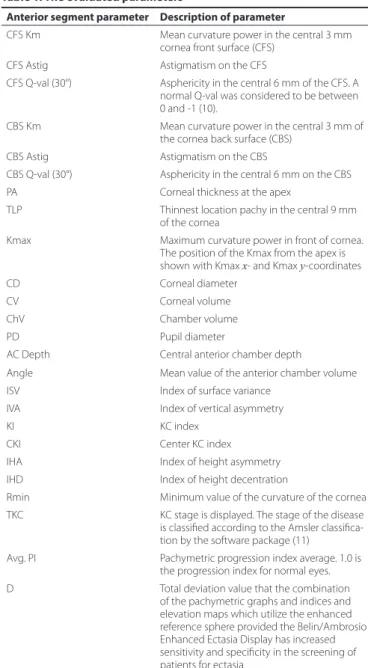
Corneal topography was initially performed with the participant in a resting position, immediately followed by repeating the procedure while the patient performed VM. The same ophthalmologist (Y.K) per-formed all the measurements. Topography images were taken using the Pentacam, which enabled images to be taken of the anterior and posterior surfaces of the cornea and iris, and of the anterior and pos-terior surfaces of the lens in a movable virtual eye(9). After positioning the participant’s head in a chin rest and head rest, the participant was asked to fixate on the center of the blue slit light. The joystick was adjusted by the examiner until appropriate alignment was obtained. A 9.00 mm diameter spherical reference, was used in elevation maps. The evaluated parameters are presented in table 1.
The results were analyzed using SPSS 16.0 software for Windows (SPSS Inc., Chicago, IL). Data are presented as mean ± standard deviation (SD). Parameters studied before and during VM were compared using paired sample t-tests, and a general linear model (repeated measures) was used for analyzing the parameters according to the stage of KC. A p-value of <0.05 was considered to indicate statistical significance.
RESULTS
The mean age of the participants with KC in both eyes was found to be 24.14 ± 6.59 years. Of the 50 participants, 27 (54%) were female and 23 (46%) were male. Both of the participants’ eyes were included in the study. Of the eyes included in the study, 7% were at stage 1 KC, 47% were at stage 2 KC, 32% were at stage 3 KC, and 14% were at stage 4 KC.
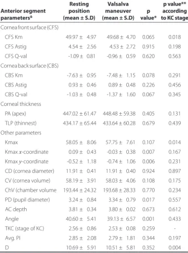
The indices of KC (ISV, IVA, KI, CKI, IHA, IHD, and Rmin) were not significantly affected by performing VM and did not change accor-ding to the stage of KC. Similarly, no statistically significant effects were found for the stage of KC, D value, and the average pachyme-tric progression index over VM. Table 2 presents results showing the effects of VM on anterior segment parameters. When the effects of the KC stage on the parameters were compared, there was a greater decline during VM in CFS Km, Kmax, and D in KC stage 4 cases. These differences were statistically significant with respect to the stage of KC (Table 2). The differences in the other parameters before and during VM did not change with the stage of the KC.
Pupil diameter increased to a statistically significant degree during VM (p=0.017). Conversely, there was a statistically significant decrease in the angle value during VM (p=0.001). Although the x- and y -coordi-nates of Kmax changed sides (p=0.007, p=0.006), the change in Kmax value was not statistically significant (p=0.107). Changes in other anterior segment parameters were also not statistically significant.
DISCUSSION
KC is a degenerative disease of thecornea characterized by stromal
thinning and conical ectasia(1). KC is usually caused to progress by
increased keratometry, irregular astigmatism, and decreased corneal
thickness(10-12). The progression of ectasia was diagnosed based on
Table 1. The evaluated parameters
Anterior segment parameter Description of parameter
CFS Km Mean curvature power in the central 3 mm cornea front surface (CFS)
CFS Astig Astigmatism on the CFS
CFS Q-val (30°) Asphericity in the central 6 mm of the CFS. A normal Q-val was considered to be between 0 and -1 (10).
CBS Km Mean curvature power in the central 3 mm of the cornea back surface (CBS)
CBS Astig Astigmatism on the CBS
CBS Q-val (30°) Asphericity in the central 6 mm on the CBS
PA Corneal thickness at the apex
TLP Thinnest location pachy in the central 9 mm of the cornea
Kmax Maximum curvature power in front of cornea. The position of the Kmax from the apex is shown with Kmax x- and Kmax y-coordinates
CD Corneal diameter
CV Corneal volume
ChV Chamber volume
PD Pupil diameter
AC Depth Central anterior chamber depth
Angle Mean value of the anterior chamber volume
ISV Index of surface variance
IVA Index of vertical asymmetry
KI KC index
CKI Center KC index
IHA Index of height asymmetry
IHD Index of height decentration
Rmin Minimum value of the curvature of the cornea TKC KC stage is displayed. The stage of the disease is classified according to the Amsler classifica-tion by the software package (11)
Avg. PI Pachymetric progression index average. 1.0 is the progression index for normal eyes. D Total deviation value that the combination
of the pachymetric graphs and indices and elevation maps which utilize the enhanced reference sphere provided the Belin/Ambrosio Enhanced Ectasia Display has increased sensitivity and specificity in the screening of patients for ectasia
KC= keratoconus.
the corneal tomography and topographic changes. These changes in cluded progressive elevation of the posterior and/or anterior surface, progressive corneal thinning at the thinnest point, and an increase in corneal anterior keratometry steepening with an irregular astigmatism in the cone area.
Ko ç l u K Y, e ta l.
3 6 5
Arq Bras Oftalmol. 2016;79(6):363-5
Although several studies have tried to investigate the factors that help determine the prognosis, it remains a challenge to predict pro-gression at the time of diagnosis in a specific patient. However, some predictors of progression include a higher cylinder and keratometry (as measured by topography), thinnest pachymetry, and shorter di sease
duration(13-15). As these topography parameters are compared between
two measurements, those measurements should be performed under ideal conditions without bias from environmental factors. An inaccu-rate corneal topography could result in mistakes in the diagnosis of progression. Some patients may perform VM involuntarily during cor-neal topography. However, in this study performing VM did not result in any statistically significant changes in the KC progression indices.
Pekel et al., in their study involving measurements with a Penta cam system, found that AC depth and central corneal thickness in healthy
individuals without KC decreased significantly with VM(8). They also
found that VM could affect corneal morphology and that it caused cer-tain reversible changes in the keratometry. Our study also investigated whether there would be changes in KC patients; we found that PD,
angle value, and the x- andy-coordinates of Kmax changed. A similar
study which evaluated normal corneas demonstrated an increase in
PD and decrease in ACD(16). The only organ related to the autonomic
nervous system in the study was the pupil(17). There was decreased
vagal and increased peripheral sympathetic stimulation during VM, and the noted dilation could be explained by this. Narrowing of the anterior chamber and iridocorneal angle could be caused even by this mild dilation(18).
With subclinical disease, anterior curvature alone may not provide enough information to detect an early corneal abnormality. The goal of the Belin/Ambrosio Enhanced Ectasia Display was to combine ele-vation and pachymetric-based corneal evaluation in an all-inclusi ve display(19). The present study found no statistically significant changes in D values, demonstrating early posterior ectatic changes during VM. However, D values in advanced KC have been found to exhibit more changes than in the other stages of KC, between, before, and during VM. These changes are not important in clinical practice because the D value is more important at the early stage of KC.
Previous studies have demonstrated the effect of VM on healthy
corneas(8,16). However, this effect has not been studied in KC patient
groups. Before the study was conducted, more anterior segment parameter changes were predicted in KC patients who had thinner and less rigid corneas than in VM studies mentioned. However, the present study found no anterior segment parameter changes that would affect stage changes and treatment plans for KC patients.
In conclusion, no changes were found in the KC indices and stage of the disease while participants were performing VM. Furthermore, it was found that there was an increase in PD and a decrease in angle, and these changes were similar to those from studies on normal corneas. The Kmax value in stage 4 KC may decrease more during VM than in other stages.
REFERENCES
1. Rabinowitz YS. Keratoconus. Surv Ophthalmol. 1998;42(2):297-319.
2. Galvis V, Sherwin T, Tello A, Merayo J, Barrera R, Acera A. Keratoconus: an inflammatory disorder? Eye (Lond). 2015;29(7):843-59.
3. Falcao M, Vieira M, Brito P, Rocha-Sousa A, Brandão EM, Falcão-Reis FM. Spectral-do main optical coherence tomography of the choroid during valsalva maneuver. Am J Ophthal-mol. 2012;154(4):687-92.
4. Sihota R, Dada T, Aggarwal A, Srinivasan G, Gupta V, Chabra VK. Does an iridotomy provide protection against narrowing of the anterior chamber angle during Valsalva maneuvre in eyes with primary angle closure. Eye (Lond). 2008;22(3):389-93. 5. Zebrowska A, Gasior Z, Jastrzebski D. Cardiovascular effects of the Valsalva maneuver du
-ring static arm exercise in elite power lifting athletes. Adv Exp Med Biol. 2013;755: 335-42. 6. Soleimani M, Hashemi H, Mehravaran S, Khabazkhoob M, Emamian MH, Shariati M,
et al. Comparison of anterior segment measurements using rotating Scheimpflug ima ging and partial coherence interferometry. Int J Ophthalmol. 2013;6(4):510-4. 7. Wang BS, Xiao L, Liu J, Dong N, Aung T. Dynamic changes in anterior segment
morpho-logy during the Valsalva maneuver assessed with ultrasound biomicroscopy. Invest Ophthalmol Vis Sci. 2012;53(11):7286-9.
8. Pekel G, Acer S, Yagci R, Kaya H, Pekel E. Impact of valsalva maneuver on corneal mor -phology and anterior chamber parameters. Cornea. 2014;33(3):271-3.
9. Huseynova T, Abdulaliyeva F, Lanza M. Comparison of Scheimpflug imaging parame-ters between steep and keratoconic corneas of Caucasian eyes. Clin Ophthalmol. 2016; 10:603-8.
10. Flynn TH, Sharma DP, Bunce C, Wilkins MR. Differential precision of corneal Pentacam HR measurements in early and advanced keratoconus. Br J Ophthalmol. 2015; pii: bjophthalmol-2015-307201.
11. Koçluk Y, Yalniz-Akkaya Z, Burcu A, Örnek F. Comparison of Scheimpflug imaging analysis of pellucid marginal corneal degeneration and keratoconus. Ophthalmic Res. 2015;53(1):21-7.
12. Steinberg J, Ahmadiyar M, Rost A, Frings A, Filev F, Katz T, Linke SJ. Anterior and pos-terior corneal changes after crosslinking for keratoconus. Optom Vis Sci. 2014;91(2): 178-86.
13. Choi JA, Kim MS. Progression of keratoconus by longitudinal assessment with corneal topography. Invest Ophthalmol Vis Sci. 2012;53(2):927-35.
14. McMahon TT, Edrington TB, Szczotka-Flynn L, Olafsson HE, Davis LJ, Schechtman KB; CLEK Study Group. Longitudinal changes in corneal curvature in keratoconus. Cornea. 2006;25(3):296-305.
15. Kanellopoulos AJ, Asimellis G. Revisiting keratoconus diagnosis and progression classi-fication based on evaluation of corneal asymmetry indices, derived from Scheimpflug imaging in keratoconic and suspect cases. Clin Ophthalmol. 2013;7:1539-48. 16. Mete A, Kimyon S, Uzun I, Kara N. Effects of Valsalva Maneuver on Ocular Biometric
Parameters: Optical Low-Coherence Reflectometry Biometer Study. Semin Ophthalmol. 2015; Aug 21:1-4.
17. Reulen JP, Marcus JT, van Gilst MJ, Koops D, Bos JE, Tiesinga G, et al. Stimulation and recording of dynamic pupillary reflex: The IRIS technique: part 2. Med Biol Eng Comput. 1988;26(1):27-32.
18. Marchini G, Pagliarusco A, Toscano A, Tosi R, Brunelli C, Bonomi L. Ultrasound biomi-croscopic and conventional ultrasonographic study of ocular dimensions in primary angle closure glaucoma. Ophthalmology. 1998;105(11):2091-8.
19. Belin MW, Khachikian SS. Elevation based topography, screening for refractive surgery: USA: Highlights of Ophthalmology International; 2008. p.83-4.
Table 2. Anterior segment parameters in the resting position and during VM in KC patients
Anterior segment parameters&
Resting position (mean ± S.D)
Valsalva maneuver (mean ± S.D)
p value*
p value** according to KC stage
Cornea front surface (CFS)
CFS Km 049.97 ± 04.97 -0 49.68 ±04.70 0.065 0.018 CFS Astig 004.54 ± 02.56 -0 04.53 ±02.72 0.915 0.198 CFS Q-val -1.09 ± 00.81 00-0.96 ± 00.59 0.620 0.563 Cornea back surface (CBS)
CBS Km 0-7.63 ± 00.95 00-7.48 ± 01.15 0.078 0.291 CBS Astig 000.93 ± 00.46 -000.89 ± 00.48 0.226 0.456 CBS Q-val 0-1.03 ± 00.48 00-1.37 ± 01.60 0.067 0.345 Corneal thickness
PA (apex) 447.02 ± 61.47 -448.48 ± 59.38 0.405 0.131 TLP (thinnest) 434.17 ± 65.44 -433.64 ± 60.28 0.679 0.439 Other parameters
Kmax 058.05 ± 08.06 -057.75 ± 07.61 0.107 0.014 Kmax x-coordinate 000.09 ± 00.43 00-0.03 ± 00.38 0.007 0.167 Kmax y-coordinate 0-0.52 ± 01.18 00-0.74 ± 01.06 0.006 0.231 CD (cornea diameter) 011.91 ± 00.41 -011.91 ± 00.40 0.924 0.897 CV (cornea volume) 058.19 ± 03.91 -058.03 ± 04.06 0.108 0.175 ChV (chamber volume 193.44 ± 24.32 -193.68 ± 28.33 0.770 0.234 PD (pupil diameter) 003.24 ± 00.84 -003.34 ± 00.79 0.017 0.557 AC depth 003.81 ± 00.34 -003.80 ± 00.02 0.673 0.612 Angle 040.60 ± 05.41 -0039.13 ±06.57 0.001 0.433 TKC (stage of KC) 002.56 ± 00.86 -002.53 ± 00.08 0.259 -Avg. PI 002.85 ± 02.08 00-2.79 ± 01.81 0.344 0.197 D 010.69 ± 05.91 -010.51 ± 05.81 0.352 0.004 *= paired samples t test; **= general linear model test for four KC stages (differences between KC stage 1, 2, 3, and 4 groups); &= see table 1 for a description of parameters.