O
RIGINALA
RTICLE Revista Brasileira de FisioterapiaThe Valsalva maneuver duration during
labor expulsive stage: repercussions on the
maternal and neonatal birth condition
Duração da Manobra de Valsalva durante o período expulsivo do parto:
repercussões maternas e nas condições neonatais de nascimento
Andrea Lemos1, Elizabeth Dean2, Armèle Dornelas de Andrade1
Abstract
Objectives: This cross-sectional study was designed toexamine the effects of the Valsalva Maneuver (VM) and its duration on the acid- base equilibrium of the neonate and its maternal repercussions during the expulsive stage of labor, after standard breathing and pushing
instructions were given. Methods: A convenience sample of women with low risk pregnancy (n=33; mean age 22.5±3.7y and gestational
age 38.1±1.12wks) and their newborns were studied during the expulsive stage of vaginal labor. Coaching consisted of standard
recommendations for breathing including prolonged VMs coordinated with pushing. Maternal outcomes included the need for uterus fundal pressure maneuver and episiotomy, perineal trauma and posture. Neonatal outcomes included blood gases sampled from the umbilical cord, and Apgar scores. Data were analyzed with the Fisher´s exact test, chi-square test, and Pearson correlation coefficient.
Results: None of the maternal outcomes were associated with VM duration. With respect to neonatal outcomes, increased VM duration was associated with reduced venous umbilical pH (r=-0.40; p=0.020), venous base excess (r=-0.42; p=0.014) and with arterial base excess
(r=-0.36; p=0.043). Expulsive stage time was negatively associated with umbilical venous and arterial pH. Conclusions: VM duration
during fetal expulsion in labor negatively affects fetal acid-base equilibrium and potentially the wellbeing of the neonate. Our results support the need to consider respiratory strategies during labor, to minimize potential risk to the mother and neonate.
Key words: Valsalva maneuver; pushing; “labour, obstetric”; umbilical cord.
Resumo
Objetivos: O presente estudo de corte transversal teve como objetivo avaliar os efeitos da Manobra de Valsalva (MV) e sua duração no equilíbrio ácido-básico fetal e nas repercussões maternas durante o período expulsivo do parto, após instrução respiratória
padronizada. Métodos: Uma amostra de conveniência em gestantes de baixo risco (n=33, média de idade 22,5±3.7 anos e idade
gestacional 38.1±1.12 semanas) e seus neonatos foi avaliada durante o período expulsivo do parto vaginal. O treinamento consistiu
em recomendações padronizadas para respiração, incluindo MV prolongadas associadas ao puxo. Os desfechos maternos incluíram o uso da manobra de pressão para o fundo do útero, episiotomia, trauma perineal e postura. Os desfechos neonatais incluíram análise dos gases sanguíneos do cordão umbilical e valores de Apgar. Os dados foram analisados por meio do teste exato de Fisher, teste
qui-quadrado e Coeficiente de Correlação de Pearson. Resultados: Nenhum dos desfechos maternos foi associado com a duração
da MV. No entanto, em relação aos desfechos neonatais, o aumento da duração da MV foi associado com redução do pH venoso umbilical (r=-0,40; p=0,020) e excesso de base (r=-0.42; p=0.014) e com o excesso de base arterial (r=-0,36; p=0,043). O tempo
do período expulsivo do parto foi negativamente associado com o pH venoso e arterial. Conclusões: A duração da MV durante esse
período do parto interfere negativamente no equilíbrio ácido-básico fetal e potencialmente no bem-estar do neonato. Esses resultados fornecem suporte para a necessidade de os fisioterapeutas considerarem estratégias de orientações respiratórias para o puxo durante o trabalho de parto para minimizar potenciais riscos para a mãe e o neonato.
Palavras-chave: Manobra de Valsalva; puxo; parto; cordão umbilical.
Received: 04/03/2010 – Revised: 10/09/2010 – Accepted: 16/11/2010
1 Physical Therapy Department, Universidade Federal de Pernambuco, Recife, PE, Brazil
2 Physical Therapy Department, Faculty of Medicine, University of British Columbia, Vancouver , BC, Canadá
Introduction
Childbirth is characterized by spontaneous alterations in the mother’s breathing coordinated with the generation of marked thoracic and abdominal pressures to assist with expulsion of the fetus. Although normally spontaneous and unconscious, breathing becomes a voluntary and controlled activity during labor, induced by the physiologic mechanisms to facilitate this process1. Breathing patterns are characterized
by periods of normal breathing interspersed with Valsalva maneuvers (VMs). VM strengths, frequencies, and durations are dependent on the stage of labor2.
During the expulsive stage of labor, the fetus is propelled caudally as a results of the high thoracic and abdominal pres-sures generated by the mother coordinated with breathing and muscle contraction. Such eforts increase the pressure on the levator ani muscles of the pelvis which generates a relex, in-voluntary urge to bear down strongly, referred to as the push stage. Associated with involuntary intra uterine contractions, a voluntary expulsive efort is manifested, through the abdo-minal and respiratory muscles, to facilitate the delivery of the neonate3. he woman may also voluntary bear down. While
in-voluntary bearing down eforts are typically short (<6 seconds), voluntary bearing down eforts directed by the care giver are often longer, and up to 10 seconds4.
Standard childbirth practices tend to promote prolonged duration of the VM (i.e., 10 s or more) which has been the focus of some studies5,6. Some investigators; however, have argued that
breathing control interventions should not be imposed. Rather, the mother should adopt a breathing pattern including the VM in accordance with what she instinctively requires7-9. When a
woman who is delivering breathes in accordance with her needs rather than having a pattern imposed, her respiratory eforts typically involve breathing with an open glottis, rather than a closed glottis, such as the case during a VM, and when she uses the closed glottis is only for a maximum of 4 to 6 seconds9.
Irrespective of the debate, evidence to support either point of view is scarce. Some physiologic evidence supports the avoi-dance of VMs of 10 seconds or more during labor and delivery6.
Despite several papers4,5,7-9 supporting its adverse maternal
and fetal efects, prolonged VMs continue to be promoted in the literature.Futhermore, no study to date has speciically examined the relationship between the VM and its duration, on maternal and neonatal outcomes, and the implications for recommendations regarding preparation for childbirth.
his study was designed to further explore the role of con-trolled VM reported in the literature by examining the rela-tionship of VM duration on maternal and neonatal outcomes. Maternal outcomes included the need for uterus fundal pres-sure maneuver, episiotomy, perineal trauma and labor posture.
Neonatal outcomes included umbilical cord blood pH, base excess, PCO2 and Apgar scores.
Methods
Participants
A cross-sectional study was conducted using a sample of convenience. his was a pilot study as there were no references in the literature of researche studies using similar methodology that could be used for sample size calculation. Inclusion cri-teria were pregnant women (both primipara and multipara), between 18 and 35 years of age, with fetus gestational age be-tween 37 and 41 weeks based on the last menstruation date, and that were admitted to the hospital at the irst stage of labor, and were classiied as low-risk. Additional criteria were cephalic presentation of the fetus, the mother’s membranes had been intact during early labor, and the presence of one live fetus. he attending obstetrician who was blind to the speciic intent of the study, established whether the inclusion criteria were met. Mothers for whom umbilical cord compression, forceps and analgesia were indicated, were excluded, because their fetus could experience acute distress (signs of hypoxia). his determination was also based on the independent assess-ment of the obstetrician. Based on normal procedures, women for whom 2% lidocaine was indicated during an episiotomy, were retained. his research was approved by the Research Ethics Committee of the Universidade Federal de Pernambuco (UFPE), Recife, PE, Brazil under protocol number: 005/2002 . Subjects were included in the study after having the study des-cribed, and providing informed signed consent.
Data collection during the expulsive stage of labor
Participating mothers were assessed by members of the obs-tetrical team during the expulsive stage of labor, with the initial and inal stages deined by 10 cm cervical dilatation and fetal body expulsion, respectively. he duration of this interval was recorded. Traditional instructions were given to the mothers by the attending nursing staf who were also blind to the speciic intent of the study. Mothers were instructed to push hard during each contraction, and at the same time take a deep breath, and hold it for as long as they could, using VM to facilitate this strong efort11. During this stage, the duration of each VM was recorded
with a calibrated chronometer (Cronobio, SW-2018, São Paulo, SP), along with the number of VMs. he average VM duration was considered as the expulsive stage VM value. A pulse oxi-meter (J.G. Moriya-1001, São Paulo, SP) positioned on the left index inger was used to assess oxygen saturation (SpO2) and
maternal heart rate during each maneuver. he body position
of the mother was recorded. he position was described as su-pine when the angle between the mother’s trunk and legs was less than 30º, and as vertical, when this angle was 30º or greater based on the elevation of the trunk. he need for oxytocin was assessed by the attending medical and nursing staf according to commonly-used delivery procedures, i.e., 1 oxytocin ampoule.
Umbilical cord blood gas analysis
Immediately after birth, the umbilical cord segment was doubly clamped and isolated for blood collection. With two 10 ml syringes lushed with .06 ml heparin 5000 UI/ml, 0.2 to 0.3 ml of blood were taken, irst from the umbilical artery and then from the umbilical vein, thereby eliminating any residual air trace. he cordblood gas analysis was conducted immedia-tely using a gasometer (GEM-Premier 3.000-Instrumentation Laboratory Company, MA-USA). he analysis of blood pH, PCO2 and base excess were performed for each of the two sam-ples (arterial and venous).
Maternal outcomes
Post delivery, the following maternal outcomes were recor-ded: whether the uterus fundal pressure maneuver was needed (i.e., an externally-applied mechanical force to the abdomen at the level of the uterus fundus), the need for an episiotomy, and the degree of perineal trauma. Perineal trauma were classiied
as irst degree when there was an injury to the skin and mu-cosa, ,second degree when there was muscle injury, third de-gree when there was injury involving the anal sphincter and fourth degree and there was injury to the rectum mucosa3.
Neonatal outcomes
he neonatal outcomes were extracted from the medical record: Apgar scores10 for the 1st and 5th minutes after birth
(normal value being greater than a score of 7), and the body weight. Normal neonatal body weight was based upon the Bra-zilian population average of 3390 g11.
Statistical analysis
he following maternal variables were analyzed as categori-cal data: parity, body position, need for the uterus fundal pres-sure maneuver, use of oxytocin, episiotomy, and the neonatal’s variables of weight and Apgar scores. Continuous data were analyzed descriptively by means and standard deviations. To examine the association between categorical variables and VM duration, the latter was classiied into two categories based on the mothers’ spontaneous pushing efortscommonly reported in the literature4: £6 s and >6 s. Chi-square tests were used to analyze
these data. When the assumptions of the chi-square test were not satisied, the Fisher’s exact test was used. To rule out a potential cumulative physiologic and psychological stress efects on the va-riables of interest, the duration of the overall labor was correlated with the acid base variables of the neonate. Associations among the continuous variables were analyzed using Pearson´s correla-tion coeicient. An α level of ≤.05 was set for all tests.
Results
hirty three mothers were included in this study. Mother’s mean age (±SD) was 22.5±3.7 years, and the mean gestational age was 38.1±1.12 weeks. here was no evidence of arterial de-saturation, deined as less than 96% oxigenation, in any mother throughout the study period. In addition, no association was observed between VM duration and maternal heart rate that was continuously monitored and recorded during each push (r=0.26; p=0.142). Only 7 women who were multipara required no episiotomy, and among them, 6 exhibited a irst degree, and one, a third degree perineal injury. No associations were obser-ved between maternal and infant’s weight, Apgar scores and VM duration (Table 1). Nine neonates exhibited an umbilical artery pH <7.2; of which 6 (66.7%) were neonates of the sub-group of women with a VM duration >6 s. Only 3 neonates had an 1st minute Apgar score of <7. All neonates had normal Apgar
scores when assessed at the 5th min.
Variable
Valsalva Maneuver
Duration (s) p value
≤6 >6
Parity of the mother n % n % 0.65ns
Primipara 11 57.9 7 50
Multipara 8 42.1 7 50
Mother’s body position 1.00 ns
≤30º 12 63.2 11 78.6
>30º 7 36.8 3 21.4
Uterus fundal pressure maneuver 0.50 ns
Performed 6 31.6 6 42.9
Not performed 13 68.4 8 57.1
Ocitocin 0.16 ns
Administered 9 47.4 10 71.4 Not administered 10 52.6 4 28.6
Episiotomy 0.67 ns
Performed 14 73.7 12 85.7 Non performed 5 26.3 2 14.3
Fetal weight (g) 1.00 ns
≤3390 13 68.4 10 71.4
>3390 6 31.6 4 28.6
Fetal Apgar score 0.62 ns
≤7 3 15.8 1 7.1
>7 16 84.2 13 92.9
Table 1. Categorical variable data for the mother and neonate separed
by the duration of the Valsalva maneuver.
Figure 4. Relationship between the duration of the expulsive stage of labor and umbilical venous pH.
R= - 0.41; p= 0.017
0 10 20 30 40 50 60
7.40 7.45 7.50
7.35 7.30 7.25 7.20 7.15 7.10 7.05
Umbilical pH V
enous Blood
Expulsive Duration (min) R= - 0.40; p= 0.020
Valsalva Maneuver Duration (s)
Umbilical pH V
enous Blood
1 3 5 7 9 11 13 15
7.50 7.45 7.40 7.35 7.30 7.25 7.20 7.15 7.10 7.05
Figure 1. Relationship between the duration of the Valsalva maneuver
during the expulsive stage of labor and umbilical venous pH (r=-0.40; p=0.020).
R = - 0.42; p= 0.014
1 3 5 7 9 11 13 15
Valsalva Maneuver Duration (s)
Umbilical Base Excess V
enous Blood
(mmol/l)
-14
-12
-10
-8
-6
-4
-2
0
Figure 2. Relationship between the duration of the Valsalva maneuver
during the expulsive stage of labor and umbilical venous base excess.
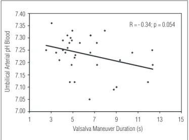
R =-0.34; p = 0.054
1 3 5 7 9 11 13 15
Valsalva Maneuver Duration (s)
Umbilical Arterial pH Blood
7.40
7.35
7.30
7.25
7.20
7.15
7.10
7.05
7.00
Figure 3. Relationship between the duration of the Valsalva maneuver
during the expulsive stage of labor and umbilical arterial pH.
A reduction was noted in venous umbilical pH (r=-0.40; p=0.020; Figure 1) and venous base excess (r=-0.42; p= 0.014; Figure 2) in relation to VM duration, but this was not observed in the venous PCO2 (r=0.25; p=0.167). here was no association
between VM duration and arterial umbilical PCO2 (r=-0.22;
p=0.219), and arterial pH (Figure 3), but an association with the arterial base excess was observed (r=-0.36; p=0.043). here was no relationship between VM duration and the expulsive stage time (r=0.10; p=0.595). However, when the expulsive stage time was compared to the umbilical venous and arterial pH (r=-0.30; p=0.092), there was a negative association with the umbilical venous pH (r=-0.41; p=0.017; Figure 4). Finally, no associations were observed between the duration of labor (1st and 2nd stages)
and the umbilical arterial pH or venous pH (r=0.31 and p=0.19; r=0.27 and p=0.176, respectively).
Discussion
his study was designed to further evaluate the evidence for breathing control recommendations that are taught by he-alth care team to expectant women, and coached during the pre-natal period and labor. he uniqueness and importance of this study, is that manipulation of the VM duration is common in perinatal instruction, yet its potential impact has not been well studied. his is the only study to our knowledge that has considered the relationship between duration of the VM and various maternal and newborn outcomes; and that considered that prolonged bearing down eforts using VM may be associa-ted with risks to the newborn.
In this study, a prolonged VM period was associated with reduced umbilical venous pH and venous base excess of the neonate. hese indings are consistent with the potential for acidosis, given there was no relationship between umbilical
venous PCO2 and VM duration. Venous blood lowing from the placenta directly to the fetus relects the maternal acid-basic status12. he gas values of this blood are similar to the
acid-base status and the oxygen content of the maternal intervilous space, because oxygen and carbon dioxide are balanced be-tween these compartments13.
he indings support that an extended period of apnea as-sociated with the mother’s muscle efort to maintain bearing down during the expulsive stage of labor, can negatively afect fetal acid-base balance, demonstrated by a reduction in base excess in the umbilical vein. Abnormal umbilical acid base balance has been associated with increased fetal stress and birth-related injuries14.
With progressive pushing by the mother with increasing intensity, duration, and frequency, the lactate concentration increases in the mother’s blood causing metabolic acidemia, and the blood reaches the umbilical vein with a low pH15.
hompson16 reported that a prolonged expulsive stage is risky
for the fetus, when the mother experiences prolonged apneic episodes, and observed a reduction in the time-related venous pH, particularly in the group characterized by prolonged VMs. Barnett and Humenick17, comparing women who used open
glottis during VM for pushing with women who used a closed glottis, reported that the umbilical venous pH remained higher in women who performed VMs with an open glottis. his is an interesting observation given that some women are spon-taneously vocal during end-stage labor. Vocalization requires that the glottis is open. Childbirth practices in some cultures that encourage women to be stoic and non vocal may not be advantageous to the neonate.
he maternal cardiorespiratory load imposed by prolonging the VM may interfere with the wellbeing of the fetus. When pushing in conjunction with a strong VM, the mother increases abdominal pressure, which can results in pressures higher than the uterus blood perfusion, reducing uterus-placental blood low18. his efect could alter the oxygen content available to
the fetus due to the development of acidemia secondary to anaerobic metabolism19.
he umbilical cord arterial blood relects the fetal acid-base status, and a pH of less than 7.2 is an indicator of fetal acidemia20. In our study, 27.2% of the neonates had an arterial
pH<7.2. Although arterial pH tended to decreases with VM duration, this inding was not statistically signiicant. When the VM duration increased, the arterial base excess decreased; however, no relationship was observed with arterial PCO2. Caldeyro-Barcia et al.8 reported higher levels of venous and
arterial pH in a sample of women in labor who used the VM with an average duration of 5 s. his inding support that VM duration is an important variable with respect to maintaining optimal acid base balance.
When the placenta function is abnormal or a maternal acid-base disturbance is present, both the arterial blood and venous blood are abnormal. Women in labor showing changes in venous pH and venous base excess also show changes in the arterial values. hus, maternal respiratory patterns may inter-fere with the fetal acid-base equilibrium12.
he primary purpose of prolonging the VM in women in end stage labor is to facilitate the expulsion of the neonate and potentially shorten labor6. In the present study, there was a
rela-tionship between the expulsive stage duration and low venous pH. However, there was no relationship between the duration of this stage with that of the VM. he sub-group of women who used prolonged VMs, hence experienced prolonged apnea episodes, did not experience shorter labor. herefore, a long expulsive stage was not associated with women using the VM for a short period of time.
here is a disagreement in the literature21-23 with respect to
the relationship between the type of pushing adopted and the fetal acid-base status and the expulsive stage duration. Group diferences across studies and breathing control strategies used to facilitate pushing may contribute to the inconsistency of in-dings. In a study of 350 women who were primiparas, Parnell et al.21 reported no diferences in the arterial pH and the base
excess when women used VM with open versus closed glottis when pushing during labor. However, the expulsive stage was longer in the group that used the closed glottis with VM and neonates with low Apgar scores at the 1st minute and low
ar-terial pH were observed in this group. Another study reported similar indings when women (primipara and multipara) pu-shed with either an open or closed glottis22. Several studies5,7,23
have compared the 10 seconds VM with spontaneous pushing, where women adopt their instinctively appropriate breathing pattern. However, umbilical blood gases were not evaluated in these studies, and no diferences in the duration of the expul-sive stage between breathing techniques were found. Caldeyro-Barcia et al.8and hompson16 reported a shorter expulsion time
in women who used VM, however; umbilical pH was negatively afected.
he Apgar score used in the clinical assessment of new-borns at birth is an easy and commonly used tool, however; its objectivity has been questioned24. In this study while umbilical
blood gas analysis detected 9 neonates with low arterial pH, the Apgar score at the 1st minute was low only for 4 neonates.
Low arterial ph and low Apgar scores were simultaneously found only in two neonates. All neonates exhibited normal Apgar scores at the 5th minute, and there was no association
between the VM duration and Apgar score. Reports from rela-ted studies have shown inconsistency between the detection of low arterial pH based on gas analysis and the Apgar index14,25.
is supported with the detection of metabolic acidemia in um-bilical cord blood.
One reason that the uterus fundal pressure maneuver is used during the late expulsive stage is to accelerate fetal expulsion26. A
push with a short duration VM relects a patient´s poor willing-ness to cooperate, thereby showing predisposition to the prac-tice of such conduct27. However, we observed that the pregnant
women who used VM of shorter duration did not present a high incidence in the use uterus fundal pressure, indicating that a long VM did not preclude the maneuver to be indicated.
An increase in maternal heart rate was noted during the VM, but there was no relationship between heart rate and VM duration. According to Sherman et al.28, the largest increases
in maternal heart rate coincide with an uterine contraction associated with pushing, and autonomically-mediated by the VM. In our study, the fetal heart rate was not recorded. We recommend that this parameter be included in future studies. Pushing in conjunction with a VM has been associated with fetal heart rate changes including prolonged deceleration and bradycardia consistent with a hypoxemic response8. hese
reports conirm the need for greater understanding about nor-mal breathing and pushing eforts in women in labor to guide the development of more reined clinical practice guidelines to support women during childbirth and delivery.
Oxytocin administration was comparable in the two sub-groups studied, namely, those who used VMs ≤6 s duration vs. >6 s duration, but in 19 women who were administered the drug, 71.4% were in the group whose VMs were greater than 6 s. Similar results were reported by Parnell et al.21 where
oxyto-cin tended to be needed by women who adopted a VM with a closed rather than an open glottis. Increases in the eiciency of contractions with oxytocin stimulation lead to an increase in the duration of the VM. Furthermore, inefective uterine con-tractions may necessitate prolonged VMs21,27.
he importance of the mother’s body position during the expulsive stage of labor should be highlighted. Adverse efects of the supine position on placenta’s circulation occur even with a 30º angle between the trunk and legs29. Additionally there is
a marked reduction of Lung Functional Residual Capacity with supine position30. he inconvenience of this position during
the expulsion has been well established, based on blood low reduction in the aorta and inferior vena cava caused by uterine compression which increases during a VM-mediated contrac-tion31. he absence of an association between VM duration
and the position adopted by women who are delivering may be due to the small sample size included in this study. In addi-tion, although women were positioned in either supine or more
upright, these positions were not markedly diferent given the constraints of the delivery table.
here were no associations between neonatal body weight and VM duration, altough it was expected that heavier babies would need longer VMs because they may be prone to com-press more the rectum/sacral plexus21. he size of the neonate
does not appear to elicit longer VMs. In a retrospective study related to the type of push used by women, Sampselle and Hines32 also observed no association between neonatal weight
and prolonged VMs.
Our data did not support an association between VM dura-tion and the status of the perineal muscles, because the need for episiotomy in this population was not selective, as there were only 7 women who were multipara who did not require it. herefore, any comparison could have added methodological bias to the results of the study as episiotomy was associated with number of previous births. Some studies4,7,32,33 however
have reported that the use of an open glottis is beneicial be-cause it reduces tears and ruptures of the perineal muscles and the incidence of episiotomy, and improves urodynamic indi-ces. his prevents the development of stretch relex occurring when the muscles are submitted to a sudden and continuous efort such as during a VM7.Spontaneous vocalization of
wo-men during childbirth could be evaluated in conjunction with maternal and neonatal outcomes of interest in a future study. Because cross cultural diferences exist with respect to vocali-zation during labor, cross cultural studies of these relationships could shed valuable insight into optimal breathing and pushing recommendations.
his study supports that duration of the VM used during the expulsive stage of labor may interfere with blood gases sampled from the umbilical cord. A prolonged VM was not associated with the use of uterus fundal pressure neither shortened the second stage of labor.
hese indings convey new information supporting strate-gies for the use of an open glottis, through vocalization, during the second stage of labor to minimize potential risk, such as hypoxia, to the neonate. Randomized controlled trials with large sample sizes are need to further evaluate VM and open glottis and to enhance the efectiveness of doctors, nurses and physical therapists in assisting delivery.
Acknowledgments
To Conselho Nacional de Desenvolvimento Cientíico e Tecnológico (CNPq).
References
1. Sampselle CM, Miller JM, Luecha Y, Ficher K, Rosten L. Provider support of spontaneous pushing during the second stage of labor. J Obstet Gynecol Neonatal Nurs. 2005;34(6):695-702.
2. Bloom SL, Casey BM, Schaffer JI, McIntire DD, Leveno KJ. A randomized trial of coached versus uncoached maternal pushing during the second stage of labor. Am J Obstet Gynecol. 2006;194(1):10-3.
3. Cunningham FG, Williams JW. Williams obstetrics. 22nd ed. New York: McGraw-Hill; 2005.
4. Thompson AM. Maternal behavior during spontaneous and directed pushing in the second stage of labour. J Adv Nurs. 1995;22(6):1027-34.
5. Yeates DA, Roberts JE. A comparison of two bearing-down techniques during the second stage of labor. J Nurse Midwifery. 1984;29(1):3-11.
6. Yildirim G, Beji NK. Effects of pushing techniques in birth on mother and fetus: a randomized study. Birth. 2008;35(1):25-30.
7. Beynon C. The normal second stage of labour; a plea for reform in its conduct. J Obstet Gynaecol Br Emp. 1957;64(6):815-20.
8. Caldeyro-Barcia R, Giussi G, Storch E, Poseiro JJ, Lafaurie N, Kettenhuber K, et al. The bearing-down efforts and their effects on fetal heart rate, oxygenation and acid base balance. J Perinat Med. 1981;9 Suppl 1:63-7.
9. Roberts JE, Goldstein SA, Gruener JS, Maggio M, Mendez-Bauer C. A descriptive analysis of involuntary bearing-down efforts during the expulsive phase of labor. J Obstet Gynecol Neonatal Nurs. 1987;16(1):48-55.
10. American Academy of Pediatrics. Use and abuse of the Apgar scores. Pediatrics. 1986;78:1148-9.
11. Baracho E. Fisioterapia aplicada à obstetrícia: aspectos de ginecologia e neonatologia. 3th ed.
São Paulo: Medsi; 2002.
12. Thorp JA, Rushing RS. Umbilical cord blood gas analysis. Obstet Gynecol Clin North Am. 1999;26(4):661-74.
13. Brar HS, Wong MK, Kerschbaum TH, Paul RH. Umbilical cord acid base changes associated with perinatal cardiac failure. Am J Obstet Gynecol. 1988;158(3Pt 1):511-8.
14. Gilstrap LC 3rd, Leveno KJ, Burris J, Williams ML, Little BB. Diagnosis of birth asphyxia on the basis of fetal pH, Apgar score, and newborn cerebral dysfunction. Am J Obstet Gynecol. 1989;161(3):825-30.
15. Nordström L, Achanna S, Naka K, Arulkumaran S. Fetal and maternal lactate increase during active second stage of labour. BJOG. 2001;108(3):263-8.
16. Thompson AM. Pushing techniques in the second stage of labour. J Adv Nurs. 1993;18(2):171-7.
17. Barnett MM, Humenick SS. Infant outcome in relation to second stage labor pushing method. Birth. 1982;9(4):221-9.
18. Bassell G, Humayun SG, Marx GF. Maternal bearing down efforts- another fetal risk? Obstet Gynecol. 1980;56(1):39-41.
19. Belai Y, Goodwin TM, Durand M, Greenspoon JS, Paul RH, Walther FJ. Umbilical arteriovenous PO2 and PCO2 differences and neonatal morbidity in term infants with severe acidosis. Am J Obstet Gynecol. 1998;178(1 Pt 1):13-9.
20. Yeomans ER, Hauth JC, Gilstrap LC 3rd, Strickland DM. Umbilical cord pH, PCO2, and bicarbonate following uncomplicated term vaginal deliveries. Am J Obstet Gynecol.1985;151(6): 798-800.
21. Parnell C, Langhoff-Roos J, Iversen R, Damgaard P. Pushing method in the expulsive phase of labor. A randomized trial. Acta Obstet Gynecol Scand.1993;72(1):31-5.
22. Paine LL, Tinker DD. The effect of maternal bearing-down efforts on arterial umbilical cord pH and length of the second stage of labor. J Nurse Midwifery. 1992;37(1):61-3.
23. Rossi MA, Lindell SG. Maternal positions and pushing techniques in a nonprescriptive environment. J Obstet Gynecol Neonatal Nurs.1986;15(3):203-8.
24. Marrin M, Paes BA. Birth asphyxia: does the Apgar score have diagnostic value? Obstet Gynecol. 1988;72(1):120-3.
25. Steer PJ, Eigbe F, Lissauer TJ, Beard RW. Interrelationships among abnormal cardiotocograms in labor, meconium staining of the amniotic fluid, arterial cord blood pH, and Apgar scores. Obstet Gynecol. 1989;74(5):715-21.
26. Cosner KR. Use of fundal pressure during second-stage labor. A pilot Study. J Nurse Midwifery. 1996;41(4):334-7.
27. Buhimschi CS, Buhimschi IA, Malinow AM, Kopelman JN, Weiner CP. Pushing in labor: performance and not endurance. Am J Obstet Gynecol. 2002;186(6):1339-44.
28. Sherman DJ, Frenkel E, Kurzweil Y, Padua A, Arieli S, Bahar M. Characteristics of maternal heart rate patterns during labor and delivery. Obstet Gynecol.2002;99(4):542-7.
29. Hanson L. Second-stage positioning in nurse-midwifery practices. Part 1: position use and preferences. J Nurse Midwifery. 1998;43(5):320-5.
30. Nørregaard O, Schultz P, Ostergaard A, Dahl R. Lung function and postural changes during pregnancy. Respir Med. 1989;83(6):467-70.
31. Gupta Janesh K, Hofmeyr GJ, Smyth Rebecca MD. Position in the second stage of labour for women without epidural anaesthesia. Cochrane Database of Systematic Reviews. The Cochrane Library. 2009;3:CD002006.
32. Sampselle CM, Hines S. Spontaneous pushing during birth: relationship to perineal outcomes. J Nurse Midwifery.1999;44(1):36-9.