Letters to the Editor
2 6 5 Arq Bras Oftalmol. 2015;78(4):265-6 http://dx.doi.org/10.5935/0004-2749.20150070
Combined 20- and 23-gauge pars plana
vitrectomy
Vitrectomia via pars plana com acesso combinado
de 20 e 23 gauge
Dear Editor:
The 23-gauge (G) transconjunctival sutureless vitrectomy (TSV) technique has advantages of decreased surgical trauma, decreased astigmatism, faster wound healing, and improved postoperative comfort(1) compared with other vitrectomy techniques. Although the efficacy of 23-G is comparable with conventional 20-G vitrectomy, some surgeons still use 20-G vitrectomy in complicated cases, such
as complex retinal detachments (RD)(2). Herein, we describe a modi-fied technique combining 20- and 23-G sclerotomies and the use of 20-G instruments that provides the advantages of both techniques in a single procedure.
Combined surgery was performed in 67 eyes of 65 patients by a single surgeon. During the follow-up period, anatomical and func-tional outcomes and operative complications were evaluated. Pos-toperative vitreous hemorrhage (VH) was classified into two groups; early postoperative VH (occurring within 1 month postoperatively) and late postoperative VH (occurring >1 month postoperatively). Transient postoperative hypotony was defined as intraocular pres-sure (IOP) <6 mmHg lasting <1 week. The degree of intraocular in-flammation was clinically defined by the presence of transient fibrin formation in the anterior chamber or vitreous cavity.
One inferotemporal 23-G transconjunctival sutureless sclero-tomy port for infusion and two superior 20-G sclerosclero-tomy ports for instrumentation, including a vitrectomy probe, were created along with two 25-G Torpedo minilight illuminations bimanual vitrectomy. A 23-G single-step TSV trocar-cannula (EyeTech) was transconjuncti-vally inserted at a 30° angle inferotemporally to the infusion line in all cases. The two superior 20-G sclerotomy ports were created with a 20-G one-step preloaded trocar cannula at a 30° angle tangential to sclera (Synergetic. Inc, Figure 1). The internal valves of the cannula minimize leakage and aid in the maintenance of IOP during the sur-gical procedure. After completing the vitrectomy, a cotton-tipped applicator was applied to the trocar entry site to prevent leakage from the sclerotomy during the removal of the trocars. The trocar entry site was inspected for wound leakage and then closed with a single 8-0 Vicryl suture.
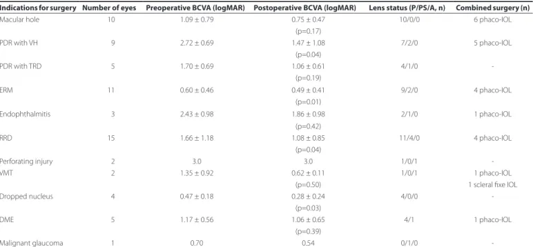
The mean age of study participants was 63.4 ± 14.2 years and 35 (54.6%) were male. The median follow-up period was 3 months (range, 1-6 months). Best corrected visual acuity increased in 39 eyes (58.2%), remained unchanged in 25 eyes (37.3%), and decreased in three eyes (4.4%) because of cataract progression in two eyes and intraoperative choroidal detachment in one eye. Surgical parameters are presented in table 1.
Figure 1. One inferotemporal 23-G TSV cannula for infusions and two superior 20-G one-step TSV cannulae with internal valves to minimize leakage.
Table 1. Surgical parameters
Indications for surgery Number of eyes Preoperative BCVA (logMAR) Postoperative BCVA (logMAR) Lens status (P/PS/A, n) Combined surgery (n)
Macular hole 10 1.09 ± 0.79 0.75 ± 0.47 10/0/0 6 phaco-IOL
(p=0.17)
PDR with VH 09 2.72 ± 0.69 1.47 ± 1.08 7/2/0 5 phaco-IOL
(p=0.04)
PDR with TRD 05 1.70 ± 0.69 1.06 ± 0.61 4/1/0
-(p=0.19)
ERM 11 0.60 ± 0.46 0.49 ± 0.41 9/2/0 4 phaco-IOL
(p=0.01)
Endophthalmitis 03 2.43 ± 0.98 1.86 ± 0.98 2/1/0 1 phaco-IOL
(p=0.42)
RRD 15 1.66 ± 1.18 1.08 ± 0.85 11/4/0 4 phaco-IOL
(p=0.04)
Perforating injury 02 3.0 3.0 1/0/1
-VMT 02 1.35 ± 0.92 0.62 ± 0.11 1/0/1 1 phaco-IOL
(p=0.50) 1 scleral fixe IOL
Dropped nucleus 04 0.47 ± 0.18 0.28 ± 0.24 4/0/0
-(p=0.03)
DME 05 1.17 ± 0.56 1.06 ± 0.65 4/1 1 phaco-IOL
(p=0.39)
Malignant glaucoma 01 0.70 0.54 0/1/0
Cartas ao Editor | Letters to the editor
2 6 6 Arq Bras Oftalmol. 2015;78(4):265-6
Suturing was not required in 60% of sclerotomies, and the remai-ning cases were closed with a single transconjunctival–scleral suture. Transient postoperative hypotony (IOP <6 mmHg) occurred in two eyes (2.9%). Increased IOP was observed in four eyes and controlled with antiglaucoma medications. Fibrin formation occurred in eight eyes (11.9%). Cataract progression was observed in six eyes and ma-naged with phacoemulsification and intraocular lens implantation. Early postoperative VH occurred in two eyes. Recurrent RD developed in three eyes and were reattached with repeated vitrectomy. Tran-sient intraoperative choroidal detachment was observed in one eye during complicated RD surgery and intraoperatively managed. No other significant complications, such as endophthalmitis; phthisis; or neovascular glaucoma, were observed.
Complicated situations during vitreoretinal surgery can be easily mastered by bimanual techniques than with conventional single-han-ded methods(3). Our technique using two superior 20-G sclerotomies for a vitrectomy probe allows bimanual surgery to be performed from a more effective approach in complicated cases.
Sutureless sclerotomy along with postoperative hypotony has been postulated to contribute to the potential risk of endophthal-mitis following TSV(4). In the present study, 20-G TSV sclerotomy ports were sutured to prevent leakage. Transient postoperative hypotony occurred in two patients, and postoperative endophthalmitis was not observed in any patients.
In a previous study, the rate of early VH was 11.4% in patients with proliferative diabetic retinopathy (PDR)(5). In our study, early postope-rative VH was observed in two eyes (2.9%), however, neither of these cases had a documented history of PDR.
In conclusion, a combined 20- and 23-G vitrectomy technique provides a feasible option in complicated cases with a reasonable com-plication rate. Future studies are required to evaluate the long-term effects of this procedure, particularly in complicated cases.
Yüksel Totan1 Emre Güler2
Submitted for publication: April 15, 2015 Accepted for publication: May 5, 2015
1 Department of Ophthalmology, Medical School, Turgut Özal University, Ankara, Turkey. 2 Department of Ophthalmology, Erciş State Hospital, Van, Turkey.
Funding: No specific financial support was available for this study.
Disclosure of potential conflicts of interest: None of the authors have any potential conflicts of interest to disclose.
Corresponding author: Emre Güler. Van Yolu Cad. No 57 - Erciş/Van 65400 - Turkey E-mail: [email protected]
REFERENCES
1. Eckardt C. Transconjunctival sutureless 23-gauge vitrectomy. Retina. 2005;25(2):208-11. 2. Spirn MJ. Comparison of 25, 23 and 20-gauge vitrectomy. Curr Opin Ophthalmol. 2009;
20(3):195-9.
3. Eckardt C. Twin lights: a new chandelier illumination for bimanual surgery. Retina. 2003;23(6):893-4.
4. Taban M, Ufret-Vincenty RL, Sears JE. Endophthalmitis after 25-gauge transconjuncti-val sutureless vitrectomy. Retina. 2006;26(7):830-1.