Is the Low-Sodium Diet Actually Indicated for All Patients with Stable
Heart Failure?
Miyoko Nakasato, Célia M. C. Strunk, Guilherme Guimarães, Marcos V. C. Rezende, Edimar A. Bocchi
Instituto do Coração (InCor) Faculdade de Medicina da Universidade de São Paulo, SP - BrazilAbstract
Background: Although a low-sodium diet is indicated for Heart Failure (HF), there is no evidence this dietary restriction is beneficial to all patients.
Objective: To prospectively study the acute effectsof a low-sodium diet in patients (pts) with heart failure (HF).
Methods: Fifty stable outpatients with mild to moderate HF who reported previously consuming 6.6 g table salt/day were studied. In Phase , all pts were submitted to a diet with g of salt during 7 days, followed by randomization in subgroups (Phase ): one to receive 6 g of salt (subgroup ) and the other, g of salt/day for 7 days (subgroup II).
Results: Phase : the diet with g of salt reduced the BMI, plasma and urinary sodium, protein consumption, iron, zinc, selenium and vitamin B; it increased plasma levels of norepinephrine, nitrate, serum aldosterone and improved quality of life. Phase : for pts with low BMI, the use of 6 g salt/day acutely decreased the levels of norepinephrine, albumin and cholesterol in plasma. No difference was observed in pts with higher BMI.
Conclusions: The diet with g salt/day for pts with HF increased the neurohormonal activation associated to HF progression. The BMI can influence the response to the neurohormonal activation in a low-sodium diet in pts with HF. Further studies to test salt restriction for longer periods are recommended.(Arq Bras Cardiol 00; 94() : 86-94)
Key Words: Diet, sodium-restricted; heart failure; sodium chloride, dietary, neurohormonal activation.
Mailing address: Miyoko Nakasato •
Rua Assungui, 50, Bl. 3 Apt. 32 - Ed. Jequitibá, Vila Gumercindo, 04.131-000, São Paulo, SP - Brazil
E-mail: [email protected]
Manuscript received January 10, 2008; revised manuscript received May 28, 2008; accepted June 12, 2008.
Introduction
Heart failure remains one of the most common, disabling, expensive and fatal medical conditions encountered by a wide range of generalist physicians and cardiologists1. It is widely accepted that pharmacological, device, and non-surgical treatments have an important role in the treatment of heart failure. The American Heart Association, in its public education site, advises limiting sodium intake; physicians often recommend to their heart failure patients to keep salt intake below 2g per day2. In fact, the restriction of dietary sodium to 2g daily or less can greatly assist heart failure patients in the maintenance of volume balance3.
Although a reduced-sodium diet is widely advocated in the management of heart failure, there is no evidence that sodium and water restrictions are beneficial to all patients1, mainly in this modern era of stable heart failure treatment with ß-blockers, ACE-inhibitors, angiotensin receptor blockers
(ARBs), and aldosterone receptor antagonists. Moreover,
the adequate amount is difficult to be established4 An undesirable consequence of this restriction is malnutrition, which may happen when patients who already have poor appetites are subjected to unpalatable diets. Furthermore, an exaggerated restriction associated with the use of diuretics can, theoretically, result in the worsening of azotemia and cardiorenal syndrome. In addition, hypovolemia can lead to neurohormonal activation, with deleterious effects to the health of heart failure patients. Furthermore, it seems clear that a more realistic target for sodium intake should be investigated to benefit heart failure patients and avoid adverse effects.
Therefore, the proposition of the present study is to evaluate the hypothesis that a low-sodium diet, associated with the standard treatment for systolic heart failure, may not be beneficial to all patients with this condition.
Methods
Study subjects
demographic, clinical, functional, and medical characteristics of the patients.
Inclusion criteria
Patients were eligible for inclusion in the study if they had been diagnosed with heart failure and: 1) were in a stable compensated phase (New York Heart Association [NYHA] functional class I, II or III), 2) had left ventricular ejection
fraction ≤ 40% measured by echocardiography in the last 6 months, 3) were aged ≥ 18 years, 4) were willing to adhere to
the low salt diet, and 5) were likely to return to the hospital.
Exclusion criteria
Patients were excluded from the study if they met the following criteria: alcoholism, acute 30-day infection, creatinine concentration > 2.5 mg/dl, systemic arterial hypertension >180/110 mmHg, body mass index > 40
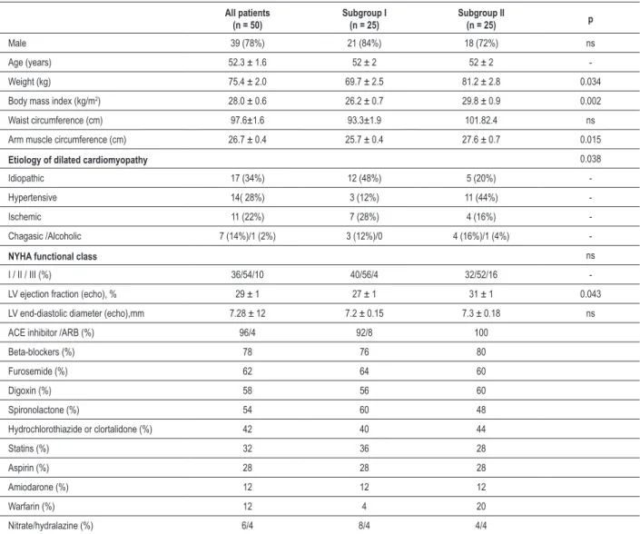
Table 1 - Baseline Characteristics of Patients at Randomization
All patients (n = 50)
Subgroup I (n = 25)
Subgroup II
(n = 25) p
Male 39 (78%) 21 (84%) 18 (72%) ns
Age (years) 52.3 ± 1.6 52 ± 2 52 ± 2
-Weight (kg) 75.4 ± 2.0 69.7 ± 2.5 81.2 ± 2.8 0.034 Body mass index (kg/m2) 28.0 ± 0.6 26.2 ± 0.7 29.8 ± 0.9 0.002
Waist circumference (cm) 97.6±1.6 93.3±1.9 101.82.4 ns Arm muscle circumference (cm) 26.7 ± 0.4 25.7 ± 0.4 27.6 ± 0.7 0.015
Etiology of dilated cardiomyopathy 0.038
Idiopathic 17 (34%) 12 (48%) 5 (20%)
-Hypertensive 14( 28%) 3 (12%) 11 (44%)
-Ischemic 11 (22%) 7 (28%) 4 (16%)
-Chagasic /Alcoholic 7 (14%)/1 (2%) 3 (12%)/0 4 (16%)/1 (4%)
-NYHA functional class ns
I / II / III (%) 36/54/10 40/56/4 32/52/16 -LV ejection fraction (echo), % 29 ± 1 27 ± 1 31 ± 1 0.043 LV end-diastolic diameter (echo),mm 7.28 ± 12 7.2 ± 0.15 7.3 ± 0.18 ns ACE inhibitor /ARB (%) 96/4 92/8 100
Beta-blockers (%) 78 76 80
Furosemide (%) 62 64 60
Digoxin (%) 58 56 60
Spironolactone (%) 54 60 48
Hydrochlorothiazide or clortalidone (%) 42 40 44
Statins (%) 32 36 28
Aspirin (%) 28 28 28
Amiodarone (%) 12 12 12
Warfarin (%) 12 4 20
Nitrate/hydralazine (%) 6/4 8/4 4/4
NYHA - “New York Heart Association”; LV - left ventricular; ACE - angiotensin converting enzyme. Continuous variables are presented as mean ± sem.
kg/m2, hypertrophic cardiomyopathy, valvular heart disease requiring surgical correction, steroid or immunosuppression for 3 months, surgery in the last 3 months, medications changes in the last 15 days or (in the case of beta-blockers) 90 days, restrictive disease, significant co-morbid conditions, such as malignancies or severe obstructive lung disease.
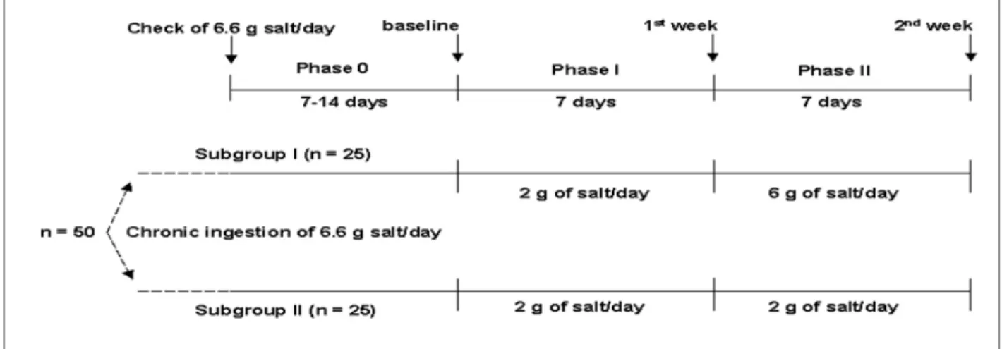
Study design
was his/her own control and a comparison was made before and after the intervention. There was no control group. In addition, we analyzed the results by dividing patients in two subgroups according to BMI and response to diet.
Data on plasma biochemistry, anthropometric measures, food consumption, and quality of life were obtained at baseline, after 7 days and again after 14 days (Figure 1).
For each individual, 24-hour urinary excretion, plasma concentration and body weight were measured at baseline and after 1 week undergoing the low-salt diet. Changes in medications or dosage were not permitted during the study. Quality-of-life questionnaires were used to study outpatients at baseline and after 1 week.
The Ethics Committee of our Institution approved the protocol of the study, and the patients agreed to participate after the explanation of its nature and purpose.
Assessment of dietary intake
All 50 patients from both groups had weekly meetings with a dietitian, who provided dietary counseling on how to decrease the intake of sodium chloride to 2 g/d or 6 g/d. These patients received instructions from the dietitian and were advised to avoid sodium-rich foods, but to otherwise keep their dietary habits.
The Burke-type dietary history method was used to provide a description of both qualitative and quantitative characteristics of the food. This information was used to estimate the usual daily food intake. To estimate energy expenditure, the Harris-Benedict equation was used to calculate basal metabolic rate; this value was then multiplied by activity factors (1.30) to determine the energy requirement for weight maintenance5. The patients were advised to maintain a fluid intake of approximately 1,000 ml during all the study phases.
Anthropometric assessment
The same observer measured the height, weight and the waist circumference. Body mass index was obtained by dividing total body weight (in kilograms) by squared height (in meters). The body mass index was classified using the
Figure 1 - Design of the study.
World’s Health Organization criteria6, that is, normal range: 18.5–24.9 kg/m2;≥ pre-obesity: 25.0–29.9 kg/m2; obesity class I: 30.0–34.9 kg/m2; obesity class II: 35.0–39.9 kg/m2;
obesity class III: ≥ 40.0 kg/m2.
Biochemical analysis
Venous blood samples were collected for norepinephrine, epinephrine, aldosterone, renin activity, B-type natriuretic peptide (BNP), IL-6, plasma nitrate, sodium, potassium, calcium, glucose, albumin, cholesterol, high-density lipoprotein cholesterol-HDL-C, triacylglycerol, urea, creatinine, iron, low-density lipoprotein cholesterol-LDL-C, and total iron-binding capacity, and transferrin measurements. Furthermore, 24-h urine collections (for sodium) were carried out in the low-salt and moderate sodium diet subgroups, at baseline and after a 7-day period.
Quality of life
Quality of life was measured by means of the Minnesota Living with Heart Failure Questionnaire (LihFE)7,8.
Statistical Analysis
All analyses were performed with the commercially available software SPSS 11.5 for Windows (SPSS Inc., Chicago, IL, USA). Continuous variables were expressed as means ± SEM, and categorical variables were presented as absolute and relative frequencies. The Student’s t test was used to analyze the comparison between groups with normal distribution. The Mann-Whitney test was used for non-parametric variables. Additionally, the analysis of variance (ANOVA) was used for repeated measures. Pearson’s Correlation Coefficients were used to assess continuous variables. A p-value less than 0.05 was considered statistically significant.
Results
and obesity class II, respectively. Only one subject complained about dizziness and weakness during the low salt diet, but remained until the end of the study.
Despite randomization, the patients from subgroup II had a higher body mass index, a bigger arm circumference and a better left-ventricular ejection fraction. The etiology of the myocardiopathy in the patients from subgroup II was predominantly hypertensive, whereas in subgroup I, it was idiopathic. Patients were divided in two subgroups based on BMI: lower BMI patients (BMI = 26.2±0.7 kg/m2) and higher BMI patients (BMI = 28±0.6 kg/m2).
There was no significant difference regarding the use of medication in either subgroup or in the biochemical variable concerning dietary intake.
Dietary intake
It was observed that salt-restriction diets induced a lower consumption of protein, phosphorus, iron, zinc, selenium, and vitamin B12.
Subgroup I patients had more intake per kg of weight of total calories, protein, fat, and selenium than those in subgroup II in both phases.
After the prescription of the low-salt diet to the patients from subgroup I, a decrease in the intake of iron, zinc and sodium was also observed (Table 2).
Anthropometric assessment
After 1 week, the low-salt diet caused weight reduction and, consequently, a decrease in the body mass index.After two weeks,patients from subgroup II continued to lose weight, whilethe patients from subgroup I stopped losing it.
Biochemical analysis
The analysis of all patients showed that the low-salt diet was responsible for an increase in serum aldosterone, plasma
Table 2 - Salt Diet Effects on Subgroups at Phases I and II
Subgroup I (BMI 26.2±0.7) Subgroup II (BMI 29.8± 0.9)
Baseline (6.6g salt) 1st week (2g salt)
2nd week
(6g salt) Baseline (6.6g salt)
1st week (2g salt)
2nd week (2g salt)
Energy (kcal/kg) 27.5 ± 2.4 24.2 ± 1.29 23.5 ± 1.30 20.1 ± 1.5 18.4 ± 1.82 18.9 ± 1.65 Protein (g/kg) 1.25 ± 0.09 1.11 ± 0.08 1.11 ± 0.08 0.92 ± 0.07 0.83 ± 0.08 0.93 ± 0.10 Fat (g) 59 ± 7 54 ± 4 51 ± 4 43 ± 3 41 ± 5 43 ± 4 Phosphorus (mg) 932 ± 78 847± 60 778 ± 60 826 ± 57 724 ± 57 780 ± 67 Magnesium (mg) 117 ± 13 130 ± 16 112 ± 10 124 ± 2.8 128 ± 15.9 123 ± 13 Iron (mg) 16.6 ± 1.4 13.8 ± 1.0* 12.8 ± 0.9 14 ± 1.3 11 ± 1.1 12 ± 1.5 Sodium (mEq) 150.3 ± 11 61.7 ± 4.2* 117.8 ± 6.2* 130.7 ± 13 67.8 ± 12.1* 59.8 ± 2.9* Zinc (mg) 15 ± 2 11 ± 2* 10 ± 1 12.2 ± 1.5 10.1 ± 1.5 11.1 ± 1.7 Selenium (mg) 101 ± 10 85 ± 8 83 ± 8 78 ± 15 72 ± 13 85 ± 13 Vit B12 (µg) 4.3 ± 0.5 2.8 ± 0.6 2.8 ± 0.5 3.8 ± 0.6 3.3 ± 0.6 3.4 ± 0.6
Data are presented as mean ± sem; *p<0.05.
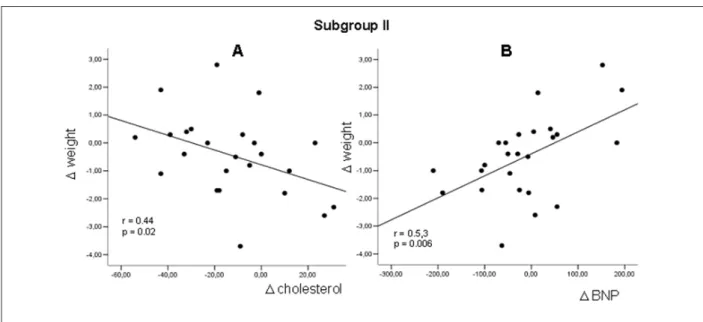
norepinephrine, plasma nitrate, serum urea, but also for a decrease in HDL-C (Figure 2). In subgroup I, the low-salt diet increased the levels of plasma norepinephrine, renin, serum aldosterone, nitrate, serum urea, serum calcium, and urine output (Figure 3). No changes were observed in 24-hour urinary sodium levels. In the patients from subgroup I, the return to the 6g-salt/day intake resulted in lower levels of plasma norepinephrine, serum calcium and total cholesterol. In subgroup II, patients receiving the low-salt diet had reduced urinary sodium, total serum cholesterol, HDL-C, and also plasma IL-6. In subgroup II, the maintenance of a low-salt diet caused an additional reduction in total serum cholesterol (Table 3-Figure 4).
Quality of life
The prescription of the low-salt diet improved the quality of life in all patients (14.78±2.09 to 11.28±1.97; p<0.05) and also in the subgroup analysis (subgroup I: 14.04 ±3.07 to 12.92±2.93; p<0.05; subgroup II: 14.64±2.90 to 9.64±2.64, p<0.05).
Discussion
The main findings of this investigation were that the recommendation to use 2g salt/day for 7 days resulted in a decrease in the intake of: a) serum sodium, b) protein , c) HDL-C, d) selenium , e) iron , f) zinc , g) and vitamin B12, as well as an increase in: h) plasma norepinephrine, i) serum aldosterone and j) serum urea.
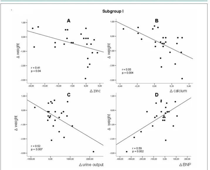
Table 3 - Anthropometric and Biochemical Data of Outpatients on a 7-day Low-Sodium Diet in Phase
Baseline (6.6 g) (n = 50)
2 g
(n = 50) p value
Anthropometric data
Weight (kg) 75.4 ± 2.0 74.9 ± 2.1 0.0052
Body mass index (kg/m2) 28.0 ± 0.6 27.8 ± 0.6 0.0049
Waist circumference (cm) 97.6±1.6 97.0±1.6 0.049 Arm muscle circumference (cm) 26.7 ± 0.4 26.5 ± 0.4 ns
Neurohormonal data
Plasma Norepinephrine (pg/ml) 551.3 ± 38.8 650.7 ± 52.4 0.0159 Plasma Epinephrine (pg/ml) 24.8 ± 4.7 28.7 ± 6.2 ns Plasma Renin activity (ng/ml/h) 16.26 ± 2.7 20.64 ± 3.1 ns Plasma BNP (pg/ml) 190.7 ± 21.1 161.8 ± 20.6 ns Plasma IL-6 (pg/ml) 7.63 ± 0.9 5.95 ± 0.6 ns Plasma Nitrate (µmol/l) 42.3 ± 6.0 44.5 ± 3.8 0.0371 Serum Aldosterone (ng/dl) 14.8 ± 2.1 19.0 ± 2.7 0.0008
Biochemical data (serum)
Sodium, mEq/l 138.2 ± 0.4 136.4 ± 0.6 0.0002
Potassium, mEq/l 4.6 ± 0.1 4.7 ± 0.1 ns
Calcium, mEq/l 4.9 ± 0.1 4.9 ± 0.03 ns
Blood glucose, mg/dl 106.7 ± 2.5 111.0 ± 3.9 ns
Albumin, g/dl 4.6 ± 0.04 4.6 ± 0.05 ns
Cholesterol, mg/dl 211.6 ± 5.7 205.8 ± 5.9 ns
LDL-C, mg/dl 137.5 ± 5.2 133.2 ± 5.6 ns
HDL-C, mg/dl 47.1 ± 1.6 45.8 ± 1.5 0.0437 Serum triacylglycerol, mg/dl 135.1 ± 9.0 134.8 ± 10.2 ns
Urea, mg/dl 45.1 ± 2.3 49.3 ± 2.7 0.0181
Creatinine, mg/dl 1.1 ± 0.04 1.2 ± 0.04 ns
Iron, mcg/dl 95.0 ± 5.9 95.5 ± 5.0 ns
Iron-binding capacity, mcg/dl 328.8 ± 8.0 331.4 ± 7.3 ns Transferrin, mg/dl 220.2 ± 6.5 223.0 ± 6.0 ns Urine output, L/day 1.319 ± 0.0656 1.380 ± 0.0752 ns Urinary sodium, mEq/day 161.2 ± 9.8 139.5 ± 9.2 0.0387
BNP - B-type natriuretic peptide; IL-6 - interleukin 6; HDL-C - high-density lipoprotein cholesterol; LDL-C - low-density lipoprotein cholesterol. Data are presented as mean ± sem; *p<0.05.
plasma IL-6, serum total cholesterol and HDL-C.
The observed reduction in serum sodium concentration is in disagreement with other publication on essential hypertension9. Mechanisms to explain the reduction in serum sodium could include restriction of sodium intake, loss of sodium due to the use of diuretics, and hemodilution10. However, the consequent hyponatremia could have potential deleterious effects, as it has been identified in several studies as a risk factor for increased morbidity and mortality in patients with heart failure. Hyponatremic patients with heart failure have poorer prognosis, significantly higher rates of major complications and mortality when compared to normonatremic
patients with heart failure11. Lower concentrations of serum sodium at admission were a predictor of an increased number of hospitalization days due to cardiovascular causes and increased mortality (within 60 days of hospital release)12. The hyponatremia observed in these studies might have been influenced by the sodium intake restriction, although a restriction of 2g of sodium per day is commonly prescribed in heart failure. Finally, our hypothesis is that the same might be happening in clinical practice.
Figure 2 -Regression Linear Plots in All Patients showing Weight Differences Between Seven-day 2g-Salt Diet (Post) and Baseline (Pre) versus Respective Differences
Between Post and Pre concerning (A) Serum Urea, (B) Serum Aldosterone, (C) Plasma Norepinephrine, and (D) Plasma BNP.
activation after sodium restriction and, consequently, hyponatremia induction, include decreased stimulation of mechanoreceptors in the left ventricle, carotid sinus, aortic arch, and renal afferent arterioles, leading to increased sympathetic and neurohormonal discharge. Moreover, a vicious cycle can be created, as increased angiotensin II and aldosterone concentrations in heart failure lead to decreased sodium and water delivery to the collecting duct. All this, combined with resistance to the natriuretic peptide action, results in the impairment of free-water excretion and hyponatremia12.
The neurohormonal activation can have deleterious effect in cases of heart failure, as the increased neurohormonal activation in hyponatremic patients may contribute to the higher mortality in this population11. Additionally, high concentrations of catecholamines, renin, angiotensin II, aldosterone, and vasopressin could contribute to the progressive remodeling and worsening of heart failure.
It seems that it has been demonstrated for the first time, as far as we know, that sodium restriction may determine a
lowered intake of protein, zinc, selenium, iron, and vitamin B12 in heart failure cases. Our reported reduction in protein intake is in accordance with a publication on the prevention of systemic hypertension16. In this study, it was demonstrated that the sodium restriction determined a lower intake of total energy, calcium, carbohydrate, protein, total fat, iron, potassium, phosphorus, zinc, riboflavin, and thiamine. We would speculate that a 2g sodium restriction could have led to an unpalatable diet. These patients have to deal with malabsorption, lack of appetite due to edema and hypomotility of the stomach and intestines, in addition to suffering from dry mouth and taste abnormalities. A direct effect of low serum sodium on appetite has not been demonstrated yet17. The reduced intake of protein and other nutrients, the increased catabolism in heart failure and the stress on the respiratory muscle18 could contribute to the development of cardiac cachexia.
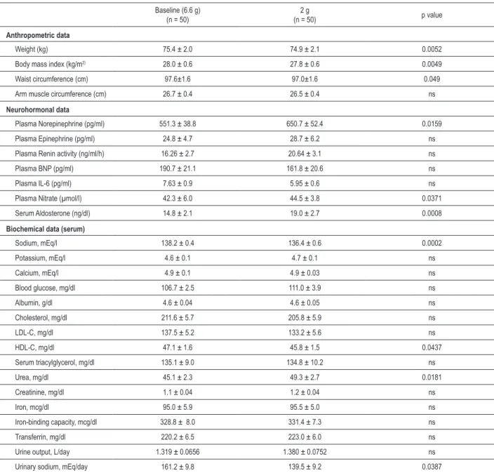
Figure 3 - Regression Linear Plots in Patients from Subgroup I showing Weight Differences Between Seven-day 2g Salt Diet (Post) and Baseline (Pre) versus Respective
Differences Between Post and Pre concerning (A) Zinc Intake, (B), Serum Calcium, (C) Urine Output, and (D) Plasma BNP.
anemia in cases of heart failure. This anemia may be associated with other factors such as: gastrointestinal malabsorption, proteinuria, chronic aspirin use, uremic gastritis, reduced intestinal iron uptake, bone marrow depression, and altered iron metabolism of chronic disease20,21. Witte et al22 demonstrated that among different groups of studied patients, 6% had vitamin B12 deficiency, 13% had iron deficiency and 8% had folate deficiency. In another study, 17% of the patients were found to be anemic and 21% had anemia caused by iron deficiency23. Anemia has been associated with functional class impairment and high mortality in heart failure24.
The reduction in selenium intake can be associated with worsening of exercise capacity. Selenium is an essential trace element and one of its main roles is to be an antioxidant in the glutathione peroxidase enzyme, in addition to being the major intracellular antioxidant. Low zinc concentrations correlate with cardiovascular medication and also with reduced nutritional protein intake. It is also associated with higher concentrations of lipid peroxidase, a marker of oxidative stress,
suggesting that zinc acts as an antioxidant25.
Our observation of the increment in nitrate levels after a 2g-sodium diet could suggest that it might be a compensatory mechanism. It would serve to counteract the increase in vasoconstriction resulting from neurohormonal activation - demonstrated by high concentrations of catecholamines and aldosterone. However, it is not known whether an increase in nitric oxide concentrations may be beneficial or aggravating to heart failure cases26. A decreased nitric oxide production is only one manifestation of endothelial dysfunction in the case of pulmonary hypertension. Nitric oxide can act via a number of additional mechanisms, such as programmed cell death, cytotoxic effects or modulation of myocyte oxygen consumption, all of which may result in impaired cardiac function and prognosis in patients with heart failure27.
Figure 4 - Regression Linear Plots in Patients from Subgroup II showing Weight Differences Between Seven-day 2g-Salt Diet (Post) and Baseline (Pre) versus Respective
Differences between Post and Pre concerning (A) serum Cholesterol and (B) Plasma BNP .
development of cardiorenal syndrome may be important, as this syndrome is related to a worsened prognosis in heart failure, followed by hyponatremia, as observed in our population4.
The observed acute improvement in quality of life could be explained by a reduction in pulmonary congestion and by protocol follow-up, which shows that monitoring can have an impact on the perception of quality of life in heart failure. However, our results are in disagreement with published data showing absence of correlation between serum sodium and quality of life response12.
Epidemiological studies have shown a strong association between obesity and increased risk of cardiovascular disease and mortality in the general population. Increased BMI is associated with an increased risk of heart failure. However, in contrast to what is observed in the general population, Curtis et al27 found out that overweight patients with heart failure have a lower risk of death, when compared to patients who have a healthy weight. These findings confirm the existence of an “obesity paradox” among patients with established heart failure27. However, if obesity is protective, then loss of weight attempts could be associated with increased mortality risk26. Our results suggest that the obese and the non-obese patients with heart failure may have different responses to interventions and, one attractive hypothesis is that it could influence the prognosis of heart failure. As evidence, in our study, the sodium restriction led to neurohormonal activation only in individuals with low body mass index.
Study limitations
Even though the short-term follow-up is limited, it is necessary for the development of a long-term safety study that would include a non-usual procedure in HF. Hence, our results justify a long-term randomized study that would test a diet with
normal salt-intake in the treatment of heart failure. Despite the fact that the short duration is a limitation of this study, it is sufficient to demonstrate the change in anthropometric, biochemical and dietary data. In addition to randomization, the other limitations were the differences regarding some characteristics, such as body mass index, left ventricular ejection fraction and etiology of dilated cardiomyopathy between the two study subgroups. Outpatients without symptoms or with mild symptoms, as long as they were clinically stable, were included in this study. The selected patients represent a typical sample of stable outpatients undergoing ambulatory follow-up.
Self-reporting of food consumption could be interpreted as a limitation. Nevertheless, this method seems to be the most suitable, as it represents the outpatients’ routine.
Conclusion
This prospective study shows that, despite the acute improvement in quality of life, the 2g-salt/day restriction diet for patients with HF:
1) increased neurohumoral activation associated with progression of HF, such as plasma norepinephrine and serum aldosterone,
2) reduced food consumption, 3) did not change the BNP.
References
1. McMurray JJV, Pfeffer MA Heart failure. Lancet. 2005; 65: 1877-89.
2. American Heart Association [Homepage on the Internet]. Learn and live: cutting down on salt. [Acessed 21 November 2005]. Available from: http:// americanheart.org/presenter.jhtml?indentifier=336
3. ACC/AHA 2005 guideline update for the diagnosis and management of chronic heart failure in the adult: a report of the American College of Cardiology/American Heart Association Task Force on Practice Guidelines for the Evaluation and Management of Heart Failure). Hunt SA. J Am Coll Cardiol. 2005; 46 (6): e1-82.
4. Swedberg K, Cleland J, Dargie H, Drexler H, Follath F, Komajda M, et al. Guidelines for the diagnosis and treatment of chronic heart failure. The task force for the diagnosis and treatment of CHF of the European Society of Cardiology. Eur Heart J. 2005; 26: 1115-40.
5. Harris JA, Benedict FG. A biometric study of basal metabolism in man. Washington: Carnegie Institute of Washington; 1919.
6. Obesity: preventing and managing the global epidemic: report of a WHO Consultation on Obesity. Geneva: World Health Organization; 1997. p. 1-276.
7. Rector TS, Kubo SH, Cohn JN. Patient self-assessment of their congestive heart failure part 2: content, reliability and validity of a new measure the Minnesota Living with Heart Failure Questionnaire. Heart Fail. 1987; 3: 198-209.
8. Rector TS, Cohn JN. Assessment of patient outcome with the Minnesota Living with Heart Failure questionnaire: reliability and validity during a randomized, double blind, placebo controlled trial of pimobendan. Am Heart J. 1992; 124: 1017-25.
9. Grassi G, Dell’Oro R, Seravalle G, Foglia G, Trevano FQ, Mancia G. Short- and long-term neuroadrenergic effects of moderate dietary sodium restriction in essential hypertension. Circulation. 2002; 106: 1957-61.
10. Issa VS, Bacal F, Mangini S, Carneiro RMD, Azevedo CHNF, Chizzola PR, et al. Solução salina hipertônica para prevenção de insuficiência renal em pacientes com insuficiência cardíaca descompensada e hiponatremia. Arq Bras Cardiol. 2007; 89 (4): 251-5.
11. Oren RM. Hyponatremia in congestive heart failure. Am J Cardiol. 2005; 95 (Suppl): 2-7.
12. Klein L, O’Connor CM, Leimberger JD, Gattis-Stough W, Pina IL, Felker GM, et al. OPTIME-CHF Investigators. Lower serum sodium is associated with increased short-term mortality in hospitalized patients with worsening heart failure: results from the outcomes of a prospective Trial of Intravenous Milrinone for Exacerbations of Chronic Heart Failure (OPTIME-CHF) study. Circulation. 2005; 111 (19): 2454-60.
13. Watson RDS, Esler MD, Leonard P, Korner PI. Influence of variation in dietary sodium intake on biochemical indices of sympathetic activity in normal man.
Clin Exp Pharmacol Physiol. 1984; 11: 163-70.
14. Alvelos M, Ferreira A, Bettencourt P, Serrão P, Pestana M, Cerqueira-Gomes M, et al. The effect of dietary sodium restriction on neurohumoral activity and renal dopaminergic response in patients with heart failure. Eur J Heart Fail. 2004; 6: 593-9.
15. Fujiwara N, Osanai T, Kamada T, Katoh T, Takahashi K, Okumura K. Study on the relationship between plasma nitrite and nitrate levels and salt sensitivity in human hypertension: modulation of nitric oxide synthesis by salt intake. Circulation. 2000; 101 (8): 856-61.
16. Morris CD. Effect of dietary sodium restriction on overall nutrient intake. Am J Clin Nutr. 1997; 65 (Suppl): 687S-91S.
17. Jacobsson A, Pihl-Lindgren E, Fridlund B. Malnutrition in patients suffering from chronic heart failure; the nurse’s care. Eur J Heart Fail. 2001; 3: 449-56.
18. Nicol SM, Carroll DL, Homeyer CM, Zamagni CM. The identification of malnutrition in heart failure patients. Eur J Cardiovasc Nurs. 2001; 1: 139-49.
19. Berger MM, Mustafa I. Metabolic and nutritional support in acute cardiac failure. Curr Opin Clin Nutr Metab Care. 2003; 6: 195-201.
20. Okonko DO, Anker SD. Anemia in chronic heart failure: pathogenetic mechanisms. J Cardiac Fail. 2004; 10 (Suppl 1): S5-9.
21. Klutstein MW, Tzivoni D. Anaemia and heart failure: aetiology and treatment. Nephrol Dial Transplant. 2005; 20 (Suppl 7): vii7-10.
22. Witte KKA, Clark AL, Cleland JGF. Chronic heart failure and micronutrients. J Am Coll Cardiol. 2001; 37: 1765-74.
23. Felker GM, Adams KF, Gattis WA, O´Connor CM. Anemia as a risk factor and therapeutic target in heart failure. J Am Coll Cardiol. 2004; 44: 959-66.
24. Sales ALF, Villacorta H, Reis L, Mesquita ET. Anemia como fator prognóstico em uma população hospitalizada por insuficiência cardíaca descompensada. Arq Bras Cardiol. 2005; 84 (3): 237-40.
25. Node K, Kitakaze M, Yoshihara F, Sasaki T, Kuzuya T, Hori M. Increased cardiac levels of nitric oxide in patients with chronic heart failure. Am J Cardiol. 2000; 86: 474-7.
26. Drexler H, Kästner S, Strobel A, Studer R, Brodde OE, Hasenfub G. Expression, activity and functional significance of inducible nitric oxide synthase in the failing human heart. J Am Coll Cardiol. 1998; 32: 955-63.
27. Curtis JP, Selter JG, Wang Y, Rathore SS, Jovin IS, Jadbabaie F, et al. The obesity paradox. Arch Intern Med. 2005; 165: 55-61.
Potential Conflict of Interest
No potential conflict of interest relevant to this article was reported.
Sources of Funding
This study was funded by FAPESP.
Study Association