https://doi.org/10.1590/0004-282X20180114
ARTICLE
Restless legs syndrome in Parkinson’s disease
and increased cardiovascular risk
Síndrome das pernas inquietas em doença de Parkinson e aumento do risco cardiovascular
Maren de Moraes e Silva
1, Cezar Henrique Lorenzi
2, Blenda Barreto Schneider
3, Catherine Enk Fischer
Seidel
4, Isabela Salomé
5, Viktor Cleto Morais Gianini
6, Renata Ramina Pessoa
1, Pilar Bueno Siqueira
Mercer
1, Maria Carolina Zavagna Witt
1Restless legs syndrome (RLS) is a disorder
character-ized by an uncontrollable need to move the legs, that occurs
during rest, and which is relieved by movement of the lower
limbs
1; however, the sensation may occur in other parts of
the body as well. In this way, RLS interferes markedly with
the individuals sleep quality. In its pathophysiology, there is
1Hospital da Cruz Vermelha Brasileira Filial do Paraná, Departamento de Neurologia, Curitiba PR, Brasil; 2Universidade Federal do Paraná,Curitiba PR, Brasil;
3Universidade Positivo, Curitiba PR, Brasil;
4Faculdade Evangélica do Paraná, Curitiba PR, Brasil; 5Pontifícia Universidade Católica do Paraná, Curitiba PR, Brasil; 6Faculdades Pequeno Príncipe, Curitiba PR, Brasil.
Correspondence: Maren de Moraes e Silva; Avenida Vicente Machado, 1310; 80420-011Curitiba PR, Brasil; E-mail: [email protected]
Support: Financial support was responsibility of the authors themselves, and there was no external source of funding for this paper.
Conflict of interest: There is no conflict of interest to declare.
Received 14 May 2018; Received in final form 01 July 2018; Accepted 29 July 2018.
ABSTRACT
Restless legs syndrome (RLS) is a disorder commonly found in patients with Parkinson’s disease, with descriptions for both conditions
impairing dopaminergic transmission in central nervous system. Previous studies in varied populations indicate an association between
the presence of RLS and increased cardiovascular risk and, so far, there are no consistent studies of this association in Parkinson’s disease.
Objective:
To analyze the influence of RLS on cardiovascular risk in patients with Parkinson’s disease.
Methods:
We reviewed the medical
records of 202 patients diagnosed with Parkinson’s disease and verified the presence of RLS, cardiovascular comorbidities, blood pressure
measurements, lipid profiles and Framingham Risk Scores.
Results:
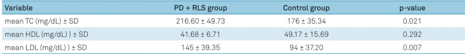
Statistically significant higher values of total cholesterol were found
for the RLS group (mean 216.6 mg/dL), as well as for LDL cholesterol (mean 145 mg/dL). No statistical difference was found among the
other factors.
Conclusion:
Patients with Parkinson’s disease and RLS have a higher prevalence of dyslipidemia than patients without RLS,
suggesting a correlation between restless legs and hyperlipidemia. It is questioned whether the dopaminergic substrate is the main factor in
the genesis of the syndrome, as even with the use of dopaminergic agonists by both groups, it was possible to observe differences between
groups. The hypothesis of the real interference of the syndrome treatment as a protective factor for cardiovascular risk was generated.
Keywords:
Parkinson disease; restless legs syndrome; cardiovascular diseases; dyslipidemias.
RESUMO
Síndrome das pernas inquietas é um distúrbio comumente encontrado em pacientes com doença de Parkinson (DP), havendo descrições
para ambas as condições de prejuízos na transmissão dopaminérgica no sistema nervoso central. Estudos prévios em populações diversas
indicam associação entre a presença da síndrome e aumento do risco cardiovascular, não havendo, até o momento, pesquisas consistentes
a respeito desta associação em DP.
Objetivo:
Analisar a influência da síndrome das pernas inquietas no risco cardiovascular em pacientes
com DP.
Métodos:
Foram revisados prontuários de 202 pacientes com diagnóstico de DP e verificada a presença de síndrome das pernas
inquietas, comorbidades cardiovasculares, aferições de pressão arterial, lipidograma e escore de Framingham.
Resultados:
Valores
maiores e estatisticamente significativos de colesterol total foram encontrados para o grupo com pernas inquietas (média de 216.6 mg/
dL), assim como para colesterol LDL (média de 145 mg/dL). Não foi encontrada diferença estatística entre os demais fatores.
Conclusões:
Pacientes com DP e síndrome das pernas inquietas têm maior prevalência de dislipidemia do que pacientes sem a síndrome, o que sugere
correlação entre síndrome das pernas inquietas e hiperlipidemia. É posto em prova o substrato dopaminérgico como principal na gênese
da síndrome, uma vez que, mesmo sob o uso de agonistas dopaminérgicos por ambos os grupos, foi possível observar diferenças entre os
estratos. Gerada a hipótese da real interferência do tratamento da síndrome como fator de proteção para o risco cardiovascular.
a decrease in central nervous system dopaminergic levels,
especially at night
2,3, with dopaminergic agonists playing a
role in improving symptoms. Other theories, such as iron
accumulation, have also been cited in literature
3.
In previous studies involving diverse populations,
RLS seems to increase several cardiovascular risk
fac-tors
4,5,6, mainly due to interference in sleep-wake cycle,
culminating in an exacerbated sympathetic activity in this
period
4,5,6,7,8. There is also evidence that RLS can lead to
impairment in cardiovascular system autonomic control,
regardless the presence of changes in sleep quality or
peri-odic leg movements
9.
Both RLS and Parkinson’s disease (PD) present with
alter-ations in dopaminergic transmission in their
pathophysiol-ogy. In addition, studies have shown that the prevalence of
RLS in patients with PD is higher than in general
popula-tion
10,11,12. Taking into account the possible deleterious effect
of RLS on cardiovascular function, it is essential that the
associations between these morbidities be established, given
the frequency of the syndrome in this population. The pres
-ent study sought to establish the prognostic impact of RLS in
Parkinson’s disease.
METHODS
This study was initiated after approval by the research
ethics committee of Universidade Positivo under the
reg-istration number CAAE 73905517.3.0000.0093 and
con-ducted according to ethical standards in accordance with
Resolution 466/2012.
Participants
The records of patients diagnosed with idiopathic PD
who attend a specialized center in a city in southern Brazil
were reviewed. We included patients diagnosed with PD
according to the UK Parkinson’s Disease Society Brain Bank
Clinical Diagnostic Criteria
13. Only patients with 50 years
or older were included in the study. Alcohol abuse patients,
smokers, patients who had diagnosed coronary artery
dis-ease before onset of RLS or who presented with a weight
gain of more than 5 kg in the last six months were excluded
from the study.
Data selection
A retrospective observational study was performed. The
patients were previously selected through medical records
analysis, according to the inclusion and exclusion criteria.
The patients were divided into two groups: PD+RLS patients
and PD patients without the syndrome (controls). The diag
-nosis of RLS was made according to the diagnostic criteria of
American Academy of Sleep Medicine
2.
Cardiovascular risk was evaluated using the Framingham
Risk Score, a previous vascular event, blood pressure
mea-surement, laboratory examination and presence of
periph-eral arterial disease.
Data analysis
Continuous data were expressed as means. Frequencies
were expressed in percentages. Numerical variables were
compared using the ANOVA test, while categorical variables
were analyzed using the chi-square test. Statistical signifi
-cance was set at p < 0.05.
RESULTS
Population characteristics
Data were analyzed for 202 patients in total: 57.43% were
male and the population mean age was 70.12 ± 9.49 years. The
mean age at onset of PD symptoms was 59.86 ± 10.64 years,
with a mean duration of symptoms of 9.64 ± 5.68 years at the
time of our evaluation. The prevalence of RLS was 7.92% (16
patients). The sample distribution in relation to demograph
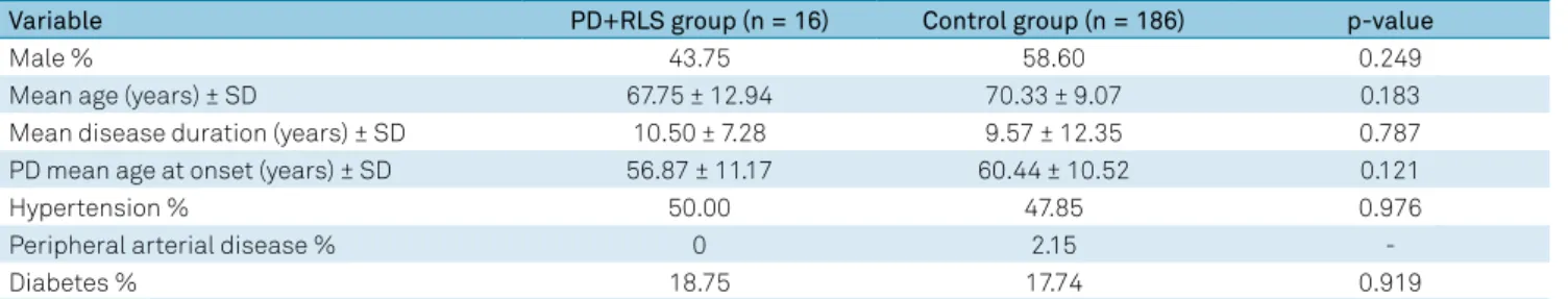
-ics and comorbidities for PD + RLS and control groups is
shown in Table 1.
Cardiovascular risk and relations
The occurrence of stroke, acute myocardial infarction and
heart failure after the diagnosis of RLS was verified, although
there was no prevalence of these morbidities in the PD+RLS
group. In the control group, the occurrence of these
condi-tions was 7.43%, 2.47% and 1.98%, respectively.
The Framingham Risk Score was calculated for
partici-pating patients, with a mean score of 15.27 ± 7.76 for
con-trol group and 12.90±4.53 for PD+RLS group. However, the
Table 1.
Characteristics and comorbidities distribution by group.
Variable
PD+RLS group (n = 16)
Control group (n = 186)
p-value
Male %
43.75
58.60
0.249
Mean age (years) ± SD
67.75 ± 12.94
70.33 ± 9.07
0.183
Mean disease duration (years) ± SD
10.50 ± 7.28
9.57 ± 12.35
0.787
PD mean age at onset (years) ± SD
56.87 ± 11.17
60.44 ± 10.52
0.121
Hypertension %
50.00
47.85
0.976
Peripheral arterial disease %
0
2.15
-Diabetes %
18.75
17.74
0.919
difference found between groups wasn’t statistically
sig-nificant (p = 0.605).
Regarding systolic blood pressure, a mean of 121.43±30.23
mmHg (median 120) was observed for control group and
121.20 ± 18.23 mmHg (median 120) for PD+RLS group. This
difference wasn’t statistically significant (p = 0.976).
Participants lipid profile of was also evaluated, with
a mean in general population of 179.50 ± 38.02 mg/dL for
total cholesterol. The separate groups analysis is shown
in Table 2. Among control group, 19.35% of patients had a
diagnosis of dyslipidemia and had received statin
prescrip-tions in previous consultaprescrip-tions, compared with 43.75% of
the PD+RLS group.
DISCUSSION
It’s believed that subcortical dopaminergic dysfunctions
play an important role in RLS genesis, as well as in PD, with
dopaminergic agonists responsible for symptoms
improve-ment in both conditions
3. In addition, a risk relation can be
established between RLS and PD
14, with a greater occurrence
of the syndrome in patients with PD
10,11,12. Impairment in
dopaminergic transmission, common in the etiology of PD
and RLS, may explain why the prevalence of RLS in patients
with PD is high.
The relation between RLS and cardiovascular risk has pre
-viously been explored, but at present, there is no confirmation
of this relation in PD population, specifically. It’s postulated
that RLS can lead to heart disease through several potential
mechanisms: its negative effect on sleep quality and duration,
coexisting sympathetic activation accompanying periodic
limb movements during sleep, or presence of common risk
fac-tors for heart disease
8. In addition, interruption of sleep,
com-mon in patients with RLS, increases the daytime heart rate
and blood pressure through elevated peripheral sympathetic
tonus
5,15. The present study, however, didn’t directly assess the
impact of this morbidity on sleep quality.
In our study, RLS prevalence was compatible with
previ-ous literature findings for general population, ranging from
5% to 10%
16,17,18,19and involving more women than men.
The difference between genders may be due to the fact that
women generally perceive and report symptoms more often
than men and because of hormonal differences. In previous
studies addressing RLS in PD exclusively, showed prevalence
rates with great variability (0% to 50%)
11,12,20,21. However, due
to the fact that the treatment of PD involves dopaminergic
compounds, which also alleviate RLS symptoms
17, the true
prevalence of RLS maybe masked in this population
22. This
risk, however, is called into question by studies that have
reported an increased prevalence of RLS, compared with
general population, in patients with long-term dopamine
use, indicating the possible contradictory correlation of RLS
increasing with the duration of previous dopaminergic drug
treatment
23,24. Because the present study showed a
statisti-cally significant difference for lipid profile changes, it’s impor
-tant to reconsider the role of dopamine as unique in genesis
of this syndrome, taking into account the probability of other
questions related to the possible increase in cardiovascular
risk of these patients, such as common risk factors. Moreover,
in our study, no statistically significant difference was found
regarding the length of PD treatment and presence of RLS.
Regarding cardiovascular risk, we analyzed the
Framingham Risk Score, blood pressure measurements and
lipid profiles, and found a statistically significant relation
only for high values of total cholesterol and LDL (low density
lipoprotein), which appeared to be higher in PD patients with
RLS. We could find no data in literature comparing RLS and
lipid profiles in PD population. There is already evidence that
the prevalence of dyslipidemia is higher in individuals with
RLS than in individuals without the syndrome
25,26. A
prospec-tive study found that elevated levels of total cholesterol were
associated with a higher risk of developing RLS, which didn’t
change significantly after exclusion of patients using statins,
indicating that the correlation didn’t occur due to drug side
effects
27. There was also a correlation between sleep quality
in patients with RLS and dyslipidemia, with an association
between poorer sleep quality and LDL levels, shorter sleep
duration and total cholesterol levels, and greater daytime
dysfunction and LDL levels in patients with RLS
28. Metabolic
disorders may occur along with activation of the
hypotha-lamic-pituitary-adrenal axis and with inflammation, which
may trigger a role in RLS genesis
29. In addition, there are
reports of RLS caused by microembolization of cholesterol
crystals
30. These data corroborate this study findings, indicat
-ing a correlation between dyslipidemia and RLS; however, the
findings described here are new as they are specific for the
PD population.
Regarding the relation between cardiovascular risk
fac-tors and RLS in other morbidities, we found conflicting evi
-dence. A prospective study in United Kingdom revealed an
association between RLS and incidence of stroke, but an
Table 2.
Lipid profile by group.
Variable
PD + RLS group
Control group
p-value
mean TC (mg/dL) ± SD
216.60 ± 49.73
176 ± 35.34
0.021
mean HDL (mg/dL) ) ± SD
41.68 ± 6.71
49.17 ± 15.69
0.292
mean LDL (mg/dL) ) ± SD
145 ± 39.35
94 ± 37.20
0.007
association wasn’t found between RLS and ischemic heart
disease
31. This finding was corroborated by another paper,
in which it was also observed that the syndrome was more
frequent in individuals with various comorbidities, including
high body mass index, hypertension and diabetes
32. Contrary
to this analysis, some studies have shown that RLS is
asso-ciated with cardiovascular disease in patients with frequent
symptoms and RLS diagnosed for more than three years
8,33. Li
et al.
8identified a significant association between long-term
RLS and coronary disease. According to their study, women
with RLS for more than three years were at increased risk for
coronary heart disease, an association that was independent
of the main risk factors for coronary disease. Other studies
have suggested a lower cardiac vagal modulation and lower
heart rate response to orthostatic stress in subjects with
RLS
34,35. The positive correlation between RLS and cardio
-vascular risk obtained by these studies maybe explained by
the reduction of cardiovascular baroreflex gain and greater
peripheral vascular resistance observed in patients with RLS
- factors not attributed to differences in sleep quality, sug
-gesting that the syndrome can directly contribute to changes
in cardiovascular system autonomic control
9. Corroborating
the above findings, another study indicated that RLS may
be related to cardiovascular disease, coronary disease and
hypertension, but with different relative risks according to
primary or secondary classification of RLS. Primary RLS
would be a risk factor for hypertension, but not for
cardio-vascular disease or coronary disease. However, when second
-ary to another morbidity, including PD, RLS would be
associ-ated with the three conditions: hypertension, cardiovascular
disease and coronary disease
36.
A previous study by Oh et al.
31failed to find a statistically
significant association between PD, RLS and cardiovascular
diseases; however, according to a study by Banno et al.
25,
noc-turnal hypertension and supine hypertension occurred more
frequently in patients with PD and RLS than in patients with
PD without RLS. In the present study, there were no significant
differences in blood pressure values between the control group
and the PD+RLS group, while other cardiovascular comorbidi
-ties were not frequent enough for an adequate comparison.
This article is limited by being retrospective, and the
sample analyzed was relatively small, which may justify not
identifying the correlation of RLS with other factors, such as
a statistically significant difference for the Framingham Risk
Score or blood pressure, already described in previous studies
with various populations. Regarding the overlap between PD
and RLS treatment as a possible factor for diagnostic
mask-ing, our results test the dopaminergic substrate as the main
factor in the RLS genesis, as even with the use of
dopami-nergic agonists in both groups, PD+RLS and control, it was
possible to observe differences between the strata, a theory
corroborated by previous findings
3,24. A hypothesis is
gener-ated, as well, of the real interference of the treatment of RLS
syndrome as a protection factor for cardiovascular risk.
Our findings, which indicated a relationship between
an unfavorable lipid profile of patients with PD (high total
cholesterol and LDL) and the presence of RLS, are new and
not previously reported in the literature, but consistent with
findings in other populations. Future research is needed to
confirm this possible relationship. Based on this, it’s possible
that new forms of primary prevention and overall care may
be found for patients with PD.
References
1. American Academy of Sleep Medicine (AASM). The international classification of sleep disorders: diagnostic and coding manual. 2nd ed. Wenchester): American Academy of Sleep Medicine; 2005. 2. Clemens S, Rye D, Hochman S. Restless legs syndrome:
revisiting the dopamine hypothesis from the spinal cord perspective. Neurology. 2006 Jul;67(1):125-30. https://doi.org/10.1212/01.wnl.0000223316.53428.c9
3. Winkelman JW. Considering the causes of RLS. Eur J Neurol. 2006 Oct;13 Suppl 3:8-14.
4. Billars L, Hicks A, Bliwise D, Sigmundsson T. Hypertension risk and PLMS in restless legs syndrome. Sleep. 2007;30:A297-8. 5. Ekstedt M, Akerstedt T, Söderström M. Microarousals during
sleep are associated with increased levels of lipids, cortisol, and blood pressure. Psychosom Med. 2004 Nov-Dec;66(6):925-31. https://doi.org/10.1097/01.psy.0000145821.25453.f7 6. Pennestri MH, Montplaisir J, Colombo R, Lavigne G, Lanfranchi
PA. Nocturnal blood pressure changes in patients with restless legs syndrome. Neurology. 2007 Apr;68(15):1213-8. https://doi.org/10.1212/01.wnl.0000259036.89411.52 7. Manconi M, Ferri R, Zucconi M, Clemens S, Rundo F, Oldani A et
al. Effects of acute dopamine-agonist treatment in restless legs syndrome on heart rate variability during sleep. Sleep Med. 2011 Jan;12(1):47-55.
8. Li Y, Walters AS, Chiuve SE, Rimm EB, Winkelman JW, Gao X. Prospective study of restless legs syndrome and coronary heart disease among women. Circulation. 2012 Oct;126(14):1689-94. https://doi.org/10.1161/CIRCULATIONAHA.112.112698 9. Bertisch SM, Muresan C, Schoerning L, Winkelman JW, Taylor
JA. Impact of Restless Legs Syndrome on Cardiovascular Autonomic Control. Sleep (Basel). 2016 Mar;39(3):565-71. https://doi.org/10.5665/sleep.5528
10. Guerreiro TM, Nishikawa DR, Ferreira LC, Melo HA, Prado RC. Restless legs syndrome in Parkinson’s disease: clinical characteristics and biochemical correlations. ArqNeuropsiquiatr. 2010 Dec;68(6):869-72. https://doi.org/10.1590/S0004-282X2010000600007
11. Braga-Neto P, Silva-Júnior FP, Sueli Monte F, Bruin PF, Bruin VM. Snoring and excessive daytime sleepiness in Parkinson’s disease. J Neurol Sci. 2004 Jan;217(1):41-5. https://doi.org/10.1016/j.jns.2003.08.010
12. Loo HV, Tan EK. Case-control study of restless legs syndrome and quality of sleep in Parkinson’s disease. J Neurol Sci. 2008 Mar;266(1-2):145-9. https://doi.org/10.1016/j.jns.2007.09.033
13. Hughes AJ, Daniel SE, Kilford L, Lees AJ. Accuracy of clinical diagnosis of idiopathic Parkinson’s disease: a clinico-pathological study of 100 cases. J NeurolNeurosurgPsychiatry. 1992
14. Grupo Brasileiro de Estudos em Síndrome das Pernas Inquietas - GBE-SPI. [Restless legs syndrome: diagnosis and treatment. Opinion of Brazilian experts]. ArqNeuropsiquiatr. 2007;65 3a:721-7. Portuguese. https://doi.org/10.1590/S0004-282X2007000400035 15. Davies RJ, Belt PJ, Roberts SJ, Ali NJ, Stradling JR. Arterial blood
pressure responses to graded transient arousal from sleep in normal humans. J Appl Physiol (1985). 1993 Mar;74(3):1123-30. https://doi.org/10.1152/jappl.1993.74.3.1123
16. Allen RP, Walters AS, Montplaisir J, Hening W, Myers A, Bell TJ et al. Restless legs syndrome prevalence and impact: REST general population study. Arch Intern Med. 2005 Jun;165(11):1286-92. https://doi.org/10.1001/archinte.165.11.1286
17. Cho YW, Shin WC, Yun CH, Hong SB, Kim JH, Allen RP et al. Epidemiology of restless legs syndrome in Korean adults. Sleep. 2008 Feb;31(2):219-23. https://doi.org/10.1093/sleep/31.2.219 18. Phillips B, Hening W, Britz P, Mannino D. Prevalence and
correlates of restless legs syndrome: results from the 2005 National Sleep Foundation Poll. Chest. 2006 Jan;129(1):76-80. https://doi.org/10.1378/chest.129.1.76
19. Vogl FD, Pichler I, Adel S, Pinggera GK, Bracco S, De Grandi A et al. Restless legs syndrome: epidemiological and clinicogenetic study in a South Tyrolean population isolate. Mov Disord. 2006 Aug;21(8):1189-95. https://doi.org/10.1002/mds.20922 20. Lang AE, Johnson K. Akathisia in idiopathic Parkinson’s disease.
Neurology. 1987 Mar;37(3):477-81. https://doi.org/10.1212/WNL.37.3.477 21. Tan EK, Lum SY, Wong MC. Restless legs syndrome in
Parkinson’s disease. J Neurol Sci. 2002 Apr;196(1-2):33-6. https://doi.org/10.1016/S0022-510X(02)00020-5 22. Bhalsing K, Suresh K, Muthane UB, Pal PK. Prevalence and
profile of Restless Legs Syndrome in Parkinson’s disease and other neurodegenerative disorders: a case-control study. Parkinsonism RelatDisord. 2013 Apr;19(4):426-30. https://doi.org/10.1016/j.parkreldis.2012.12.005
23. Angelini M, Negrotti A, Marchesi E, Bonavina G, Calzetti S. A study of the prevalence of restless legs syndrome in previously untreated Parkinson’s disease patients: absence of co-morbid association. J Neurol Sci. 2011 Nov;310(1-2):286-8. https://doi.org/10.1016/j.jns.2011.08.012 24. Lee JE, Shin HW, Kim KS, Sohn YH. Factors contributing to
the development of restless legs syndrome in patients with Parkinson disease. Mov Disord. 2009 Mar;24(4):579-82. https://doi.org/10.1002/mds.22410
25. Banno K, Delaive K, Walld R, Kryger MH. Restless legs syndrome in 218 patients: associated disorders. Sleep Med. 2000 Jul;1(3):221-9. https://doi.org/10.1016/S1389-9457(00)00023-X
26. Schlesinger I, Erikh I, Avizohar O, Sprecher E, Yarnitsky D. Cardiovascular risk factors in restless legs syndrome. Mov Disord. 2009 Aug;24(11):1587-92. https://doi.org/10.1002/mds.22486 27. De Vito K, Li Y, Batool-Anwar S, Ning Y, Han J, Gao X. Prospective
study of obesity, hypertension, high cholesterol, and risk of restless legs syndrome. Mov Disord. 2014 Jul;29(8):1044-52. https://doi.org/10.1002/mds.25860
28. Winkelman JW, Shahar E, Sharief I, Gottlieb DJ. Association of restless legs syndrome and cardiovascular disease in the Sleep Heart Health Study. Neurology. 2008 Jan;70(1):35-42. https://doi.org/10.1212/01.wnl.0000287072.93277.c9 29. Schilling C, Schredl M, Strobl P, Deuschle M. Restless legs
syndrome: evidence for nocturnal hypothalamic-pituitary-adrenal system activation. MovDisord. 2010 Jun;25(8):1047-52. https://doi.org/10.1002/mds.23026
30. Harvey JC. Cholesterol crystal microembolization: a cause of the restless leg syndrome. South Med J. 1976 Mar;69(3):269-72. https://doi.org/10.1097/00007611-197603000-00007
31. Oh YS, Kim JS, Park IS, Song IU, Son YM, Park JW et al. Association between nocturnal/supine hypertension and restless legs syndrome in patients with Parkinson’s disease. J Neurol Sci. 2014 Sep;344(1-2):186-9. https://doi.org/10.1016/j.jns.2014.06.056
32. Winkelman JW, Finn L, Young T. Prevalence and correlates of restless legs syndrome symptoms in the Wisconsin Sleep Cohort. Sleep Med. 2006 Oct;7(7):545-52. https://doi.org/10.1016/j.sleep.2006.01.004 33. Cikrikcioglu MA, Hursitoglu M, Erkal H, Kınas BE, Sztajzel J, Cakirca
M et al. Oxidative stress and autonomic nervous system functions in restless legs syndrome. Eur J Clin Invest. 2011 Jul;41(7):734-42. https://doi.org/10.1111/j.1365-2362.2010.02461.x
34. Izzi F, Placidi F, Romigi A, Lauretti B, Marfia GA, Mercuri NB et al. Is autonomic nervous system involved in restless legs syndrome during wakefulness? Sleep Med. 2014 Nov;15(11):1392-7. https://doi.org/10.1016/j.sleep.2014.06.022
35. Van Den Eeden SK, Albers KB, Davidson JE, Kushida CA, Leimpeter AD, Nelson LM et al. Risk of Cardiovascular Disease Associated with a Restless Legs Syndrome Diagnosis in a Retrospective Cohort Study from Kaiser Permanente Northern California. Sleep (Basel). 2015 Jul;38(7):1009-15. https://doi.org/10.5665/sleep.4800