Original Article
Experience with Heterotopic Heart Transplantation in Patients with
Elevated Pulmonary Vascular Resistance. Late Follow-up
Jose Henrique Andrade Vila, José Pedro da Silva, Luciana da Fonseca, José Francisco Baumgratz, Américo Tangari Jr,
Weverton Ferreira Leite, Claudia Jesus Guilhen, Egas Armelin
Hospital Beneficência Portuguesa de São Paulo, São Paulo, SP - Brazil
Abstract
Background: Along the past few years the number of papers on heterotopic cardiac transplant has been very scarce in the medical literature, including at the international level; this is particularly true in reference to the long term follow-up of these patients and the reason which led to the presentation of our report.
Objective: To report the initial clinical experience and late evolution of 4 patients undergoing heterotopic heart transplantation, indications for this procedure and its major complications.
Methods: The surgeries were performed between 1992 and 2001, and all had as indication for heterotopic transplantation the PVR, which ranged from 4.8 WU to 6.5WU, with a transpulmonary gradient above 15mmHg. In the 3rd patient, a direct anastomosis between the pulmonary arteries was performed without the use of a prostetic tube, and a mitral valvuloplasty and a LV aneurysmectomy were performed in the native heart.
The immediate immunosuppressive regimens were double, with cyclosporine and azathioprine in the first 3 patients, and cyclosporine and mycophenolate mofetil in the 4th patient.
Results: One immediate death occurred from graft failure, one death occurred after 2 ½ years, from endocarditis in an intraventricular thrombus in the native heart, and a third death occurred 6 years after transplantation, from post-operative complications of the aortic valve surgery in the native heart. The remaining patient is well, 15 years after the transplantation. This patient is in functional class II (NYHA), 6 years after a surgical occlusion of the native heart aortic valve.
Conclusion: Heterotopic heart transplantation results are inferior to those of orthotopic heart transplantation because they present higher RVP. The intraventricular thrombi, in the native heart, which require prolonged anticoagulation, and aortic valve complications, also in the native heart, may require surgical treatment. However, a patient’s 15-year survival has demonstrated a long-term effectiveness of this option for selected patients. (Arq Bras Cardiol 2010;94(2): 244-249) Key Words: Heart transplantation; hypertension, pulmonary / complications; follow-up studies.
Mailing address: Jose Henrique Andrade Vila •
Rua Gaivota, 222/81, Indianópolis - 0422-030, São Paulo, SP - Brazil E-mail: [email protected]
Manuscript received on August 22, 2008; revised manuscript received on October 31, 2008; accepted on November 11, 2008.
Introduction
The high pulmonary vascular resistance (PVR) which occurs in the final stage of cardiomyopathies has been a major obstacle to the performance of orthotopic heart transplantation, due to the risk of early failure of the right ventricle (RV) of the transplanted organ, limiting its use in patients with PVR below 5 Wood units (WU)1,2, or 4 Wood
units, currently in preference.
The clinical application of the heterotopic heart transplantation (HetTrx) by Barnard, in 1974, increased the possibility of performing cardiac transplantation in patients with a PVR above 4 WU3,4, and it was widely used by the
South African Group, especially with the additional argument that in case of graft failure, in a scenario when cyclosporine
was not yet available, the patients would survive thanks to the native organ. Among other indications of this procedure, we can mention its use in potentially reversible ventricular dysfunctions, and in cases of size disproportion between donor and recipient. This last condition is a possibility, particularly in patients with ischemic cardiomyopathy, even with the possibility of conventional surgical treatment, when the heterotopic graft could be implanted for greater security in the early evolution of these patients3-5.
The aim of this article is to report the experience of four procedures performed in our department, emphasizing the details of the procedures, a modification in the original operative technique, and the later follow-up of the patients, up to 15 years.
Methods
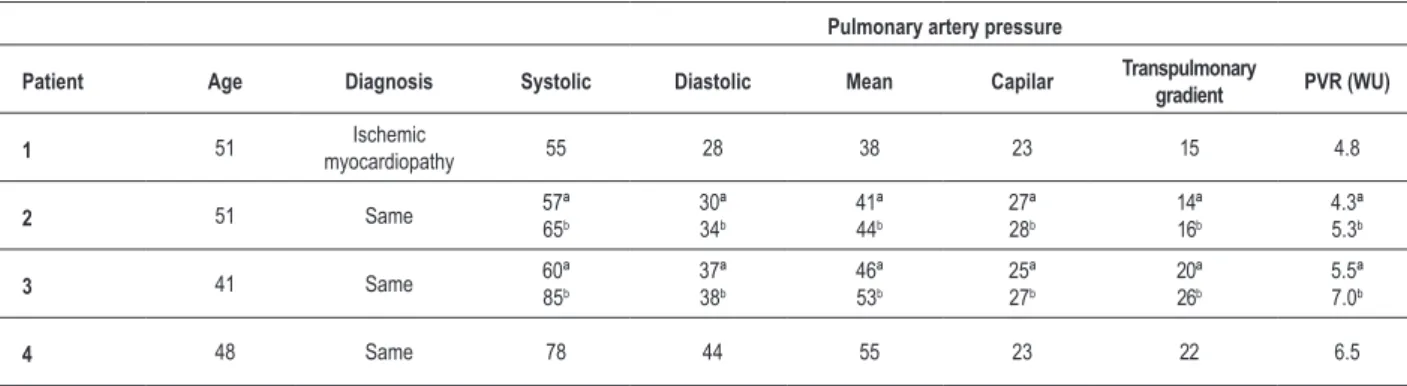
re-Table 1 – Preoperative clinical characteristics
Pulmonary artery pressure
Patient Age Diagnosis Systolic Diastolic Mean Capilar Transpulmonary
gradient PVR (WU)
1 51 Ischemic
myocardiopathy 55 28 38 23 15 4.8
2 51 Same 57ª
65b
30ª 34b
41ª 44b
27ª 28b
14ª 16b
4.3ª 5.3b
3 41 Same 60ª
85b
37ª 38b
46ª 53b
25ª 27b
20ª 26b
5.5ª 7.0b
4 48 Same 78 44 55 23 22 6.5
A - previous measurement, B - immediate preoperative measurement.
transplants. In this experiment, in 4 patients, we opted for the heterotopic transplantation, according to the marked increase of PVR. The clinical characteristics of these 4 patients are described in Table 1, which emphasizes the significantly high levels of PVR.
From a clinical perspective, the patients had severe ischemic cardiomyopathy. All of them had extensive myocardial infarction, and their coronary arteries were not suitable to surgical treatment. They were in the ICU, receiving inotropic and vasodilator drugs, and were on the waiting list for heart transplantation. The first patient had undergone a previous myocardial revascularization surgery, with occlusion of the grafts, and the last patient had undergone coronary angioplasty on two occasions prior to the transplantion. In the latter patient, who underwent surgery in November 2001, in addition to a graft implant in a heterotopic position, the correction of a left ventricle aneurysm and a valvuloplasty of the mitral valve were performed. The other two patients were also in functional class IV (NYHA), and they had been frequently admitted to the ICU to receive vasodilator and inotropic drugs intravenously.
The surgical procedure was carried out with distant donor hearts in patients 1, 3 and 4, and with the heart from a donor in the adjacent room in patient 2. The donor organs were removed by sectioning the aorta above the brachiocephalic trunk and the pulmonary artery (PA) in the proximal part of the pulmonary branches. For myocardial protection, we used the St. Thomas cardioplegic solution, with antegrade infusion via native aortic valve in the heart of the receiver, except in the last case, in which we used retrograde coronary sinus infusion. In the donor heart we also used the antegrade cardioplegia administered via aortic infusion. We associated topical hypothermia by the immersion of the organs in cold Ringer’s solution, and by packaging them for local or distant transportation. In the heart of the receiver, myocardial protection was performed with a cold blood cardioplegic solution.
The implantation surgical techniques were similar in the first two cases - the pulmonary artery of the donor heart was connected to the trunk of the native PA, using a bovine pericardium graft to facilitate the anastomosis. In patients 3 and 4 the original technique was used, with a direct anastomosis of the donor’s PA to the right branch of the receiver’s PA,
without any interposition graft, because the distance between them was much shorter5. To make this procedure feasible,
the superior vena cava of the receiver was transected near the right atrium (RA), its lower end was sutured and its upper end was mobilized, fully exposing the right branch of the PA. To make this possible, we needed a wide dissection of the donor’s pulmonary artery and its left branch, when the organ was removed. After the anastomoses of the left atria and the PAs were performed, the upper end of the superior vena cava of the receiver was anastomosed to the superior vena cava of the donor. Another right atrial connection was made in the lower portion of the receiver RA, near the interatrial septum, allowing the venous return of the receiver to reach the donor RA. The long ascending aorta of the donor was anastomosed as far as possible from the ascending aorta of the recipient, so as to facilitate the access to the PA in case of bleeding.
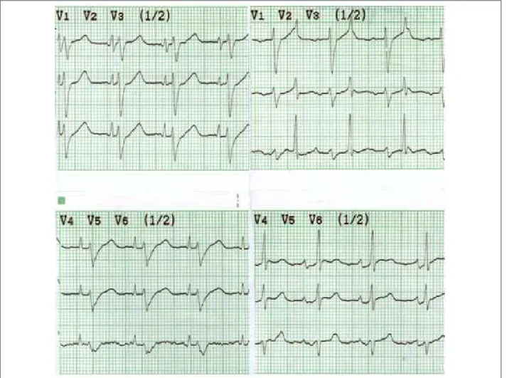
As an external pacemaker, temporary electrodes were placed in the RA of both hearts and in the RV of the transplanted heart, enabling a synchronized cardiac pacing through a sequential pacemaker, connecting the atrial poles to the atrial electrodes of the native heart (of higher frequency) and the atrial electrodes of the transplanted heart to the ventricular poles of the pacemaker. Thus, the pacemaker feels the P-wave of the native heart and, after an adjustable pause, stimulates the atrium of the transplanted heart, producing synchronized beats of both hearts, in the same logical sequence of the intra-aortic balloon which is important in optimizing the cardiac output in the first postoperative days6-8. After discharge from
the ICU and the removal of epicardial wires of the pacemaker, there was no more electrical synchronization between the two hearts; however, a spontaneous tendency to an equalization of the heart beats occurred, which could be attributed to the lower adrenergic stimulation provided by an improved cardiac output, due to the implanted heart. (Figure 1)
Original Article
Vila et al
Heterotopic heart transplantation, high PVR
Figure 1 -On the left column, classic precordial leads showing the implanted heart beats and the native heart beats, with large QRS. In the right column, inverse precordial
leads, in a different time period, showing the native heart beats and the implanted heart beats. Notice the similar heart rates with no pacemaker resynchronization.
responsible for the worsening of the myocardial function. After the first episode, we started triple immunosuppression with prednisone 0.5 mg/kg/d, which was gradually reduced to 0.1 mg/kg/d after an intravenous pulse for three days.
The third and fourth patients had a single grade II rejection episode each, which was controlled by an oral regimen of prednisone for two months. We also maintained oral anticoagulant in patients 3 and 4, who had late survival, and diuretics, enzyme antagonists and, then, the beta-blocker carvedilol, in order to prevent or delay the progressive myocardial dysfunction of the native heart.
This practice, however, was not successful; on the contrary, the deterioration of the left ventricular function of the native heart was relentless, and even both patients with late survival showed progressive aortic insufficiency with marked clinical worsening, which required surgical correction, as mentioned in the literature9.
Results
The early and late results of the surgeries are summarized in Table 2. Patient 2 died at the hospital, 12 hours after
transplantation, from graft and native heart failure.
Patient 1 had good evolution in the first year and then showed progressive heart failure, after two episodes of acute rejection and coronary artery disease in the graft. A retransplantation was performed two years after the transplantation, which resulted in death from low cardiac output and acute sepsis on the seventh postoperative day.
Initially the 3rd patient had an adequate evolution, with
good physical capacity—functional class II (NYHA)—practicing sports, such as swimming, regularly. Figure 2 shows chest X-rays features, with the conventional surgical technique and the new technique developed by the group5. In this patient,
who underwent the new surgical technique, the pulmonary angiography showed good anatomical and functional results (Figure 3).
Table 2 – Results
Patient Evolution 1 Evolution 2
1
Retransplantation after 2 years due to rejection and coronary disease in the graft
Death from low cardiac output and acute sepsis on the 7th day after the
retransplantation 2 In-hospital death
3 aortic valve after 6 yearsSurgical oclusion of the in functional class II (NYHA)15-year evolution, currently
4 Surgical oclusion of the aortic valve after 7 years
Death after 7 years and 4 months
Figure 2 -Chest X-rays (PA view) with the conventional technique. At left: with the use of a prosthetic tube; at right: with a direct anastomosis between the pulmonary
arteries. Notice in this case the lower compression of the right lung.
Figure 3 - Pulmonary angiography using the technique of a direct anastomosis between the pulmonary arteries without the use of a prosthetic tube. There is a good
distribution of low between the two lungs.
with a deterioration of the clinical conditions, despite the increase in the clinical treatment with enzyme antagonists (captopril), spironolactone, carvedilol and diuretics. We opted for the surgical correction of the aortic valve
insufficiency, which was preferred for technical reasons to the native organ explantation, and this resulted in a significant functional improvement of the patient, who returned to functional class II (NYHA). The ventricular function of the graft in heterotopic position in this patient had always been normal, and there was no significant rejections detected by non-invasive tests, such as myocardial mapping with Gallium and echocardiogram.
Original Article
Vila et al
Heterotopic heart transplantation, high PVR
with a total survival of 7 years and 4 months after the transplant surgery.
Discussion
In reviewing our results with orthotopic heart transplantation, we observed that the pulmonary hyperresistance was an important direct or indirect cause of hospital mortality. This is in line with the experience of the Stanford University, which reported a mortality rate of 42% in the first three months, when the receivers’ PVR was more than 2.5 WU, despite the use of sodium nitroprusside (Nipride R), and a mortality rate of 25 % for those whose PVR was reduced to less than 2.5 WU with Nipride R, though at the expense of systemic hypotension10.
The indications for HetTrx are still controversial, but the patients with PVR between 4.5 and 7 WU benefit from this technique, according to the majority of transplant centers10,11. However, in cases of PVR of 7 or more fixed
units, a cardiopulmonary transplantation is preferred to the HetTrx because, besides the higher surgical risk, this level of PVR is associated with irreversible and probably progressive anatomical changes in the pulmonary vasculature1. In the
latter group, a unilateral heart and lung block transplantation is also possible, with the possibility of reverting the anatomical and functional changes in the pulmonary vasculature due to the immediate reduction in the pulmonary pressure and the consequent stimulus for pulmonary vascular reaction12.
A more simple, practical and accurate criterium was proposed by the group of Pittsburg13, based on the
transpulmonary gradient (TPG), which is the difference between the mean pulmonary artery pressure and the mean pulmonary capillary pressure. This study demonstrated that patients with TPG greater than 15 mmHg had higher mortality rates than those with TPG from 10 to 15; the mortality was even lower when the gradient was less than 10 mmHg. Furthermore, this study does not define what is the gradient that renders the cardiopulmonary transplant necessary.
It is not always easy to work based on hemodynamic data as proposed by most authors, especially in Brazil where there is a long delay between the preoperative hemodynamic study and the day when the transplant is performed, which often results in the progression of the vascular lung disease. Therefore, we have routinely used a hemodynamic monitoring with vasodilator drugs on the day of the transplant, in the ICU or in the operating room (before anesthetic induction), using a Swan-Ganz catheter, while we awaited the arrival of the donor. It may be surprising (Table 1) to observe that the 2nd
patient had 1 WU above the initial preoperative measure. This assessment allows us, in cases of higher PVR, to use pulmonary vasodilator drugs preventively, including via inhalation, and orally, after discharge from the ICU.
In patient 1, we noticed that the PVR was a little less than 5 WU, but the TPG was above 10. The severe clinical evolution of this patient in the last 3 months of waiting stressed the indication for a HetTrx, which was reinforced by the finding of a donor of inappropriate size (weight 15% lower).
As to the new technique, which was used in 3 patients and avoids the use of the Dacron graft between the pulmonary
arteries, it represents the first major innovation in the last 20 years and has the advantages of preventing the theoretically possible complications with the artificial graft (infection, thrombosis, pulmonary embolism and obstruction by fibrous panus), and it also facilitates the endomyocardial biopsy, because the anastomosis of the cava superior easily leads the biopsy forceps to the RV of the transplanted heart5.
As to the late results, the HetTrx showed lower survival than the orthotopic transplantation (one-year actuarial survival of 61.4%, and 56% for 2 years), but this comparison is valid only as an information source, since the HetTrx patients have different clinical characteristics in comparison to the orthotopic heart transplantation patients14.
In this small experiment, we observed that the patients with global cardiomyopathy, involving also the RV, as our patient 2, suffer higher risk during surgery, because the receiver’s diseased RV would have to maintain the pulmonary circulation after the aortic clamping, which is usually long, during the transplantation. Beyond this more immediate risk, there is the problem of progression of the cardiomyopathy, which made the presence of this heart disadvantageous due to potential complications. However, the patients who have ischemic cardiomyopathies that are restricted to the LV, with only RV hypertrophy, as in cases 1, 3 and 4 of our experience, are ideal candidates, because they will have a better initial performance, and their RV will perform well its role of overcoming the high PVR.
However, in patients 3 and 4, the long-term follow up demonstrated the complications mentioned in the literature15,16, mainly progressive deterioration of the left
ventricular function and aortic insufficiency due to severe anuloaortic ectasia, in the native heart. This latter complication required corrective surgery in the third patient 6 years ago, resulting in better clinical conditions, with the exclusion of the native heart from circulation, which was maintained satisfactorily by the implanted heart, and the patient was in good clinical conditions, 15 years after surgery. In the fourth patient, during the surgical correction of the aortic valve insufficiency, an attempt of total explantation of the native heart was complicated by an episode of severe stroke, and the patient died at the hospital.
The causes of progressive ventricular dysfunction in the native heart could be summarized as follows: 1) Progression of myocardial disease, due to arrhythmias and progression of coronary disease in the native heart; 2) elevation of the post-load due to dyssynchrony of the implanted heart with the native heart, which may lead to the simultaneous occurrence of heart beats, determining an important elevation of the impedance during the systole of the diseased heart; 3) anullo-aortic dilatation by the mechanisms mentioned above, determining progressive aortic insufficiency9.
Cowell et al17 and Morris-Thurgood et al18 demonstrated that
References
1. Griepp RB, Stinson EB, Doog E, Clark DA, Shumway NE. Determinants of operative risk in human heart transplantation. Am J Surg. 1971; 122 (2): 192-7.
2. Barnard CN. The present status of heart transplantation. S Afr Med J. 1975; 49 (7): 213-7.
3. Barnard CN, Losman JG. Left ventricular bypass. S Afr Med J. 1975; 49 (9): 303-12.
4. Novitzky D, Cooper DK, Barnard CN. The surgical technique of heterotopic heart transplantation. Ann Thorac Surg. 1983; 36 (4): 476-82.
5. Da Silva JP, Cascudo MM, Baumgratz JF, Vila JH, Wafae Filho M, de Carvalho Neto DO, et al. Heterotopic heart transplantation: a direct pulmonary artery anastomosis technique. J Thorac Cardiovasc Surg. 1994; 108 (4): 795. 6. Melvin KR, Pollick C, Hunt Sa, McDougall R, Goris ML, Oyer P, et al.
Cardiovascular physiology in a case of heterotopic cardiac transplantation. Am J Cardiol. 1982; 49 (5): 1301-7.
7. Beck W, Gersh BJ. Left ventricular bypass using a cardiac allograft: hemodynamic studies. Am J Cardiol. 1976; 37 (7): 1007-12.
8. Losman JG, Barnard CN. Heterotopic heart transplantation: a valid alternative to orthotopic transplantation: results, advantages and disadvantages. J Surg Res. 1982; 32 (4): 297-312.
9. Akasaka T, Lythall D, Cheng A, Yoshida K, Yoshikawa J, Mitchell A, et al. Continuous aortic regurgitation in severely dysfunctional native heart after heterotopic cardiac transplantation. Am J Cardiol. 1989; 63 (20): 1483-8. 10. Costard A, HilI I, Schroder J, Fowler M. Response to nitroprusside L predictor
of early post-transplant mortality. J Am Coll Cardiol. 1989; 13: 62A. 11. Kirklin JK, Naftel DC, Kirklin JW, Blackstone EH, White-Williams C, Bourge
RC. Pulmonary vascular resistance and the risk of heart transplantation. J Heart Transplant. 1988; 7 (5): 331-6.
12. Da Silva JP, Vila JH, Cascudo MM, Baumgratz JF, Saraiva PA, Netto CD. Heart and unilateral lung transplantation for cardiomyopathy with high pulmonary vascular resistance. Ann Thorac Surg. 1992; 53 (4): 700-2.
13. Kormos RL, Thompson M, Hardesty RL, Griffith BP, Trento A, Urestik BF, et al. Utility of preoperative right heart catheterization data as a predictor of survival after heart transplantation. J Heart Transplant. 1986; 5: 391.
14. Kaye MP. The Registry of the International Society for Heart Transplantation: Fourth Official Report -1987. J Heart Transplant. 1987; 6 (2): 63-7. 15. Sivaratnam DA, Kelly MJ, Esmore D, Richardson M, Kalff V. Demonstrating
time sequence and extent of sustained decrease in native heart ejection fraction after heterotopic transplantation. J Heart Lung Transplant. 2004; 23 (6): 690-5.
16. Hildebrandt A, Reichenspurner H, Gordon GD, Horak A, Odell J. Heterotopic heart transplantation: mid-term hemodynamic and echocardiographi analysis: the concern of arteriovenous-valve incompetence. J Heart Transplant. 1990; 9: 675-82.
17. Cowell RP, Morris-Thurgood J, Coghlan JG, Ilsley CD, Mitchell AG, Khaghani A, et al. Effects of paced counterpulsation on exercise capacity and hemodynamics after heterotopic heart transplantation. Am J Cardiol. 1995; 75 (5): 415-7.
18. Morris-Thurgood J, Cowell RP, Paul V, Kalsi K, Seymour AM, Ilsley C, et al. Hemodynamic and metabolic effects of paced linkage following heterotopic cardiac transplantation. Circulation. 1994; 90 (5): 2342-7.
19. Mulligan MS, Shearon TH, Weill D, Pagani FD, Moore J, Murray S. Heart and lung transplantation in the United States, 1997-2006. Am J Transplant. 2008; 8 (4 Pt 2): 977-87.
20. Keenan RJ, Konishi H, Kawai A, Paradis IL, Nunley DR, Iacono AT, et al. Clinical trial of tacrolimus versus ciclosporine in lung transplantation. Ann Thorac Surg. 1995; 60 (3): 580-4.
result, this could lead to a better functional preservation of the left ventricle of the native heart and perhaps the prevention of valve dysfunctions of the organ.
In conclusion, this experience with heterotopic heart transplantation was illustrative, because despite a late survival of 15 years, with the patient in good condition, even in this case, a surgical procedure for functional exclusion of the native heart was necessary. The other patients died, one at hospital, other two years after surgery, and the remaining 7 years after surgery. We believe, however, that the progressive ventricular dysfunction in the native heart, and the annulo-aortic ectasia and its consequent aortic insufficiency could be postponed by the employment of permanent synchronization by pacemaker, which was not used at the time in our patients. With it, this procedure could compare itself favorably with, for instance, cardio-pulmonary transplantation, another option for the
treatment of patients with high PVR, in which a survival of 15 years is peculiarly rare19-20.
Potential Conflict of Interest
No potential conflict of interest relevant to this article was reported.
Sources of Funding
There were no external funding sources for this study.
Study Association