INTRODUCTION
Gastroesophageal reflux disease (GERD) is the end results of involuntary gastric contents relux into the esophagus, causing heartburn and acid regurgitation symptoms or injury to esopha-geal tissue(2,13). Thus, esophagitis is considered as a complication
of GERD(25). GERD includes two types; erosive esophagitis and
non-erosive reflux disease (NERD) (endoscopy-negative reflux disease) (1). GERD classiied into four grades (A, B, C and D)
ac-cording to Los Angeles classiication(22). Other classiication was
Savary – Miller that classiied it into ive grades (I, II, III, IV and V)(4). The prevalence of GERD symptoms varies between 9% and
42%(15). In spite of high prevalence of GERD symptoms, its aetiology
is still not completely understood(9). One of the important factors in
the causation of GERD is environmental factors(3). These include
life style factors like body weight, nutrition, alcohol consumption, smoking, the intake of non-steroidal anti-inlammatory drugs, and sleeping position(16). Regarding genetic factors; there is an evidence
for the role of genetic component to GERD as conirmed by familial aggregation of GERD symptoms(21). Other studies done in Sweden
and UK in monozygotic and dizygotic twins revealed considerable genetic contribution to the aetiology of GERD(3,6). Prevalence of
gastrointestinal relux symptoms in Asian population are affected by racial and ethnic factors(8,14). All these suggest a genetic etiology
of GERD in addition to environmental factors. Other genetic factor is Human Leukocyte Antigen (HLA) system, which has an extensive polymorphism and considered as an excellent marker for population genetic analyses and disease association. Rajendra et al.(18) found
that inheritance of the HLA-B*07 gene confers an increased risk for
Human leukocyte antigen HLADRB1 determinants
susceptibility to gastroesophageal reflux disease
Batool Mutar
MAHDI
1, Riyadh Mohamad
HASAN
2and Wafaa Hazim
SALIH
1Received 25/8/2016 Accepted 20/9/2016
ABSTRACT – Background – Gastroesophageal relux disease (GERD) is characterized by diverse symptoms. There is an evidence for a genetic component to GERD as supported by familial aggregation of this disease. Objective – To investigate whether certain human leucocyte antigen genes HLA-DRB1 are associated with GERD. Methods – Patients and controls were prospectively recruited from GIT center at Al-Kindy Teaching Hospital (Baghdad-Iraq) between January 2014 and July 2016. Sixty Iraqi Arab Muslims patients with a history of heartburn and dyspepsia compared with 100 Iraqi Arab Muslims controls. All study patients and control groups underwent upper gastrointestinal endoscopic examinations and their serums were analyzed for CagA antibodies Immunoglobulin G (IgG) for H. pylori. HLA-DRB1 genotyping were done to both groups. Results – A total of 60 patients with erosive gastritis; GERD (Grade II and III) were evaluated, together with 100 controls. There is a signiicant increase of H. pylori infection (P=0.0001) in GERD patients than control group. HLA-DRB1* 15:01 was signiicantly increased in GERD patients in comparison with control group and an increased frequency of HLADRB1*11:01 in control group compared with patients group. Conclusion – There is an association between HLA-DRB1 *15:01 in GERD patients with H. pylori positive patients.
HEADINGS – HLA-DR1 antigen. Gastroesophageal relux. Helicobacter pylori.
Declared conflict of interest of all authors: none Disclosure of funding: no funding received
1 Department of Microbiology, Al-Kindy College of Medicine, Baghdad University, Iraq; 2 Department of Surgery, Al-Kindy College of Medicine, University of Baghdad, Iraq.
Correspondence: Batool Mutar Mahdi. Director of HLA typing Research Unit. Department of Microbiology. Al-kindy college of Medicine. Baghdad University. AL-Nahda Square – Baghdad-Iraq. E-mail: [email protected]
Barrett’s esophagus in south Asians (mostly south Indians) but not Orientals (Malays and Chinese). They also showed that HLA-B*07 positive patients with Barrett’s esophagus had a signiicantly higher family history of GERD symptoms, compared with their HLA-B*07 negative counterparts. HLA molecules perform a central function in the regulation of the immune response in many diseases. HLA alleles might predispose some individuals to particular diseases and malignancies(11). Many studies found the associations between HLA
alleles and susceptibility or resistance to certain diseases.
In this study, we examine if the human leucocyte antigen HLA-DRB1 alleles, important for immune responsiveness, may be a sus-ceptibility locus for GERD disease in Iraqi Arab Muslims patients.
METHODS
Patients and controls were prospectively recruited from GIT center at Al-Kindy Teaching Hospital (Baghdad-Iraq) between January 2014 and July 2016. The demographic details of all patients and control groups were recorded. Written informed consent was obtained from all patients and control group for this study. The study protocol was reviewed and approved by the Scientiic and Ethical Committee of Al-kindy medical college and Al-Kindy Teaching Hospital. The patient group and control groups were sex and age matched.
Exclusion criteria included those patients with Barrett’s eso phagus and esophageal varices. Patients with secondary causes of gastro-oesophageal relux disease, patients who had consumed antacids, H2 blockers, proton pump inhibitors (PPIs), non-steroidal anti-inlammatory drugs (NSAIDs), alcohol, history of Helicobac-ter pylori eradication, subjects with a history of gastrointestinal surgery, peptic ulcer, and gastric cancer or with systemic disease requiring chronic medication were excluded.
The 100 Iraqi Arab Muslims controls consisted of people un-dergoing upper gastrointestinal endoscopy for reasons other than relux symptoms, Barrett’s esophagus or any form of dyspepsia and heartburn. This group included those with normal OGD and being investigated for anaemia or faecal occult blood positive stools, chronic diarrhoea for unknown reason requiring small bowel biopsy, irritable bowel syndrome and screening for familial adenomatous polyposis.
Oesophagogastric examinations
All studied patients and control groups underwent upper gastrointestinal endoscopic examinations using gastroscope: GIF-H260; Olympus, Tokyo, Japan and Display screen; Olympus OEV-261H liquid crystal display monitor; Olympus, Tokyo, Japan. The gastroesophageal junction was deined as the squamoco-lumnar junction and the proximal margin of gastric folds. The endoscopic indings of erosive esophagitis in the lower esophagus were classiied using the Savary and Miller classiication. All of them either Grade II which is conluent erosive or exudative mu-cosal lesions which do not extend around the entire esophageal circumference or Grade III which is erosive or exudative mucosal lesions which cover the entire esophageal circumference and lead to inlammation of the wall without stricture (according to Savary and Miller, 1979) (23). Histopathological study was done by taking
specimens from gastric mucosa to conirm the diagnosis and pres-ence of H. pylori.
Serological tests
Blood samples (3 mL) were drawn into plain tube. Separated serums were analyzed for CagA antibodies Immunoglobulin G (IgG) for H. pylori using an immunological test (immunochroma-tography test) (ACON, USA).
HLA Class II genotyping (HLA-DRB1)
Two mL of venous blood were collected in EDTA containers for DNA extraction from human blood using blood kit (QIAmp DNA blood Mini Kit, QIAGEN INC- Germany).
DNA concentration and puriication product was estimated using Nanodrop –South Korea. DNA was veriied by electropho-resis in a 2% agarose gel containing ethidium bromide and was visualized under UV light.
Locus- and allele-speciic ampliication of genomic patients and control DNA was performed for DRB1. DNA Ampliica-tion and HybridizaAmpliica-tion was performed using a sequence-speciic oligonucleotide probes (SSOP) by HLA-DRB1 ampliication and hybridization kits (SSO HLA type DRB1 plus and Mastermix for HLA type DRB1 Amp plus kits -Innogenetics-Belgium) by AutoLipa – 48Innogenetics-Belgum. The results were interpreted using LiRas version-5.0 software- Innogenetics-Belgium.
Statistical analysis
HLA-DRB1 frequencies were determined by direct counting. The frequency of each allele was compared between patients and control group using chi-square test Fisher exact test using MiniTab version. 3.0 software. In each comparison, the Odds ratio (OR) along with the 95% conidence interval (95% CI) was used. Gene frequencies for both groups were calculated. P-value less than 0.05 were considered statistically signiicant.
RESULTS
A total of 60 patients with erosive gastritis; GERD (Grade II and III) were evaluated, together with 100 controls. The mean age of patients was 45.67±5.54, as compared with 44±15.22 for the controls. The male to female sex ratio was 1.0 in the patients group versus also 1.0 in controls. About 50% of them were GERD II and the rest were GERD III by endoscopy.
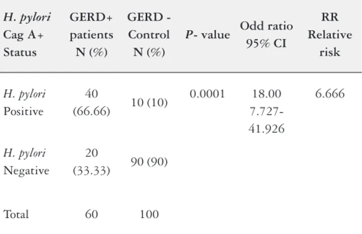
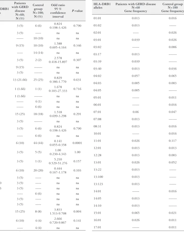
There is a signiicant increase of H. pylori infection (P=0.0001) in GERD patients than control group. The Odd ratio (OD) =18.00 with 95% CI = from 7.727-41.926. The relative risk =6.666 that indicates an association between H. pylori and disease as shown in Table 1. Control and patients were typed for identifying the DRB1* alleles using DNA based methodology (PCR-SSOP). There was an increased frequency of HLADRB1*11:01 in control group compared with patients group (P=0.0001, Odds ratio=0.141, 95% CI: 0.055-0.358). Other allele like HLA-DRB1* 15:01 was significantly increased in GERD patients in comparison with control group (P=0.004, Odds ratio=3.833, 95% CI: 1.513-9.708) as shown in Table 2.
The highest genotype frequency in GERD patients was 15:01 which is equal to 0.065 while in the control group was 11:01 which is 0.117 as shown in Table 3.
The distribution of HLA polymorphism HLA*DRB1 was in-vestigated in the control and patients groups of Iraqi Arab Muslims. The observed and expected phenotypes of all loci for the patients group as demonstrated in Table 4 were in a good agreement with Hardy-Weinberg equilibrium as shown in Table 5.
TABLE 1. Helicobacter pylori in GERD (Grade II and III) patients compared with control group
H. pylori
Cag A+ Status
GERD+ patients
N (%)
GERD - Control
N (%)
P- value Odd ratio 95% CI
RR Relative
risk
H. pylori Positive
40
(66.66) 10 (10)
0.0001 18.00 7.727-41.926
6.666
H. pylori Negative
20
(33.33) 90 (90)
Total 60 100
TABLE 2. Frequencies of HLA-DRB1 alleles in patients with GERD disease compared with control group
HLA-DRB1 alelles
Patients with GERD
disease N=60;
N (%)
Control group N=100;
N (%)
Odd ratio 95 % conidence
interval
P-value
01:01 3 (5) 6 (6) 0.824
0.198-3.426 0.790
01:02 3 (5) --- na na
02:01 --- 10 (10) na na
03:01 9 (15) 10 (10) 1.588
0.605-4.164 0.346
03:02 --- 14 (14) na na
03:17 3 (5) 2 (2) 2.578
0.418-15.897 0.307
03:39 9 (15) --- na na
03:40 3 (5) --- na na
04:02 13 (21.66) 25 (25) 0.829
0.386-1.770 0.631
04:03 1 (1.66) 1 (1) 1.678
0.103-27.333 0.716
04:05 1 (1.66) --- na na
05:01 --- 4 (1) na na
06:01 --- 6 (6) na na
07:01 15 (25) 18 (18) 1.518
0.699-3.298 0.291
07:08 3 (5) --- na na
08:31 3 (5) 6 (6) 0.824
0.198-3.426 0.790
10:01 --- 6 (6) na na
11:01 6 (10) 44 (44) 0.141
0.055-0.358 0.0001
12:01 3 (5) 5 (5) 1.00
0.230-4.343 1.00
12:28 3 (5) 1 (1) 5.210
0.529-51.276 0.157
13:01 6 (10) 20 (20) 0.444
0.167-1.178 0.103
13:22 3 (5) --- na na
13:100 3 (5) --- na na
13:123 3 (5) --- na na
14:01 --- 6 (6) na na
14:05 3 (5) --- na na
14:10 3 (5) --- na na
15:01 15 (25) 8 (8) 3.833
1.513-9.708 0.004
16:01 6 (10) 4 (4) 2.666
0.720-9.867 0.141
17:01 --- 4 (4) na na
GERD: gastroesophageal relux disease; na: not applicable.
TABLE 3. Genotypes frequencies of HLA-DRB1 alleles in patients with GERD disease and control group
HLA-DRB1 alleles
Patients with GERD disease N=60
Gene frequency
Control group N=100 Gene frequency
01:01 0.013 0.016
01:02 0.013
---02:01 --- 0.026
03:01 0.039 0.026
03:02 --- 0.006
03:17 0.013
---03:39 0.039
---03:40 0.013 0.036
04:02 0.057 0.065
04:03 0.005 0.003
04:05 0.005
---05:01 --- 0.011
06:01 --- 0.016
07:01 0.06 0.047
07:08 0.013
---08:31 0.013 0.016
10:01 --- 0.016
11:01 0.026 0.117
12:01 0.013 0.013
12:28 0.013 0.003
13:01 0.026 0.052
13:22 0.013
---13:100 0.013
---13:123 0.013
---14:01 --- 0.016
14:05 0.013
---14:10 0.013
---15:01 0.065 0.021
16:01 0.026 0.011
17:01 --- 0.011
DISCUSSION
Erosive type of Gastroesophageal relux disease (GERD) pre-disposes to Grade IV Barrett’s esophagus that leads to esophageal adenocarcinoma(7). Any abnormal cell express new antigens as a
result of the multiple genetic changes that are associated with cell inlammation or transformation(11), that recognized by T helper or T
cytotoxic cells presented by human leucocyte antigen (HLA) class I or class II molecules(24). HLA system is highly polymorphism system
and excellent marker for population genetic analyses and disease as-sociation studies. HLA molecules perform a crucial and important function in the regulation of the immune response In this study, HLA-DRB1* 15:01 was signiicantly increased in GERD patients in comparison with control group (P=0.004, Odds ratio=3.833, 95% CI: 1.513-9.708). Thus, this allele considered as predisposing factor for GERD while HLADRB1*11:01 is a protective factor because there is an increased frequency of HLADRB1*11:01 in control group compared with patients group (P=0.0001, Odds ratio=0.141, 95% CI: 0.055-0.358). The expression of HLA-DR antigens is more complex. The squamous epithelium of the esophagus is devoid of HLA class II expression, like lung, stomach and breast epithelium(5,17). The
in-creased expression of class II may be due to infection with H. pylori that constitutes 66.66% of GERD patients and there is a signiicant increase of H. pylori infection (P=0.0001) in GERD patients than control group. HLA class II antigens appear in pathological cir-cumstances, like in inlammation, infection, tumour transformation and autoimmunity(20). Other study demonstrated the association
between Barrett’s esophagus in Asians, particularly Indians, with HLA-B7; reinforcing a genetic component to gastroesophageal relux disease(18). The prevalence of GERD is differed in different
ethnic countries across the world, there is a higher prevalence rates in Western countries compared with the Far East. Among oriental countries, GERD may be more common in Japan compared with Hong Kong, Singapore and Korea. Studies suggest that GERD is relatively frequent in the Middle East, although a comparison of Turkish and ethnically Dutch patients in Holland showed a lower prevalence of relux esophagitis in Turkish individuals compared with the ethnically Dutch(10). Ethnic differences in the prevalence of
GERD with familial aggregation suggest the possibility of a genetic component to GERD in addition to environmental factors, e.g. Helicobacter pylori infection, abdominal adiposity and metabolic syndrome. The HLA-B07 gene commonly found in South Asian and Caucasian populations, but not Orientals, and the high prevalence of H. pylori in South Asians and the consequent atrophic gastritis and hypochlorhydria may partially ameliorate this genetic predisposition to disease(19). Other study found increase frequency of GERD with
gastrointestinal malformation in children that supporting a genetic part to gastro-oesophageal relux disease(12). A larger sample size and
different ethnic populations Other than Arab populations should be genotyped to further conirm this association and identify possible additional risk factors in the human leucocyte antigen locus.
CONCLUSION
There is an association between HLA-DRB1 *15:01 in GERD patients with H. pylori positive patients.
Authors’ contributions
Mahdi BM: design the research, did the tests, write the manu-script, analysis data. Hasan RM did the gastroendoscopy to the patients and revise the manuscript. Salih WH did the tests and collect data and results.
TABLE 4. Observed and expected numbers and percentages of HLA-DRB1 alleles in patients with GERD disease
HLA-DRB1 alelles
Patients with GERD disease Observed (N=60)
N (%)
Patients with GERD disease Expected (N=60)
N (%)
01:01 3 (5) 3.09 (5.15)
01:02 3 (5) 3.09 (5.15)
02:01 ---
---03:01 9 (15) 9.17 (15.28)
03:02 ---
---03:17 3 (5) 3.09 (5.15)
03:39 9 (15) 9.17 (15.28)
03:40 3 (5) 3.09 (5.15)
04:02 13 (21.66) 13.28 (22.13)
04:03 1 (1.66) 1.19 (1.98)
04:05 1 (1.66) 1.19 (1.98)
05:01 ---
---06:01 ---
---07:01 15 (25) 15.09 (25.15)
07:08 3 (5) 3.09 (5.15)
08:31 3 (5) 3.09 (5.15)
10:01 ---
---11:01 6 (10) 6.07 (10.11)
12:01 3 (5) 3.09 (5.15)
12:28 3 (5) 3.09 (5.15)
13:01 6 (10) 6.07 (10.11)
13:22 3 (5) 3.09 (5.15)
13:100 3 (5) 3.09 (5.15)
13:123 3 (5) 3.09 (5.15)
14:01 ---
---14:05 3 (5) 3.09 (5.15)
14:10 3 (5) 3.09 (5.15)
15:01 15 (25) 15.09 (25.15)
16:01 6 (10) 6.07 (10.11)
17:01 ---
---GERD: gastroesophageal relux disease
TABLE 5. Hardy – Weinberg equilibrium in HLA-DRB1 locus of patients with GERD disease
HLA locus Chi 2 DF P
DRB1 0.106 22 Not signiicant
REFERENCES
1. Barlow WJ, Orlando RC. The pathogenesis of heartburn in nonerosive relux disease: a unifying hypothesis. Gastroenterology. 2005;128:771-8.
2. Bredenoord AJ, Weusten BL, Curvers WL, Timmer R, Smout AJ. Determinants of perception of heartburn and regurgitation. Gut. 2006;55:313-8.
3. Cameron AJ, Lagergren J, Henriksson C, Nyren O, Locke GR 3rd, Pedersen NL. Gastroesophageal relux disease in monozygotic and dizygotic twins. Gas-troenterology. 2001;122: 55-9.
4. Dent J. Endoscopic grading of relux esophagitis: The past, present and future. Best Pract Res Clin Gastroenterology. 2008;22:585-99.
5. Garrido F, Cabrera T, Concha A, Glew S, Ruiz-Cabello F, Stern PL. Natural history of HLA expression during tumour development. Immunol Today. 1993;14:491-9.
6. Gold BD. Review article: epidemiology and management of gastro-esophageal relux in children. Aliment Pharmacol Ther. 2004;19(Suppl.1.):22-7.
7. Hameeteman W, Tytgat GNJ, Houthoff HJ, van den Tweel JG. Barrett’s esophagus: development of dysplasia and adenocarcinoma. Gastroenterology. 1989;96:1249-56.
8. Ho KY, Kang JY, Seow A. Prevalence of gastrointestinal symptoms in a multi-racial Asian population, with particular reference to relux-type symptoms. Am J Gastroenterol. 1998;93:1816-22.
9. Kahrilas PJ. GERD pathogenesis, pathophysiology, and clinical manifestations. Cleve Clin J Med. 2003; 70(Suppl. 5):S4-19.
10. Kang JY. Systematic review: geographical and ethnic differences in gastro-oe-sophageal relux disease. Aliment Pharmacol Ther. 2004;20:705-17.
11. Little AM, Stern PL. Does HLA type predispose some individuals to cancer? Mol Med Today 1999;5:337-42.
12. Marseglia L, Manti S, D’Angelo G, Gitto E, Salpietro C, Centorrino A, Scalfari G, Santoro G, Impellizzeri P, Romeo C. Gastroesophageal relux and congenital gastrointestinal malformations. World J Gastroenterol. 2015;28:8508-15. 13. Moss SF, Armstrong D, Arnold R, et al. GERD 2003 – a consensus on the way
ahead. Digestion. 2003;67:111-7.
Mahdi BM, Hasan RM, Salih WH. Antígeno leucocitário humano HLADRB1 é determinante de susceptibilidade para a doença do reluxo. Arq Gas-troenterol. 2017,54(1):41-5.
RESUMO – Contexto – A doença do reluxo gastroesofágico (DRGE) caracteriza-se por diversos sintomas. Há evidências de um componente genético para a doença de reluxo suportado pela agregação familiar desta doença. Objetivo – Investigar se certos genes de antígeno de leucócito humano HLA-DRB1 são associados à DRGE. Métodos - Pacientes e indivíduos controles foram recrutados prospectivamente do centro GIT no Al-Kindy Hospital (Bagdá-Iraque) entre de 2014 janeiro e julho de 2016. Sessenta pacientes muçulmanos árabes iraquianos com uma história de azia e dispepsia foram comparados com 100 indivíduos controles. Todos os pacientes do estudo e grupos de controle foram submetidos a exames de endoscopia gastrointes-tinal alta e seus soros foram analisados para anticorpos CagA imunoglobulina G (IgG) para H. pylori. Genotipagem HLA-DRB1 foram feitas para ambos os grupos. Resultados – Um total de 60 pacientes com gastrite erosiva; GERD (grau II e III) foram avaliados, em conjunto com 100 controles. Houve aumento signiicativo de infecção pelo H. pylori (P=0,0001) em pacientes com DRGE em relação ao grupo controle. O HLA-DRB1* 15:01 aumentou signiicativamente em pacientes com DRGE em comparação com o grupo controle e houve uma maior frequência de HLADRB1* 11:01 no grupo controle em comparação com o grupo de pacientes com DRGE. Conclusão – Há uma associação entre HLA-DRB1* 15:01 em pacientes com DRGE positivos para a infecção por H. pylori.
DESCRITORES – Antígeno HLA-DR1. Reluxo gastroesofágico. Helicobacter pylori.
14. Neumann CS, Cooper BT. Ethnic differences in gastrooesophageal relux disease. Eur J Gastroenterol Hepatol. 1999;11:735-9.
15. Nilsson M, Johnsen R, Ye W, Hveem K, Lagergren J. Prevalence of gastro-oesoph-ageal relux symptoms and the inluence of age and sex. Scand J. Gastroenterol. 2004;39:1040-5.
16. Nocon M, Labenz J, Willich SN. Lifestyle factors and symptoms of gastro-oesoph-ageal relux – a population-based study. Aliment Pharmacol Ther. 2006;23, 169-74. 17. Rajendra S, Ackroyd R, Karim N, Mohan C, Ho JJ, Kutty MK. Loss of human leucocyte antigen class I and gain of class II expression are early events in carcino-genesis: clues from a study of Barrett’s oesophagus. J Clin Pathol. 2006;59:952-7. 18. Rajendra S, Ackroyd R, Murad S, Mohan C, Ho JJ, Goh KL, Azrena A, Too CL. Human leucocyte antigen determinants of susceptibility to Barrett’s oesophagus in Asians – a preliminary study. Aliment Pharmacol Ther. 2005;21:1377-83. 19. Rajendra S. Barrett’s oesophagus in Asians-are ethnic differences due to genes
or the environment? J Intern Med. 2011;270:421-7.
20. Rockett JC, Darnton SJ, Crocker J, Matthews HR, and A G Morris AG. Ex-pression of HLA-ABC, HLA-DR and intercellular adhesion molecule-1 in oesophageal carcinoma. J Clin Pathol. 1995;48:539-44.
21. Romero Y, Cameron AJ, Locke GR, Schaid DJ, Slezak JM, Branch CD, Melton LJ. Familial aggregation of gastroesophageal relux in patients with Barrett’s esophagus and esophageal adenocarcinoma. Gastroenterology.1997; 113:1449-56.
22. Sami SS and Ragunath K. The Los Angeles classiication of Gastroesopgageal relux disease. Video journal and Encyclopedia of GI Endoscopy. 2013;1:103-4. 23. Savary M, Miller G. The Esophagus. Handbook and Atlas of Endoscopy.
Solo-thurn: Gassmann Verlag, AG, 1978.
24. Townsend A, Ohlen C, Bastin J, Ljunggren HG, Foster L, Karre K. Association of class I major histocompatibility heavy and light chains induced by viral peptides. Nature. 1989;340:443-8.